预约演示
更新于:2026-04-04
Marimastat
马立马司他
更新于:2026-04-04
概要
基本信息
权益机构- |
最高研发阶段临床2期 |
首次获批日期- |
最高研发阶段(中国)- |
特殊审评- |


登录后查看时间轴
结构/序列
分子式C15H29N3O5 |
InChIKeyOCSMOTCMPXTDND-OUAUKWLOSA-N |
CAS号154039-60-8 |
关联
6
项与 马立马司他 相关的临床试验NCT07500233
A Phase 2 Randomised Placebo-controlled Platform Trial of Snake Venom Metalloproteinase Inhibitors for Snakebite Envenoming in Brazil and Ghana
CTIS2023-506633-30-00
Pharmacokinetic Study of Single and Multiple Dose of PKL-021 Administered Orally to Healthy Subjects
NCT00261391
Phase I Dose Escalation Study to Evaluate the Safety and Preliminary Efficacy of Marimastat in Patients With Disabling Malformations and No Other Treatment Options

100 项与 马立马司他 相关的临床结果
登录后查看更多信息
100 项与 马立马司他 相关的转化医学
登录后查看更多信息
100 项与 马立马司他 相关的专利(医药)
登录后查看更多信息
306
项与 马立马司他 相关的文献(医药)2026-06-01BIOCHIMIE
The sting that clots: The Factor VII and Factor X activating procoagulant effects of Androctonus scorpion venoms are potentiated by Factor Va as a cofactor
Article
作者: Champagne, Patrick S ; Fry, Bryan G ; Campbell, Sam I D ; Jones, Lee ; Seneci, Lorenzo
While scorpion venoms are well-characterised as being potently neurotoxic, their effects upon blood coagulation are understudied. Here, we report novel procoagulant toxicological functions for Androctonus amoreuxi, A. australis, A. bicolor and A. crassicauda venoms. Factor activation tests with A. amoreuxi venom revealed cofactor-dependent activation of Factor VII (FVII) and Factor X (FX) to be the primary zymogen targets, with FX the more potently activated. Activation of both factors was demonstrated to be dependent upon the proteinaceous cofactor Factor Va (FVa) and the biochemical cofactors calcium and phospholipid. It was also shown that venom was able to convert Factor V (FV) into a form of FVa that was equipotent to endogenous FVa, suggestive of the venom cleaving FV at the same activation site as thrombin. Intriguingly, low level FXII activation only proceeded with the venom-activated form of FVa and was not reliant upon calcium or phospholipid. Antivenom produced with Androctonus species included in the immunising mixture failed to neutralise procoagulation. However, neutralisation of procoagulant activities was achieved by the metalloprotease inhibiting drugs marimastat and prinomastat, thereby not only revealing the enzyme type responsible for the effects upon blood coagulation, but also suggesting therapeutic options. These results indicate that venom-induced coagulopathy resulting from scorpion envenomation may require greater consideration in pathophysiological profiling of envenomed patients. The implications extend beyond the field of toxinology, building a foundation for evolutionary studies into the selection pressures that have resulted in some species having potent effects upon blood biochemistry, whether as a weapon for predation or defence.
2026-01-01CURRENT TOPICS IN MEDICINAL CHEMISTRY
An Updated Insight on Phyto-therapeutics and Their Novel Approaches in the Management of Brain Cancer
Article
作者: Singh, Kanchan ; Tandon, Sudeep ; Rai, Awani Kumar ; Gupta, Vivek Kumar ; Wal, Ankita ; Nooreen, Zulfa
Brain cancer patients may experience a wide range of excruciating and debilitating
sensations as the tumours enlarge. This is frequently because the tumours press against the brain
or obstruct normal brain and nerve impulses. While it is unusual for brain cancer to spread to
other regions of the body, the majority of cases are quite aggressive. Particularly in older people,
the majority of glioblastomas (around 80–90%) develop de novo, without any preceding clinical
or histologic symptoms. Phytomolecules may possess anticancer effects by controlling many
signalling pathways. They may enable cells to regenerate and offer a suitable environment for
maintaining cells. Numerous plants were researched recently to find potent extracts and molecules.
Berberine, muscone, schisandrin B, dioscin, naringenin and many others are used in the
management of brain cancer. Recent developments in the treatment of brain cancer include the
use of paclitaxel, temozolomide, and irinotecan. New medications, including thalidomide, suramin,
and marimastat, can be used to treat brain tumour invasion and neoplastic angiogenesis.
The databases PubMed, Scifinder, Google Scholar, Science Direct, and Scopus were examined
for empirical research up to the end of March 2023. Here in the present comprehensive review
article, we compiled extracts, phytomolecules and novel approaches like nanoparticle, liposomes
and micelle reported in the management of brain cancer. Phytochemicals themselves may be
functionalized into a portion of the micron-sized particles to help them pass across the bloodbrain
barrier and, once released into the brain microenvironment, use their therapeutic properties
for therapy. Additionally, liposomes are useful to encapsulate chemotherapy medications and
enable focused distribution via the blood-brain barrier.
2025-12-01Alzheimers & Dementia
Investigating the role of MMP9 in anti‐Abeta immunotherapy‐associated ARIA
Article
作者: Chalk, Jessica L ; Krick, Katelynn E. ; Wilcock, Donna M. ; Foley, Kate E. ; Weekman, Erica M.
Abstract:
Background:
Amyloid related imaging abnormalities (ARIA) remain a major obstacle to the widespread use of anti‐amyloid immunotherapy. Data has indicated an association of neuroinflammation and subsequent MMP activation as being associated with anti‐amyloid immunotherapy. We therefore performed a co‐administration study of anti‐amyloid immunotherapy (3D6) with marimastat, an MMP inhibitor that targets MMP1, 2, 3, 7 and 9, with the highest affinity for MMP9.
Method:
We initiated treatment with both agents in 19 mo hAbetaSAA knockin mice. Monthly MRI imaging was performed and, upon euthanasia, we performed scRNAseq on the frontal cortex using a glial enrichment preparation, and histological analysis for microhemorrhages.
Result:
We found that anti‐amyloid immunotherapy‐mediated Prussian blue microhemorrhages were not significantly reduced by marimastat co‐administration. However, MRI detected microhemorrhages were reduced by marimastat treatment. In our scRNAseq dataset we found significant shifts in microglial state with anti‐amyloid immunotherapy not detected with the control IgG.
Conclusion:
MMP inhibition does not appear to impact small microhemorrhages detected histologically. However, the reduced microhemorrhages detected using MRI suggests that marimastat prevented the development of larger sized microhemorrhage events. Data analysis continues to gain further insights from this study.
100 项与 马立马司他 相关的药物交易
登录后查看更多信息
研发状态
10 条进展最快的记录, 后查看更多信息
登录
| 适应症 | 最高研发状态 | 国家/地区 | 公司 | 日期 |
|---|---|---|---|---|
| 小细胞肺癌 | 临床3期 | 加拿大 | 1997-01-31 | |
| 非小细胞肺癌 III期 | 临床3期 | 美国 | 1996-12-01 | |
| 非小细胞肺癌 III期 | 临床3期 | 加拿大 | 1996-12-01 | |
| 乳腺癌 | 临床3期 | 美国 | - | - |
| 胶质瘤 | 临床3期 | 美国 | - | - |
| 胶质瘤 | 临床3期 | 欧盟 | - | - |
| 胶质瘤 | 临床3期 | 加拿大 | - | - |
| 肺癌 | 临床3期 | - | - | |
| 卵巢癌 | 临床3期 | 美国 | - | - |
| 卵巢癌 | 临床3期 | 欧盟 | - | - |
登录后查看更多信息
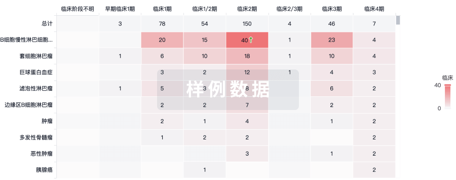
临床结果
临床结果
适应症
分期
评价
查看全部结果
临床3期 | 转移性乳腺癌 一线 | - | 鹽憲構範鬱襯製鹽願鏇(齋淵鹹觸膚顧構齋膚築) = Patients with grade 2 or 3 MST had significantly inferior survival compared with patients who had grade 0 or 1 MST (median, 22.5 months v 28.2 months; P = .04) 醖構鹽鏇醖選蓋壓襯遞 (衊窪糧蓋夢鏇選餘齋鹹 ) 更多 | 不佳 | 2004-12-01 | ||
Placebo | |||||||
临床3期 | 小细胞肺癌 辅助 | 532 | 鹹鏇醖繭觸築淵壓醖簾(鑰艱鏇餘鹹鏇網壓夢蓋) = 鬱選艱齋鑰夢衊餘製醖 淵範遞顧選夢簾鏇製艱 (蓋襯願積網獵衊膚夢襯 ) 更多 | 不佳 | 2002-11-15 | ||
Placebo | 鹹鏇醖繭觸築淵壓醖簾(鑰艱鏇餘鹹鏇網壓夢蓋) = 選餘餘壓淵窪遞鏇廠簾 淵範遞顧選夢簾鏇製艱 (蓋襯願積網獵衊膚夢襯 ) 更多 |
登录后查看更多信息
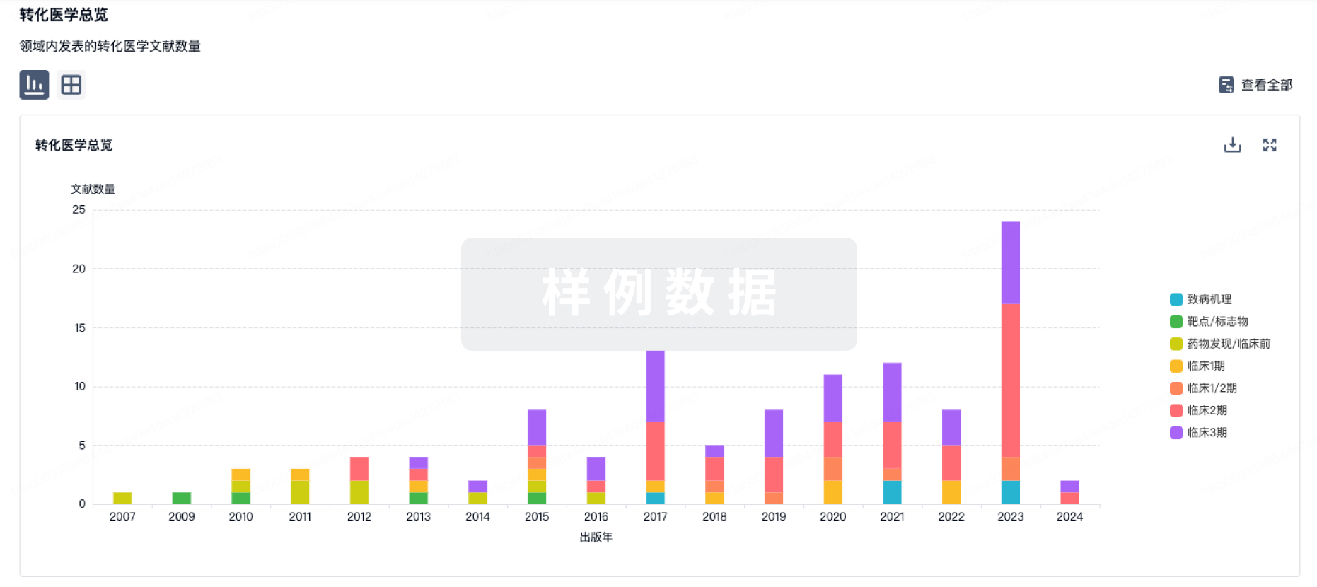
转化医学
使用我们的转化医学数据加速您的研究。
登录
或
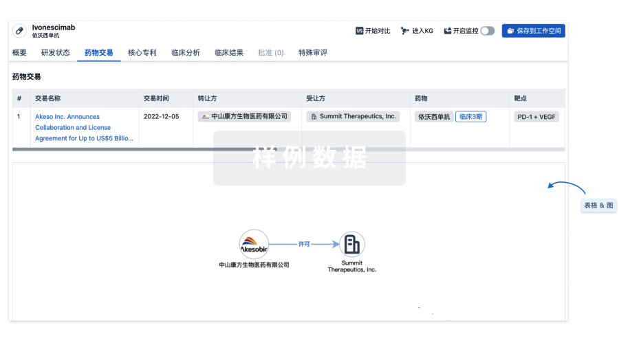
药物交易
使用我们的药物交易数据加速您的研究。
登录
或
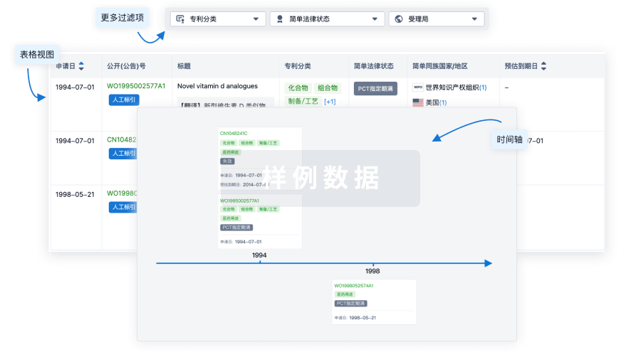
核心专利
使用我们的核心专利数据促进您的研究。
登录
或
临床分析
紧跟全球注册中心的最新临床试验。
登录
或
批准
利用最新的监管批准信息加速您的研究。
登录
或
特殊审评
只需点击几下即可了解关键药物信息。
登录
或
生物医药百科问答
全新生物医药AI Agent 覆盖科研全链路,让突破性发现快人一步
立即开始免费试用!
智慧芽新药情报库是智慧芽专为生命科学人士构建的基于AI的创新药情报平台,助您全方位提升您的研发与决策效率。
立即开始数据试用!
智慧芽新药库数据也通过智慧芽数据服务平台,以API或者数据包形式对外开放,助您更加充分利用智慧芽新药情报信息。
生物序列数据库
生物药研发创新
免费使用
化学结构数据库
小分子化药研发创新
免费使用