预约演示
更新于:2025-09-09
Tokyo Dental College
更新于:2025-09-09
概览
标签
神经系统疾病
其他疾病
内分泌与代谢疾病
小分子化药
疾病领域得分
一眼洞穿机构专注的疾病领域

技术平台
公司药物应用最多的技术
靶点
公司最常开发的靶点
关联
US20240041733
专利挖掘靶点 |
作用机制 |
在研机构 |
原研机构 |
在研适应症 |
非在研适应症 |
最高研发阶段 |
首次获批国家/地区 |
首次获批日期 |
JPRN-UMIN000051907
Visual function of eyes implanted with non-diffractive extended depth of focus intraocular lens - Visual function of non-diffractive EDOF IOL
JPRN-UMIN000051512
Comparison of the preventive effect of ondansetron and dexamethasone on nausea and vomiting after orthognathic surgery between remimazolam and propofol anesthesia. - Comparison of the preventive effect of ondansetron and dexamethasone on nausea and vomiting after orthognathic surgery between remimazolam and propofol anesthesia.
JPRN-UMIN000051727
Elucidation of gene polymorphisms that contribute to nausea and vomiting after administration of ondansetron - Elucidation of gene polymorphisms that contribute to nausea and vomiting after administration of ondansetron
100 项与 Tokyo Dental College 相关的临床结果
登录后查看更多信息
登录后查看更多信息
2025-12-31Journal of Oral Microbiology
Article
作者: Miura, Nobuaki ; Imamura, Kentaro ; Okuda, Shujiro ; Takahashi, Naoki ; Ono, Shigeru ; Yamazaki, Kazuhisa ; Sasaki, Nobuo ; Miyauchi, Eiji ; Mizuno, Kentaro ; Tsuboi, Yuuri ; Yamazaki, Kyoko ; Kikuchi, Jun ; Morita, Hidetoshi ; Ohno, Hiroshi ; Nakajima, Takako
Objectives:
Animal studies suggest that periodontopathic bacteria induce gut dysbiosis and related pathology, possibly connecting periodontitis to non-oral diseases. However, the effects on the gut ecosystem in periodontitis patients are not fully understood.
Methods:
We conducted a comprehensive analysis of the salivary and gut microbiota using 16S rRNA sequencing in periodontitis patients before and after treatment, comparing them to healthy participants. Serum metabolites were also analyzed.
Results:
Periodontitis patients showed high alpha diversity in both salivary and gut microbiota with a strong correlation. Significant differences were also observed in the gut microbiota composition between patients before treatment and healthy participants, irrespective of the ectopic colonization of periodontitis-associated bacteria in the gut. Co-abundance group analysis demonstrated that the gut microbiota of healthy participants was enriched with short-chain fatty acid producers. Changes in the gut microbiota coincided with alterations in the serum metabolite profile. While periodontal therapy improved salivary microbiota, it did not significantly affect gut microbiota.
Conclusions:
Gut dysbiosis of periodontitis patients may impact systemic metabolite profiles. Given that periodontal therapy alone did not substantially improve the gut microbiota, adjunctive strategies targeting the gut microbiome may be effective in reducing the risk of periodontitis-associated diseases.
2025-10-01AMERICAN JOURNAL OF OPHTHALMOLOGY
TFOS DEWS III Editorial
Review
作者: Craig, Jennifer P ; Chen, Wei ; Dogru, Murat ; Sullivan, David A ; Wolffsohn, James S ; Stapleton, Fiona ; Jones, Lyndon ; Perez, Victor L
2025-10-01ANNALS OF ANATOMY-ANATOMISCHER ANZEIGER
Article
作者: Murakami, Gen ; Abe, Shin-Ichi ; Yoshihashi, Yuki ; Miyamoto, Eri ; Kitamura, Kei ; Tanaka, Tomohito ; Hirano-Kawamoto, Ai
BACKGROUND:
Macrophages and interdigitating dendritic cells (DCs) are key professional antigen-presenting cells. However, DCs appear to be absent in healthy nasal mucosa, despite the extensive ciliated respiratory epithelium being highly exposed to various antigens.
METHODS:
Using histological specimens from 20 elderly cadavers, we examined the distribution of immunoreactive cells in the nasal vestibular skin, mucocutaneous junction, and ciliated mucosa. CD1a, CD83 and DC-SIGN were used as DC markers, with the latter two being typically employed in lymphatic tissue studies.
RESULTS:
Macrophages and CD8-positive lymphocytes were widely distributed throughout the subcutaneous and submucosal tissues at all epithelial depths. These cells were occasionally found embedded within both the mucocutaneous junction epithelium and basal layer of the mucosal epithelium. In contrast, CD4-positive lymphocytes were scarce across all examined sites. CD169-positive macrophages, considered the first-line gatekeepers in lymphatic tissues, were localized along deep vessels and glands. CD1a-positive DCs (Langerhans cells) were absent from both the cytokeratin 14-negative squamous epithelium and ciliary epithelium but were abundant in the basal layer of the cytokeratin 14-positive stratified squamous epithelium. CD1a-positive cells, which exhibit either a dendritic or round morphology, were occasionally scattered through the elastic fiber-rich subcutaneous tissue. A few DC-SIGN- or CD83-positive DCs were seen in glands and along deep vessels in subcutaneous and submucosal tissues CONCLUSION: Hair follicles at the nasal vestibule were likely accompanied by a cluster of CD1a-positive cells and CD8-positive lymphocytes. Macrophages, rather than DCs, were likely the primary antigen-presenting cells for CD8-positive lymphocytes in aged nasal respiratory mucosa.
100 项与 Tokyo Dental College 相关的药物交易
登录后查看更多信息
100 项与 Tokyo Dental College 相关的转化医学
登录后查看更多信息
组织架构
使用我们的机构树数据加速您的研究。
登录
或
管线布局
2026年07月04日管线快照
管线布局中药物为当前组织机构及其子机构作为药物机构进行统计,早期临床1期并入临床1期,临床1/2期并入临床2期,临床2/3期并入临床3期
药物发现
1
1
其他
登录后查看更多信息
当前项目
登录后查看更多信息
药物交易
使用我们的药物交易数据加速您的研究。
登录
或
转化医学
使用我们的转化医学数据加速您的研究。
登录
或
营收
使用 Synapse 探索超过 36 万个组织的财务状况。
登录
或
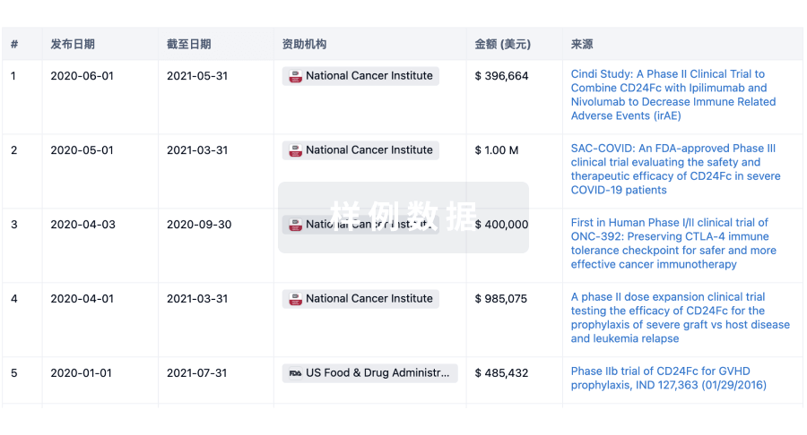
科研基金(NIH)
访问超过 200 万项资助和基金信息,以提升您的研究之旅。
登录
或
投资
深入了解从初创企业到成熟企业的最新公司投资动态。
登录
或
融资
发掘融资趋势以验证和推进您的投资机会。
登录
或
生物医药百科问答
全新生物医药AI Agent 覆盖科研全链路,让突破性发现快人一步
立即开始免费试用!
智慧芽新药情报库是智慧芽专为生命科学人士构建的基于AI的创新药情报平台,助您全方位提升您的研发与决策效率。
立即开始数据试用!
智慧芽新药库数据也通过智慧芽数据服务平台,以API或者数据包形式对外开放,助您更加充分利用智慧芽新药情报信息。
生物序列数据库
生物药研发创新
免费使用
化学结构数据库
小分子化药研发创新
免费使用