预约演示
更新于:2026-06-04

Nihon University
更新于:2026-06-04
概览
标签
内分泌与代谢疾病
神经系统疾病
其他疾病
蛋白水解靶向嵌合体(PROTAC)
小分子化药
预防性疫苗
疾病领域得分
一眼洞穿机构专注的疾病领域

技术平台
公司药物应用最多的技术
靶点
公司最常开发的靶点
关联
靶点 |
作用机制 |
在研机构 |
原研机构 |
在研适应症 |
非在研适应症 |
最高研发阶段 |
首次获批国家/地区 |
首次获批日期 |

靶点 |
作用机制 |
在研机构 |
原研机构 |
在研适应症 |
非在研适应症 |
最高研发阶段 |
首次获批国家/地区 |
首次获批日期 |


靶点 |
作用机制 |
在研机构 |
原研机构 |
在研适应症 |
非在研适应症 |
最高研发阶段 |
首次获批国家/地区 |
首次获批日期 |
JPRN-UMIN000060737
A Multicenter Observational Study of Pacemaker Implantation in Patients With Advanced Atrioventricular Block and Preserved or Moderately Reduced Left Ventricular Ejection Fraction (>35%) - REAL-AV Study
JPRN-UMIN000060021
Evaluation of Oral Function Improvement Through the Combined Use of Oral Motor Exercises and Eating/Drinking - Evaluation of Oral Function Improvement
JPRN-UMIN000058718
Efficacy of Comprehensive Therapy, Including Pharmacotherapy and Dietary/Lifestyle Guidance, on Renoprotection in Patients with Chronic Kidney Disease - Efficacy of Comprehensive Therapy in Patients with Chronic Kidney Disease
100 项与 Nihon University 相关的临床结果
登录后查看更多信息
登录后查看更多信息
2026-10-01PARASITOLOGY INTERNATIONAL
First record of the invasive American tapeworm Ophiotaenia saphena (Cestoda: Proteocephalidae) in the Tokyo daruma pond frog Pelophylax porosus porosus in Japan
Article
作者: Ishikawa, Takanori ; Sasaki, Mizuki
Adult proteocephalid tapeworms of the genus Ophiotaenia (Cestoda: Proteocephalidae) were recovered from the endemic Japanese amphibian species, Pelophylax porosus porosus (Amphibia: Ranidae) from Tochigi, Japan. As a result of molecular analyses based on the 28S ribosomal RNA and cytochrome c oxidase subunit I genes, these worms were identified as Ophiotaenia saphena, a species native to North America. Their morphological features were also consistent with those of O. saphena. This species is presumed to have been introduced into Japan with the American bullfrog, Lithobates catesbeianus (Ranidae). This study presents the first record of O. saphena in Japan and documents a new host record for this species.
2026-06-01Cannabis and Cannabinoid Research
How Has Japan’s Cannabis Control Act Been Amended?
Review
作者: Mikami, Ayako ; Masataka, Yuji ; Katayama, Munenori ; Umemura, Futaba ; Akino, Kozo ; Takumi, Ichiro ; Shibata, Kosuke ; Akahoshi, Yoshiyuki ; Nakazawa, Ryota ; Miki, Naoko ; Yoshida, Chikako ; Matsumoto, Toshihiko
Background::
In 2023, Japan’s Cannabis Control Act underwent its first major revision since its establishment in 1948. The legal framework surrounding cannabis had long remained rigid, with limited scope for medical or industrial applications.
Methods::
This review examines the content and implications of the 2023 legal amendments based on governmental documents, legislative records, and secondary analyses of regulatory shifts. The assessment focuses on three key domains: medical application, industrial use, and drug control.
Results::
Under the revised law, cannabis-derived products intended for medical use were brought under the same regulatory framework as opioid analgesics, theoretically enabling physicians to prescribe them. Simultaneously, the longstanding restriction limiting industrial use to mature stalks and seeds was lifted. However, this liberalization was counterbalanced by the introduction of a stringent THC threshold. On the criminal side, cannabis continues to be regulated as an illicit substance, and new penalties for use have been introduced. The revised law came into effect on December 12, 2024.
Conclusion::
The 2023 amendment represents a significant shift in Japan’s cannabis policy, aiming to balance expanded medical and industrial opportunities with continued drug control. Its practical implications remain to be seen and warrant close monitoring in the coming years.
2026-06-01BIOGERONTOLOGY
A gain-of-function screening reveals dAnkmy2 as a potential mediator of lifespan extension and oxidative stress resistance in Drosophila melanogaster
Article
作者: Aigaki, Toshiro ; Tsuda, Manabu ; Akita-Tanaka, Mihoko ; Matsuo, Takashi ; Togawa, Toru
Gain-of-function screening in Drosophila melanogaster provides a powerful approach for identifying genes that modulate lifespan; however, induction strength and environmental stress can substantially influence phenotypic outcomes. Here, we performed a pilot Gene Search (GS)-based overexpression screen using a heat-inducible hs-GAL4 driver and compared lifespan analyses at 25 °C and 30 °C to evaluate the impact of induction conditions on the detectability of lifespan-modulating genes. Induction at 30 °C caused uniformly shortened lifespans across genotypes and did not reveal robust lifespan-extending candidates. In contrast, screening at 25 °C, where moderate hs-GAL4 induction produces robust and detectable transgene expression, revealed multiple longevity-promoting lines. Lifespan measurements at the two temperatures were poorly correlated, indicating that elevated temperature imposes a dominant physiological burden that masks gene-specific effects. Using this strategy, we characterized a candidate line overexpressing Drosophila Ankyrin repeat and MYND domain-containing protein 2 (dAnkmy2). Overexpression of dAnkmy2 significantly extended adult lifespan and enhanced resistance to oxidative stress without detectable changes in canonical antioxidant gene expression. In contrast, loss of dAnkmy2 caused larval lethality, indicating an essential developmental function. Given the conserved role of Ankmy2 in ciliary biology, our results raise the possibility that cilia-associated processes may be involved in lifespan regulation. Collectively, this study establishes a proof-of-principle framework for detecting subtle genetic modulators of aging in genetically robust systems.
2025-12-15
并购抗体药物偶联物
2025-12-14
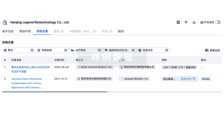
100 项与 Nihon University 相关的药物交易
登录后查看更多信息
100 项与 Nihon University 相关的转化医学
登录后查看更多信息
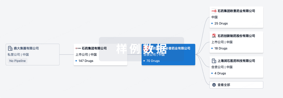
组织架构
使用我们的机构树数据加速您的研究。
登录
或
管线布局
2026年07月21日管线快照
管线布局中药物为当前组织机构及其子机构作为药物机构进行统计,早期临床1期并入临床1期,临床1/2期并入临床2期,临床2/3期并入临床3期
临床前
13
1
临床2期
登录后查看更多信息
当前项目
登录后查看更多信息
药物交易
使用我们的药物交易数据加速您的研究。
登录
或
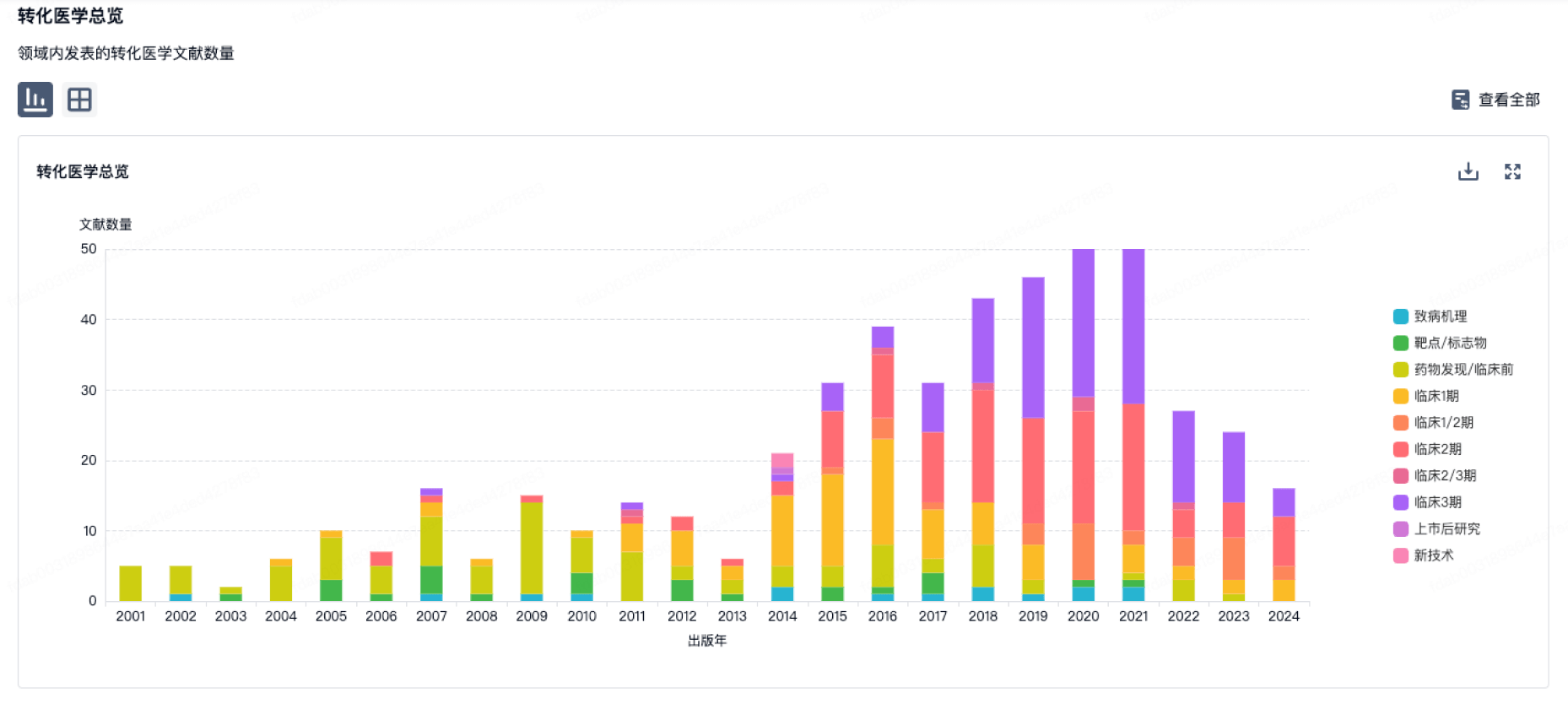
转化医学
使用我们的转化医学数据加速您的研究。
登录
或
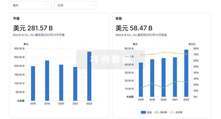
营收
使用 Synapse 探索超过 36 万个组织的财务状况。
登录
或
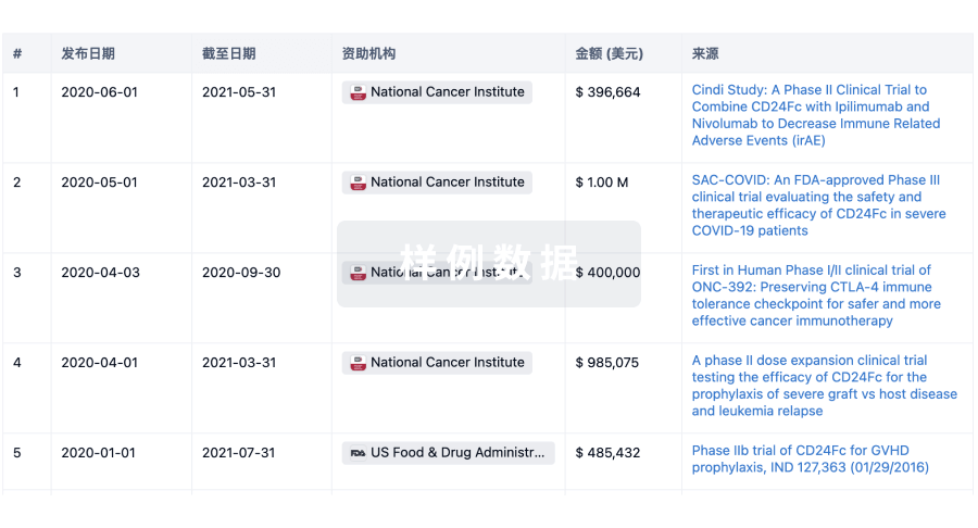
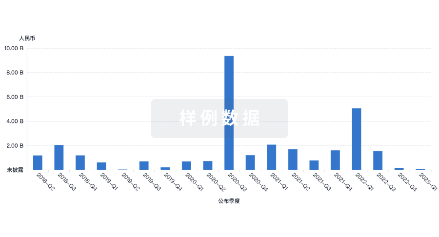
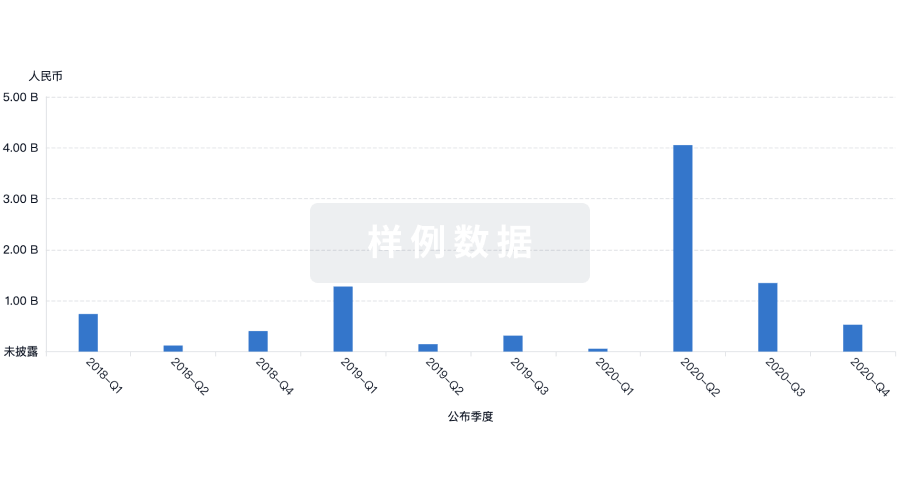
科研基金(NIH)
访问超过 200 万项资助和基金信息,以提升您的研究之旅。
登录
或
投资
深入了解从初创企业到成熟企业的最新公司投资动态。
登录
或
融资
发掘融资趋势以验证和推进您的投资机会。
登录
或
芽仔
全新生物医药AI Agent 覆盖科研全链路,让突破性发现快人一步
立即开始免费试用!
智慧芽新药情报库是智慧芽专为生命科学人士构建的基于AI的创新药情报平台,助您全方位提升您的研发与决策效率。
立即开始数据试用!
智慧芽新药库数据也通过智慧芽数据服务平台,以API或者数据包形式对外开放,助您更加充分利用智慧芽新药情报信息。
生物序列数据库
生物药研发创新
免费使用
化学结构数据库
小分子化药研发创新
免费使用