预约演示
更新于:2026-06-09

Jubilant DraxImage, Inc.
更新于:2026-06-09
概览
标签
其他疾病
呼吸系统疾病
泌尿生殖系统疾病
诊断用放射药物
小分子化药
核素偶联药物
疾病领域得分
一眼洞穿机构专注的疾病领域
技术平台
公司药物应用最多的技术
靶点
公司最常开发的靶点

暂无数据
关联
靶点 |
作用机制 |
在研机构 |
原研机构 |
在研适应症 |
非在研适应症 |
最高研发阶段 |
首次获批国家/地区 |
首次获批日期 |
靶点 |
作用机制 |
在研机构 |
原研机构 |
在研适应症 |
非在研适应症 |
最高研发阶段 |
首次获批国家/地区 |
首次获批日期 |
靶点 |
作用机制 |
在研机构 |
原研机构 |
在研适应症 |
非在研适应症 |
最高研发阶段 |
首次获批国家/地区 |
首次获批日期 |
NCT07261241
NANT 2021-02: A Randomized Phase 2 Study of 131I-MIBG With Vorinostat VS. 131I-MIBG With Dinutuximab vs. 131I-MIBG With Dinutuximab and Vorinostat for Relapsed or Refractory Neuroblastoma

NCT05037799
Optimization of Image Quality for Myocardial Perfusion Imaging With Rubidium-82 PET in Patients With Known or Suspected Ischemic Heart Disease (RUBY-DOSE)
NCT03561259
A Phase II, Open Label, Two-Arm Study of Therapeutic Iobenguane (131I) as Single Agent or in Combination With Vorinostat for Recurrent or Progressive High- Risk Neuroblastoma Subjects (OPTIMUM TRIAL)
100 项与 Jubilant DraxImage, Inc. 相关的临床结果
登录后查看更多信息
登录后查看更多信息
2023-11-01Seminars in nuclear medicine
The Past, Present, and Future Role of Artificial Intelligence in Ventilation/Perfusion Scintigraphy: A Systematic Review
Review
作者: Eugene Leung ; Amir Jabbarpour ; Grégoire Le Gal ; Ran Klein ; Siraj Ghassel ; Jochen Lang ; Eric Moulton
Ventilation-perfusion (V/Q) lung scans constitute one of the oldest nuclear medicine procedures, remain one of the few studies performed in the acute setting, and are amongst the few performed in the emergency setting. V/Q studies have witnessed a long fluctuation in adoption rates in parallel to continuous advances in image processing and computer vision techniques. This review provides an overview on the status of artificial intelligence (AI) in V/Q scintigraphy. To clearly assess the past, current, and future role of AI in V/Q scans, we conducted a systematic Ovid MEDLINE(R) literature search from 1946 to August 5, 2022 in addition to a manual search. The literature was reviewed and summarized in terms of methodologies and results for the various applications of AI to V/Q scans. The PRISMA guidelines were followed. Thirty-one publications fulfilled our search criteria and were grouped into two distinct categories: (1) disease diagnosis/detection (N = 22, 71.0%) and (2) cross-modality image translation into V/Q images (N = 9, 29.0%). Studies on disease diagnosis and detection relied heavily on shallow artificial neural networks for acute pulmonary embolism (PE) diagnosis and were primarily published between the mid-1990s and early 2000s. Recent applications almost exclusively regard image translation tasks from CT to ventilation or perfusion images with modern algorithms, such as convolutional neural networks, and were published between 2019 and 2022. AI research in V/Q scintigraphy for acute PE diagnosis in the mid-90s to early 2000s yielded promising results but has since been largely neglected and thus have yet to benefit from today's state-of-the art machine-learning techniques, such as deep neural networks. Recently, the main application of AI for V/Q has shifted towards generating synthetic ventilation and perfusion images from CT. There is therefore considerable potential to expand and modernize the use of real V/Q studies with state-of-the-art deep learning approaches, especially for workflow optimization and PE detection at both acute and chronic stages. We discuss future challenges and potential directions to compensate for the lag in this domain and enhance the value of this traditional nuclear medicine scan.
2020-07-01European Journal of Nuclear Medicine and Molecular Imaging
Radioaerosols and the updated EANM guideline in ventilation/perfusion imaging
Letter
作者: Fournier, France ; LaFrance, Norman
2026-06-01Journal of Nuclear Medicine Technology
Development and Evaluation of Automatic Pipeline for Patient-Specific Registration to a Bronchopulmonary Segment Atlas for Planar Perfusion Scintigraphy
Article
作者: Klein, Ran ; Ansari, Mohsen ; Lang, Jochen ; Jabbarpour, Amir ; Moulton, Eric
Relating planar lung scintigraphic image features to bronchopulmonary anatomy is a mental task requiring specialized medical experience. This study aimed to accurately normalize spatial data to overlay patient images onto a bronchopulmonary segment atlas (BSA), enhancing image interpretation for nonexperts and enabling quantification. Methods: This study evaluates the efficacy of 3 spatial normalization techniques: naïve registration, cost function masking, and perfusion defect removal with convolutional autoencoders. Autoencoders were trained for each of 6 projection angles using a large cohort of healthy patients (n = 660). Perfusion planar population templates for each projection, with its corresponding BSA, were constructed using a random subset sample of these patients (n = 149). Synthetic perfusion defects were applied on 60 projections from 10 patients with normal perfusion, allowing a comprehensive assessment of each spatial normalization technique's performance and effect on defect size in the template space. Results: The results reveal that autoencoder preprocessing significantly outperforms the naïve method and exhibits comparable or superior performance to cost function masking, particularly in preserving defect size and minimizing registration error to the population template within the defect. Visual comparisons further support the efficacy of autoencoder preprocessing in preserving anatomic features. Conclusion: Autoencoder preprocessing is a fully automatic and reliable method for reducing distortions during spatial normalization in perfusion scintigraphy, highlighting its potential for enhancing registration accuracy in clinical practice for BSA overlay and defect quantification.

100 项与 Jubilant DraxImage, Inc. 相关的药物交易
登录后查看更多信息
100 项与 Jubilant DraxImage, Inc. 相关的转化医学
登录后查看更多信息
组织架构
使用我们的机构树数据加速您的研究。
登录
或
管线布局
2026年07月21日管线快照
管线布局中药物为当前组织机构及其子机构作为药物机构进行统计,早期临床1期并入临床1期,临床1/2期并入临床2期,临床2/3期并入临床3期
批准上市
8
2
其他
登录后查看更多信息
当前项目
登录后查看更多信息
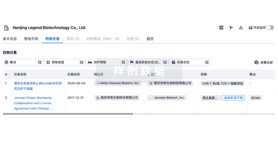
药物交易
使用我们的药物交易数据加速您的研究。
登录
或
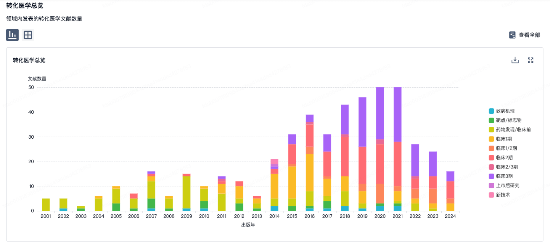
转化医学
使用我们的转化医学数据加速您的研究。
登录
或
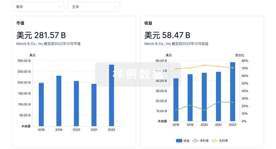
营收
使用 Synapse 探索超过 36 万个组织的财务状况。
登录
或
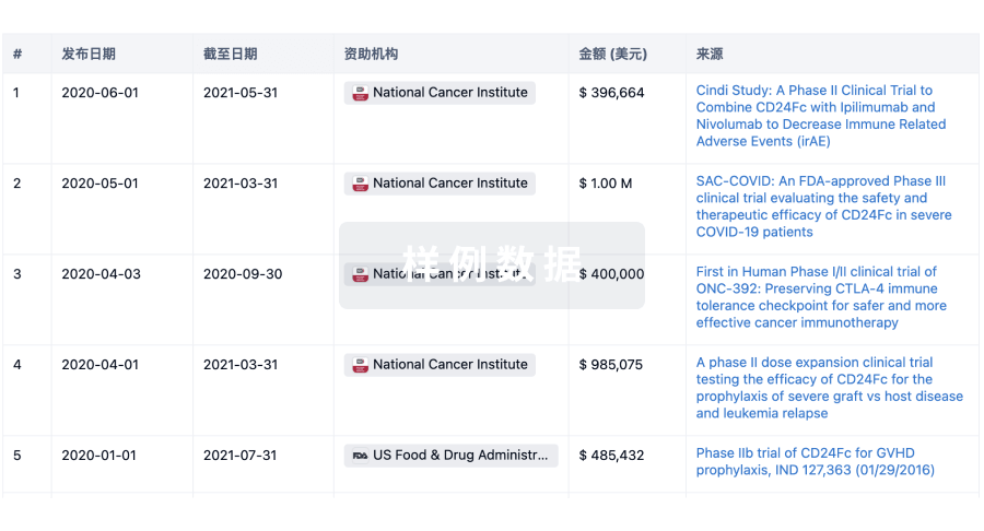
科研基金(NIH)
访问超过 200 万项资助和基金信息,以提升您的研究之旅。
登录
或
投资
深入了解从初创企业到成熟企业的最新公司投资动态。
登录
或
融资
发掘融资趋势以验证和推进您的投资机会。
登录
或
芽仔
全新生物医药AI Agent 覆盖科研全链路,让突破性发现快人一步
立即开始免费试用!
智慧芽新药情报库是智慧芽专为生命科学人士构建的基于AI的创新药情报平台,助您全方位提升您的研发与决策效率。
立即开始数据试用!
智慧芽新药库数据也通过智慧芽数据服务平台,以API或者数据包形式对外开放,助您更加充分利用智慧芽新药情报信息。
生物序列数据库
生物药研发创新
免费使用
化学结构数据库
小分子化药研发创新
免费使用