预约演示
更新于:2026-09-18
Mifamurtide
米伐木肽钠
更新于:2026-09-18
概要
基本信息
最高研发阶段批准上市 |
首次获批日期 欧盟 (2009-03-06), |
最高研发阶段(中国)- |
特殊审评- |






登录后查看时间轴
结构/序列
分子式C59H111N6NaO20P |
InChIKeyJSQUZKOPTKLMDG-MHTUOZSYSA-N |
CAS号838853-48-8 |
关联
12
项与 米伐木肽钠 相关的临床试验NCT07622836
A Prospective, Multicenter, Phase II Clinical Study of Postoperative Chemotherapy Combined With QL1706 for High-risk Triple-negative Breast Cancer.
This study is a prospective, multicenter, phase II clinical trial designed to evaluate the efficacy and safety of postoperative chemotherapy combined with QL1706 in patients with high-risk triple-negative breast cancer.
After enrollment, participants will receive 8 cycles of chemotherapy combined with QL1706. The standard chemotherapy regimen is the AC-T regimen (4 cycles of epirubicin plus cyclophosphamide, followed by 4 cycles of a taxane) - a Category I recommendation in the 2025 CSCO guidelines. The final choice of chemotherapy regimen is at the investigator's discretion. Starting from cycle 9, participants will receive QL1706 monotherapy as maintenance treatment. Dosing will continue until protocol-defined treatment discontinuation criteria are met, the participant experiences intolerable toxicity, or the participant withdraws informed consent. The maximum number of QL1706 dosing cycles is 17.
After completing treatment, participants will continue to undergo post-treatment safety follow-up and survival follow-up. For participants who discontinue treatment for reasons other than disease progression or death, tumor progression follow-up will also be conducted after treatment ends.
After enrollment, safety assessments will be performed every 3 weeks, and imaging evaluations will be performed every 12 weeks (±7 days) until confirmed disease progression per RECIST v1.1, initiation of another new anti-cancer therapy, withdrawal of informed consent, or death, whichever occurs first.
After enrollment, participants will receive 8 cycles of chemotherapy combined with QL1706. The standard chemotherapy regimen is the AC-T regimen (4 cycles of epirubicin plus cyclophosphamide, followed by 4 cycles of a taxane) - a Category I recommendation in the 2025 CSCO guidelines. The final choice of chemotherapy regimen is at the investigator's discretion. Starting from cycle 9, participants will receive QL1706 monotherapy as maintenance treatment. Dosing will continue until protocol-defined treatment discontinuation criteria are met, the participant experiences intolerable toxicity, or the participant withdraws informed consent. The maximum number of QL1706 dosing cycles is 17.
After completing treatment, participants will continue to undergo post-treatment safety follow-up and survival follow-up. For participants who discontinue treatment for reasons other than disease progression or death, tumor progression follow-up will also be conducted after treatment ends.
After enrollment, safety assessments will be performed every 3 weeks, and imaging evaluations will be performed every 12 weeks (±7 days) until confirmed disease progression per RECIST v1.1, initiation of another new anti-cancer therapy, withdrawal of informed consent, or death, whichever occurs first.
开始日期2026-06-05 |
申办/合作机构  常州市第二人民医院 常州市第二人民医院 [+1] |
NCT07787429
Evaluation of the Safety and Efficacy of Mifamurtide Versus Standard Treatment With Sorafenib in Patients With High-risk Osteosarcoma (DRAGONFLY)
Prospective, open, interventional, randomized, non-commercial trial
开始日期2026-04-01 |
申办/合作机构 |

ChiCTR2600120465
Efficacy and Mechanism of Camrelizumab in Combination with Risedronate and Chemotherapy for Triple-Negative Breast Cancer: An Exploratory Clinical Study
开始日期2026-03-04 |
申办/合作机构 |

100 项与 米伐木肽钠 相关的临床结果
登录后查看更多信息
100 项与 米伐木肽钠 相关的转化医学
登录后查看更多信息
100 项与 米伐木肽钠 相关的专利(医药)
登录后查看更多信息
144
项与 米伐木肽钠 相关的文献(医药)2026-06-01·JCO Precision Oncology
Prediction of Anthracycline Benefit in Hormone Receptor–Positive, Human Epidermal Growth Factor Receptor 2–Negative Early-Stage Breast Cancer by the MammaPrint 70-Gene Signature for Patients Enrolled in the FLEX Study
Article
作者: Giffoni, Danilo ; Layeequr Rahman, Rakhshanda ; Ramaswamy, Harshini ; Chmielewski-Stivers, Nicole ; Brufsky, Adam M. ; Audeh, William ; Gold, Linsey P. ; Graham, Cathy Lynne ; Weber, Raquel ; Menicucci, Andrea R. ; Johnson, Nathalie ; Alberty-Oller, J. Jaime ; Brown, Eric ; Elkhanany, Ahmed ; O'Shaughnessy, Joyce ; Osborne, Cynthia R. ; Mahtani, Reshma
PURPOSE:
Randomized trials have not demonstrated clear benefit from anthracyclines for patients with hormone receptor–positive, human epidermal growth factor receptor 2–negative (HER2−) early breast cancer (EBC). The MammaPrint 70-gene signature, specifically the High-Risk 1 (H1) and High-Risk 2 (H2) subgroups, was evaluated in predicting benefit from anthracycline- and taxane-based chemotherapy (AC-T) compared with taxane/cyclophosphamide (TC) in hormone receptor–positive HER2− EBC.
MATERIALS AND METHODS:
This analysis used prospectively collected real-world data from patients enrolled in the FLEX study with stage I-III hormone receptor–positive HER2− EBC, MammaPrint High-Risk, and BluePrint Luminal B tumors who received adjuvant AC-T or TC. Inverse probability of treatment weighting (IPTW) was performed to balance clinical characteristics between treatment groups. The association between the chemotherapy regimen and 3-year invasive disease-free survival (IDFS) was assessed using Kaplan-Meier estimates and Cox proportional hazards models.
RESULTS:
Among patients with H1 cancers (n = 1,106), 3-year IDFS did not differ between AC-T and TC (95.9%
v
95.9%; adjusted hazard ratio [HRs], 1.14 [95% CI, 0.64 to 2.06];
P
= .70). By contrast, patients with H2 cancers (n = 153) demonstrated significantly improved 3-year IDFS with AC-T versus TC (100%
v
94.8%; adjusted HRs, 0.10 [95% CI, 0.01 to 0.75]; absolute benefit 5.2%;
P
= .025). A significant treatment-by-MammaPrint interaction demonstrated that AC-T benefit increased with higher genomic risk (adjusted HRs, 0.09 [95% CI, 0.01 to 0.85];
P
= .036). Clinical and pathologic variables did not predict anthracycline benefit.
CONCLUSION:
Patients with MammaPrint H2 tumors had a clinically meaningful benefit from anthracycline-containing chemotherapy, whereas patients with H1 tumors did not derive more benefit from AC-T than from TC despite overlapping high-risk features. These findings support the use of MammaPrint to inform chemotherapy regimen selection and warrant further validation.
2026-04-10·JOURNAL OF CLINICAL ONCOLOGY
Reply to: Mifamurtide in Nonmetastatic High-Grade Osteosarcoma
Letter
作者: Broto, Javier Martín ; Ibrahim, Toni ; Serra, Massimo ; Palmerini, Emanuela
2026-01-01·Journal of clinical and experimental dentistry
Risk factors for dyseugia during chemotherapy in breast cancer women: A cohort STROBE-guided
Review
作者: de Barros Silva, Paulo Goberlânio ; Bastos de Moura, José Fernando ; Nóbrega Malta, Cássia Emanuella ; Barbosa, Jennifer Vianna ; Crispim, André Alves ; Aragão Matos Carlos, Anna Clara ; Gonzaga Silva, Lúcio Flávio ; Silva Marques Araújo, Ana Beatriz ; Fontes Borges, Marcela Maria
Background:
To evaluate the taste function of breast cancer patients undergoing Doxorubicin-Cyclophosphamide-Taxanes (AC-T) chemotherapy.
Material and Methods:
68 breast cancer patients treated with AC-T were objectively (taste function) and subjectively (scales) evaluated in each chemotherapy cycle. Quality-of-life (QoL), other side effects, clinical-pathological and sociodemographic data, hematological test, general health scores, and the Body Mass Index (BMI) were additionally evaluated. ANOVA-RM/Bonferroni, Friedman/Dunn, chi-square/Fisher's exact tests were applied (SPSS 20.0, p<0.05).
Results:
There was a reduction in gustatory sensitivity (p<0.001), salivary flow (p<0.001), periodontal health (p<0.001), and QoL (p<0.001) from the third AC-T cycle and an increase in all side effects. There was an association between low taste sensitivity and lower BMI (p=0.008) and general health scores (p=0.006). Low gustatory sensitivity events was directly associated with the number of AC-T cycles (p<0.001), reduced salivary flow (p=0.019), poorer QoL (p<0.05), and a higher incidence of nausea and anorexia (p<0.05). In addition, age (p<0.001) and trastuzumab (p<0.001) were potential risk factors for dysgeusia.
Conclusions:
The incidence of dysgeusia is high in breast cancer patients treated with AC-T; it is cycle-dependent and is associated with weight loss, poorer QoL, and impaired oral and general health. The mechanisms involved in the use of trastuzumab should be investigated.
27
项与 米伐木肽钠 相关的新闻(医药)2026-09-03
The osteosarcoma treatment pipeline is gradually expanding, with emerging therapies such as OST-HER2 (OS Therapies), DANYELZA (naxitamab-gqgk; SERB Pharmaceutical), Vactosertib (MedPacto), Risvutatug rezetecan (GSK), Olaparib with Ceralaserti (AstraZeneca), OMO-103 (Peptomyc), Eflornithine (DFMO) and AMXT 1501 (Aminex Therapeutics), PEEL-224 (Peel Therapeutics), ADCE-D01 (ADCendo), Dendritic cell-based vaccine (CellxLife), LNTH-2403 (Lantheus), and other investigational agents representing promising assets that could reshape the future treatment landscape, particularly in relapsed and metastatic settings.
LAS VEGAS, Sept. 3, 2026 /PRNewswire/ -- Osteosarcoma management generally relies on a multimodal treatment strategy that integrates surgery with chemotherapy and, in selected cases, radiotherapy. According to the NCCN Guidelines, commonly recommended chemotherapeutic agents include
methotrexate, doxorubicin, cisplatin, and ifosfamide. For patients with relapsed, refractory, or metastatic osteosarcoma, second-line options recommended by the NCCN include high-dose ifosfamide, etoposide, regorafenib, sorafenib, radium-223 dichloride, and everolimus.
Despite these treatment options, the osteosarcoma therapeutic landscape remains relatively limited, with few therapies specifically approved for the disease. In the United States,
levoleucovorin (KHAPZORY) has received FDA approval, whereas
mifamurtide (MEPACT) has been authorized by the European Commission for use across the European Union.
Find out the major differences between osteosarcoma vs Ewing sarcoma @
The emerging osteosarcoma pipeline is increasingly exploring targeted therapies and immunotherapeutic strategies. Key candidates include
OST-HER2, which employs HER2-directed cellular immunotherapy to stimulate antitumor immune responses;
naxitamab-gqgk (DANYELZA), which targets GD2-expressing tumor cells through antibody-mediated cytotoxicity;
Vactosertib, an inhibitor of TGF-β signaling associated with tumor progression and metastasis; and
Risvutatug Rezetecan, an antibody-drug conjugate (ADC) designed to deliver targeted cytotoxic activity to tumor cells.
However, osteosarcoma drug development remains predominantly concentrated in early-stage clinical trials, particularly Phase II studies. The limited number of late-stage trials restricts the availability of large, randomized datasets capable of demonstrating statistically significant improvements in overall survival, event-free survival, and durable response rates. Consequently, the lack of robust confirmatory evidence continues to present a major challenge to regulatory approval and contributes to differences in treatment availability across regions.
Aparna Thakur, an oncology expert at DelveInsight, said that increasing research efforts in targeted therapies, immunotherapies, tyrosine kinase inhibitors, and cell-based therapies are expanding the future therapeutic landscape and creating opportunities for innovation.
Reflecting ongoing therapeutic innovation and increasing unmet medical needs, DelveInsight estimates that the osteosarcoma market across the seven major markets, comprising the United States, EU4 (Germany, France, Italy, and Spain), the United Kingdom, and Japan, was valued at approximately
USD 60 million in 2025. The market is projected to expand at a
CAGR of 9% during the forecast period through 2036, driven by the development of novel targeted therapies, advances in multimodal treatment approaches, increasing investment in osteosarcoma research, and growing efforts to address treatment-resistant and relapsed disease.
Below, we highlight 8 promising emerging therapies poised to reshape the future of the osteosarcoma market.
OS Therapies' OST-HER2
HER2-targeted cancer vaccine
OST-HER2 is an investigational
HER2-targeted immunotherapy being developed for HER2-expressing malignancies, including osteosarcoma. The therapy is designed to stimulate an anti-tumor immune response by activating HER2-specific T cells, enabling them to recognize and eliminate cancer cells. Following intravenous administration, the OST-HER2 vector is rapidly taken up by antigen-presenting cells (APCs), which subsequently initiate a targeted immune response against HER2-positive tumors. Translational studies, including research in canine osteosarcoma, have demonstrated the therapy's potential to reduce disease recurrence and suppress metastatic progression. OST-HER2 is currently being evaluated as a second-line treatment in a Phase IIb clinical trial, with OS Therapies planning to pursue a Biologics License Application (BLA) under the U.S. FDA's
Accelerated Approval pathway in the second half of 2026.
In August 2026, OS Therapies, Inc. announced that the U.S. FDA had granted its request for a
Type C Statistical Methods meeting to discuss 2.5-year overall survival data from the Phase IIb trial evaluating OST-HER2 for recurrence prevention in patients with fully resected pulmonary metastatic osteosarcoma. The meeting is expected to take place in mid-September 2026 with representatives from the Center for Biologics Evaluation and Research (CBER), alongside a pending Biomarker Data Review Meeting with members of the Center for Drug Evaluation and Research (CDER) Biomarker Qualification Program (BQP). CBER is responsible for the regulatory review and approval of gene-edited products such as OST-HER2. The company also expects to report interim 3-year overall survival data in early September 2026.
Earlier, in March 2026, OS Therapies announced that the European Medicines Agency's (EMA) Committee for Advanced Therapies (CAT) had granted
Advanced Therapy Medicinal Product (ATMP) designation to OST-HER2 for the treatment of pulmonary recurrence in patients with resected osteosarcoma in the European Union. The designation followed the EMA's earlier
Fast Track Designation for the same indication. In the United States, OST-HER2 has also received both
Orphan Drug Designation and Fast Track Designation from the FDA for the treatment of osteosarcoma.
The osteosarcoma market is expected to evolve gradually, with OST-HER2 projected to emerge as a potential key asset, generating approximately USD 37 million across the 7MM by 2036, as per Thakur.
Iterion Therapeutics' Tegavivint
TBL1X protein inhibitors
Tegavivint targets
TBL1, an essential co-activator of Wnt-driven gene transcription, thereby inhibiting the oncogenic activity of β-catenin while potentially avoiding the toxicities associated with upstream Wnt inhibition. This differentiated mechanism supports its development as a targeted therapeutic approach. Tegavivint, in combination with gemcitabine, is being investigated in pediatric osteosarcoma and has received
Orphan Drug Designation (ODD) and Rare Pediatric Disease Designation (RPDD), with topline results anticipated in the second half of 2027.
In February 2026, Iterion Therapeutics announced the dosing of the first patient in a clinical study evaluating tegavivint in combination with gemcitabine in patients with relapsed or refractory osteosarcoma. The study is sponsored by Emory University and is being conducted at Children's Healthcare of Atlanta.
Discover more about the new treatment options for osteosarcoma @
Osteosarcoma Bone Cancer
SERB Pharmaceutical's DANYELZA
Antibody-dependent cell cytotoxicity (ADCC)
DANYELZA (naxitamab-gqgk) is a fully
humanized anti-GD2 monoclonal antibody developed to provide favorable pharmacokinetic characteristics and enhanced cytotoxic activity. Early clinical studies in pediatric patients have shown a manageable safety profile and encouraging therapeutic activity. However, its safety and tolerability in older patients with osteosarcoma remain insufficiently characterized. In September 2025, SERB Pharmaceuticals completed the acquisition of
Y-mAbs Therapeutics, broadening its rare oncology portfolio through the addition of DANYELZA and reinforcing its capabilities in developing targeted therapies for rare cancers.
Sadaf Javed, Functional Head of Forecasting and Analytics at DelveInsight, stated that the asset carries a "selective responder" profile rather than broad efficacy, with commercial potential constrained by small eligible populations and uncertain tolerability in older osteosarcoma patients. Overall, it is viewed as a niche orphan oncology product with modest expansion upside rather than a high-growth oncology franchise.
Aminex Therapeutics' AMXT 1501 + DFMO
Polyamine uptake inhibitors
Aminex Therapeutics' immuno-oncology candidate is an oral, small-molecule therapy that combines
AMXT 1501, the company's proprietary polyamine uptake inhibitor, with
DiFluoroMethylOrnithine (DFMO, eflornithine), an off-patent inhibitor of polyamine synthesis. By targeting complementary pathways, the AMXT 1501 and DFMO combination is designed to work synergistically to enhance immune-mediated antitumor activity across a broad range of solid tumors.
To know more about osteosarcoma drug development, visit @
Osteosarcoma Treatment Pipeline
MedPacto's Vactosertib
TGF-β1 kinase inhibitor
Vactosertib (TEW-7197) is a
TGF-β1 signaling inhibitor developed to modulate the tumor microenvironment. Although TGF-β signaling plays an important role in regulating cell proliferation and apoptosis under normal physiological conditions, its dysregulated activity in tumors can suppress antitumor immune responses, promote metastasis, and contribute to resistance to cancer therapies. TGF-β can also drive fibroblast proliferation within and surrounding tumor tissues, leading to excessive extracellular matrix deposition. This dense matrix can create a physical barrier around tumors, limiting immune-cell infiltration and reducing therapeutic access. By inhibiting stromal formation, vactosertib may improve the penetration of anticancer therapies and enhance their activity against tumor cells.
In January 2023, MedPacto announced that the U.S. FDA had granted
Fast Track Designation (FTD) to vactosertib as a monotherapy for patients with relapsed/refractory metastatic osteosarcoma. This followed the agency's grant of
Rare Pediatric Disease Designation (RPDD) for the same indication in September 2022. In July 2023, the European Commission granted
Orphan Drug Designation (ODD) to vactosertib for the treatment of osteosarcoma. The candidate had previously received
ODD from the U.S. FDA in August 2021.
According to Javed, vactosertib is a high-upside but still unproven platform asset, with valuation and outlook hinging on whether later-stage studies can confirm consistent response rates and clinically meaningful durability in a broader population.
GSK's Risvutatug rezetecan
ADC targeting B7-H3
Risvutatug rezetecan (Ris-Rez; GSK5764227; HS-20093) is an investigational
antibody-drug conjugate (ADC) designed to selectively target B7-H3, a cell-surface protein highly expressed in numerous solid tumors. By binding to B7-H3-positive cancer cells, the ADC facilitates the targeted delivery of its cytotoxic payload to tumor cells, with the aim of maximizing antitumor efficacy while limiting exposure to healthy tissues. The therapy is being evaluated across multiple tumor types under
GSK's global EMBOLD clinical development program. In 2025, the US FDA granted risvutatug rezetecan
Breakthrough Therapy Designation (BTD) for patients with late-line relapsed or refractory osteosarcoma, underscoring its potential as a treatment option for this challenging and underserved patient population.
Explore more about recent advancements in osteosarcoma treatments @
Osteosarcoma Treatment Market
Peel Therapeutics' PEEL-224
DNA topoisomerase I inhibitors
PEEL-224 is a polymer-based nanocarrier incorporating four small molecules and is being developed as a potential cancer therapy. Its active component is derived from camptothecin, a naturally occurring compound found in the Chinese Happy Tree, which is believed to have evolved as a plant defense mechanism.
Camptothecin and its derivatives target topoisomerase 1 (TOP1), an enzyme involved in DNA replication, leading to DNA damage and ultimately the death of rapidly dividing cancer cells. By modifying this naturally derived molecule and conjugating it to a synthetic polymer, PEEL-224 is designed to enhance therapeutic efficacy while potentially reducing toxicity.
Preclinical findings for PEEL-224 have been encouraging, with studies demonstrating sustained complete responses in some highly aggressive solid tumors, in certain cases lasting for more than six months. Importantly, the molecular design of PEEL-224 is intended to prevent its efflux from cancer cells through the ABCG2 multidrug transporter. This mechanism may help reduce the development of treatment resistance and potentially enable PEEL-224 to overcome resistance to irinotecan.
ADCendo's ADCE-D01
DNA topoisomerase I inhibitors
ADCE-D01 is a first-in-class
antibody-drug conjugate (ADC) designed to target uPARAP, an endocytic receptor that is highly expressed across a broad range of mesenchymal cancers, including soft tissue sarcoma, osteosarcoma, gastrointestinal stromal tumors (GIST), mesothelioma, and glioblastoma. uPARAP represents a promising ADC target due to its rapid internalization, constitutive recycling, and distinct expression pattern between tumor and healthy tissues. ADCE-D01 incorporates a uPARAP-binding antibody engineered to preserve the receptor's interaction with its natural ligand, collagen, together with a clinically validated protease-cleavable linker and a potent deruxtecan payload.
Preclinical studies have demonstrated robust antitumor activity across multiple in vivo models, along with an encouraging toxicology profile, supporting ADCE-D01's potential as a first- and best-in-class treatment option for patients with mesenchymal cancers characterized by significant unmet medical needs.
ADCE-D01 is currently being evaluated in a multicenter Phase I/II clinical trial (NCT06797999) to assess its safety, tolerability, and optimal dose in patients with soft tissue sarcoma. The study is being conducted at clinical trial sites across the United States and Europe. The U.S. FDA has granted ADCE-D01 both
Fast Track Designation and Orphan Drug Designation (ODD) for the treatment of soft tissue sarcoma.
Download the report to understand the top emerging therapies in osteosarcoma treatment @
Osteosarcoma Clinical Trials
Source:
Osteosarcoma Market Report
Osteosarcoma Market Insights, Epidemiology, and Market Forecast – 2036 report delivers an in-depth understanding of the disease, historical and forecasted epidemiology, as well as the market trends, market drivers, market barriers, and key osteosarcoma companies, including
OS Therapies, SERB Pharmaceutical, MedPacto, GSK, AstraZeneca, Peptomyc, Iovance Biotherapeutics, Aminex Therapeutics, US WorldMeds, Peel Therapeutics, CellxLife, Lantheus, ADCendo, Iterion Therapeutics, iOncologi, Lisata Therapeutics, and others.
Related Reports
Osteosarcoma Clinical Trial Analysis
Osteosarcoma
Pipeline Insight
–
2026 report provides comprehensive insights about the pipeline landscape, pipeline drug profiles, including clinical and non-clinical stage products, and the key osteosarcoma companies, including
Zentalis Pharmaceuticals, MedPacto, Salarius Pharmaceuticals, NextPoint Therapeutics, Base Therapeutics, EMD Serono, Hansoh BioMedical R&D Company, Cellectar Biosciences, OS Therapies, AlaMab Therapeutics
, and others.
Ewing Sarcoma Market
Ewing Sarcoma Market Insights, Epidemiology, and Market Forecast – 2036 report delivers an in-depth understanding of the disease, historical and forecasted epidemiology, as well as the market trends, market drivers, market barriers, and key Ewing sarcoma companies, including
Salarius Pharmaceuticals, Jazz Pharmaceuticals, Pharmamar, Eli Lilly, Pfizer, Bioatla, Cellectar Biosciences, Sumitomo Pharma Oncology, Inhibrx, and others.
Soft Tissue Sarcoma Market
Soft Tissue Sarcoma Market Insights, Epidemiology, and Market Forecast – 2036 report delivers an in-depth understanding of the disease, historical and forecasted epidemiology, as well as the market trends, market drivers, market barriers, and key STS companies, including
Advenchen Laboratories, Philogen, Gradalis, Epizyme, Chugai Pharma France, CytRx, Taiho Pharmaceuticals, KaryoPharm Therapeutics, Nanobiotix, Apexigen, Lytix Biopharma, Incyte Corporation, Iovance Biotherapeutics, Aadi Bioscience, Inc., AVEO Pharmaceuticals, Bayer, VasGene Therapeutics, Mirati Therapeutics, Novartis Pharmaceuticals, Incyte Corporation, Tracon Pharmaceuticals, Jiangsu Hengrui Medicine, Exelixis, Qbiotics, AstraZeneca, Loxo Oncology, ImmunityBio, Monopar Therapeutics, Chipscreen Biosciences, Ltd., Agenus, C4 Therapeutics, Inc., Noxopharm Limited, Moleculin Biotech, Inc., Sunshine Guojian Pharmaceutical (Shanghai) Co., Ltd., Tracon Pharmaceuticals Inc., Guangdong Xiangxue Precision Medical Technology Co., Ltd., Cornerstone Pharmaceuticals, Takara Bio Inc., Jazz Pharmaceuticals, Lyell Immunopharma, Telix Pharmaceuticals, and others.
Chondrosarcoma Market
Chondrosarcoma Market Insights, Epidemiology, and Market Forecast – 2036 report delivers an in-depth understanding of the disease, historical and forecasted epidemiology, as well as the market trends, market drivers, market barriers, and key chondrosarcoma companies, including
Aadi Bioscience Inc., Acrotech Biopharma LLC, ADC Therapeutics SA, Advenchen Laboratories LLC, Alkermes Plc, Astex Pharmaceuticals Inc., Atlanthera, Bayer AG, BioAtla Inc., BioMed, Valley Discoveries Inc., Bristol-Myers Squibb Co., Chia Tai Tianqing Pharmaceutical Group Co., Ltd., Eli Lilly and Co., ENB Therapeutics LLC, Epizyme Inc., Horizon Therapeutics Plc, Incyte Corp, Inhibrx Inc., Iovance Biotherapeutics Inc., Jiangsu Hengrui Medicine Co., Ltd., Karyopharm Therapeutics Inc., Les Laboratoires Servier SAS, Merck & Co., Inc., Nanode Therapeutics Inc., Nerviano Medical Sciences SRL, NKGen Biotech Inc., Novartis AG, Oncorus Inc., Ono Pharmaceutical Co. Ltd., Pfizer Inc., Pharma Mar SA, Rigel Pharmaceuticals Inc., Shanghai Affinity Biomedical Technology Co., Ltd, Shanghai De Novo Pharmatech Co., Ltd., Taiho Pharmaceutical Co., Ltd., and others.
About
DelveInsight
DelveInsight is a leading Business Consultant and Market Research firm focused exclusively on life sciences. It supports pharma companies by providing comprehensive end-to-end solutions to improve their performance. Get hassle-free access to all the healthcare and pharma market research reports through our subscription-based platform PharmDelve
.
Contact Us
Shruti Thakur
[email protected]
+14699457679
SOURCE DelveInsight Business Research, LLP
21%
more press release views with
Request a Demo
快速通道免疫疗法孤儿药临床2期
2026-08-20
5 抗转移治疗
NICOLE S. AMATO*Ethos Veterinary Health, Woburn, Massachusetts, USA杜宏超 ph.D 翻译自:Therapeutic Strategies in Veterinary Oncology引言
在大多数人类和兽医肿瘤患者中,癌细胞发生转移(metastasis)的能力是实体瘤相关发病与死亡的首要原因(Guan, 2015; Abu-Helil and van der Weyden, 2019)。传统上,恶性实体瘤的治疗思路是通过手术和/或放射治疗控制原发肿瘤,随后使用细胞毒性药物靶向微转移灶(micro metastases);或在复发时,采用类似的细胞毒性方案治疗大转移灶(macro metastatic lesions)(DeVita and Chu, 2008; Palumbo et al., 2013)。这种治疗策略在特定组织学类型的肿瘤中取得了可量化的获益;然而,进一步的临床突破不太可能再沿同一方向实现(Crawford, 2013; Anderson et al., 2019)。事实上,目前亟需新的药物研发策略与方案,以满足大多数肿瘤患者这一持续未被满足的需求(Khanna et al., 2014; Anderson et al., 2019)。具体而言,需要研发作用机制有别于现有药物的新型抗转移进展治疗药物(即靶向转移进展而非使显性肿瘤缩小的药物)(Khanna et al., 2014; Fontebasso and Dubinett, 2015)。
本章将探讨抗转移进展治疗的概念,重新审视经典的转移级联反应(metastatic cascade)、转移前微环境(premetastatic niche, PMN)、肿瘤休眠(dormancy)以及转移耐受(metastatic endurance)的概念(Stewart et al., 2019),以此理解潜在的疾病靶点/过程,为研发这些亟需的新型治疗药物提供依据。本章将综述这些潜在药物的基本分类及其研发面临的重要挑战,随后讨论比较肿瘤学(comparative oncology)作为降低这一高难度药物研发领域投资风险的手段。事实上,比较肿瘤学已被提出可作为解决所有物种抗转移治疗研究与开发中部分公认难题的潜在方案(Paoloni and Khanna, 2007; Gordon et al., 2009; Fan et al., 2020; van der Weyden et al., 2020)。最后,尽管本章重点关注转移进展的全身性预防与治疗,但也将探讨抗转移治疗与已形成可测量转移灶的治疗之间的交叉点,包括手术在已确立转移瘤管理中的潜在作用与风险,并以肺转移瘤切除术(pulmonary metastasectomy, PM)和淋巴结清扫与切除术为例(图5)。抗转移治疗:满足未被满足的临床需求
历史上,大多数细胞毒性抗肿瘤药物均基于其抑制细胞增殖或诱导凋亡的生物学活性而开发,而非针对转移的生物学机制(Gandalovičová et al., 2017; Falzone et al., 2018)。即使这类药物在缩小可测量肿瘤病灶方面效果显著,其在预防转移——肿瘤患者总体死亡的首要原因——方面的附加价值却十分有限(Falzone et al., 2018)。因此,当前大多数抗肿瘤药物的研发方向与肿瘤患者最迫切的需求并不匹配,患者最需要的是能够解决转移进展问题的药物(Khanna et al., 2014; Fontebasso and Dubinett, 2015; Anderson et al., 2019)。尽管如此,由于转移进展的一小部分过程确实涉及细胞增殖与凋亡逃逸,这种方向错位的研发方案也偶有活性迹象。例如,在患有局限性骨肉瘤(osteosarcoma, OSA)的人和犬中,辅助性使用细胞毒性化学治疗已被证实可通过延缓转移进展带来获益(Mauldin et al., 1988; Berg et al., 1992, 1995; Thompson and Fugent, 1992; Jafari et al., 2020; Marconato et al., 2021)。然而,有理由认为,针对更广泛转移进展生物学机制的药物研发努力,相比仅靶向可测量肿瘤形成与转移进展之间狭窄生物学重叠区(即凋亡与细胞增殖)的药物,将为患者带来更有意义的临床价值(Khanna et al., 2014)。
随着我们对转移进展生物学机制认识的不断深入,目前已有新的机会开发或重新定位多种治疗药物,使其可能特异性地靶向这一过程(Anderson et al., 2019)。例如,近年来免疫治疗在肿瘤治疗中的价值,正是源于其兼具缩小可测量肿瘤与靶向转移的双重作用(Borcoman et al., 2018)。类似的偶然性药物研发将缓慢地为存在转移风险的患者提供更多治疗选择;然而,直到最近,在人或动物中几乎没有针对性地、有目的地开发专门靶向转移进展生物学机制的抗转移药物(Lu et al., 2018; Koons et al., 2021)。与大量发生转移的肿瘤相比,该领域的进展相对匮乏,这推动了研究者去发现非细胞毒性的抗转移治疗药物,以抑制转移进展、改善患者预后,并使抗肿瘤药物研发与患者最重要的需求相一致。转移的难题
良性肿瘤通常生长缓慢且呈非侵袭性,往往可通过手术切除或放射治疗实现治愈性治疗(Talmadge and Fidler, 2010)。相比之下,恶性肿瘤生长相对较快,对周围组织呈现不同程度的侵袭,且并非总能通过手术切除和治愈性方案处理(Talmadge and Fidler, 2010)。这些肿瘤在组成上具有内在异质性,同时包含肿瘤细胞与非肿瘤细胞成分,共同促使特定转移细胞亚群产生转移倾向(Mendoza and Khanna, 2009)。
肿瘤转移从各个角度而言都是一项临床挑战。由于尚未完全阐明的原因,治疗可测量转移灶与仅治疗原发肿瘤相比具有独特性且难度更大(Qian et al., 2017)。目前尚不清楚转移灶是否本身对原发肿瘤有效的治疗方案(手术或放射治疗)具有耐药性,也不清楚某些转移性肿瘤是否能从类似的局部区域治疗中获益——即重新将手术及其他可测量肿瘤控制方案用于已确立的转移灶(Hellman and Weichselbaum, 1995; Weichselbaum and Hellman, 2011)。儿童骨肉瘤是一类特殊的肿瘤,其特点是相当比例的患者在发生肺转移后,通过肺转移瘤切除术可基本获得治愈(Croteau and Heaton, 2019)。目前尚不清楚但值得研究的是,这一策略在大转移灶形成后是否同样适用于其他类型的肿瘤,以及阐明可解释并预测转移瘤切除术在其他肿瘤中价值的生物学机制。
尽管存在上述已知的例外情况,但对于大多数肿瘤而言,治愈性治疗(手术和放射治疗)的价值与合理性在转移性疾病患者中均有所下降,且不再作为优先方案。因此,临床医师必须在更大程度上依赖全身性治疗。遗憾的是,无论是由于固有耐药还是获得性耐药,大多数大转移灶对当前的全身性治疗均无反应(Qian et al., 2017; Wang et al., 2019),预防转移进展对患者而言因而变得愈发重要。转移进展药物研发的挑战
显然,肿瘤转移与转移性进展问题是肿瘤患者最大的未满足需求。尽管这一事实显而易见,但靶向转移进展的药物需求与持续聚焦于研发靶向癌细胞增殖或诱导凋亡药物之间的错位依然存在(Fan et al., 2020)。这种方向错位在多个层面上都难以克服。首先,专门用于抗转移药物的临床前检测方法与药物筛选平台直到最近才得以开发和应用(Koons et al., 2021)。其次,即使这些药物得以开发并满足转移性肿瘤进展这一未被满足的需求,它们也不太可能成功缩小可测量肿瘤,因此难以顺利通过传统的人类早期临床试验(Sleeman and Steeg, 2010; Khanna et al., 2014; Anderson et al., 2019)。进而,如果这些药物在早期临床试验中失败,它们就不太可能被优先推进至后期临床试验(Anderson et al., 2019)——而靶向转移进展的抗肿瘤药物恰恰最有可能在后期试验中取得成功。因此,在传统的抗肿瘤药物研发模式下,最亟需的药物注定会失败。
从这一角度来看,未来要取得成功,必须发展替代性的抗肿瘤药物研发与测试方案(Khanna et al., 2014)。某些类型的人类肿瘤已经认识到这些药物研发挑战,并进行了相应调整。最值得注意的是,在儿童骨肉瘤中,近期设计并成功完成了可规避这些挑战的创新性II期临床试验(Bishop et al., 2016)。此外,利用犬骨肉瘤抗转移药物试验的平行数据来降低人类II期试验决策风险的机会已得到认可与支持,目前尚需进一步优化(Khanna et al., 2014)。靶向转移进展的历史方案
历史上,抗肿瘤药物研发走过了一条多少有些随意而迂回的道路,其中也涉及转移问题。这些努力取得的成功十分有限。事实上,由此产生的治疗药物虽具有抗转移活性,但属于偶然发现,而非有目的研发的结果。侵袭抑制剂
癌细胞获得侵袭表型(invasive phenotype)的能力被认为是恶性肿瘤的重要特征(Hanahan and Weinberg, 2011),且历来是肿瘤治疗的靶点。由于细胞水平的证据支持侵袭在转移中的作用,许多靶向细胞侵袭的早期药物研发项目相继启动,包括对基质金属蛋白酶(matrix metalloproteinase, MMP)抑制剂的研究(Martin and Matrisian, 2007)。尽管这类药物在临床前试验中初显前景,但由于诸多已明确的原因,其在II期和III期临床试验中表现不佳(Vandenbroucke and Libert, 2014)。回顾来看,基于基质侵袭本身并不足以独立促成转移这一新认知,如今可以预见这类药物为何无法有效靶向转移进展(Martin and Matrisian, 2007)。此外,临床试验设计也可能是失败原因之一:这些药物是在晚期、可测量的转移性疾病患者中进行测试的,而临床前试验考察的是在转移过程早期用药的效果(Vandenbroucke and Libert, 2014)。这类药物要取得成功,必须在侵袭发生之前而非确立之后给药;但由于休眠微转移灶很可能在当前给药方案开始治疗前早已存在,这一情景颇具挑战。
这类药物的失败确实为当前及未来旨在靶向并抑制转移进展的药物研发路径提供了重要教训。除了关于侵袭与肿瘤转移的认知不足之外,侵袭抑制剂的研发还凸显了一个难题:如何在细胞毒性药物传统的最大耐受剂量方案之外,合理确定这类药物的剂量与给药方案(Li et al., 2013)。此外,这类药物的失败也指向了识别最可能对治疗产生反应的患者亚群(即MMP靶点表达水平或活性)的必要性与挑战(Martin and Matrisian, 2007)。靶向侵袭药物研发的重新兴起需要解决这些挑战,并深化对转移级联反应中关键依赖环节的疾病靶点与生物标志物的认识。血管生成与抗血管生成治疗
一般而言,血管生成(angiogenesis)是指从已有血管形成新毛细血管的过程(Patan, 2004)。这一过程在出生后发育阶段很少发生,成年血管系统相对稳定,生理条件下极少增殖(Eelen et al., 2020)。然而在某些病理状态下,例如肿瘤中,确实会发生血管生成,部分原因是这些病理性扩增的肿瘤团块(原发或继发肿瘤)对氧气及生存所需营养物质的需求不断增加(Nishida et al., 2006; Muz et al., 2015)。
抗血管生成治疗作为一种肿瘤治疗策略,其作用不仅限于转移,因为抑制血管生成具有双重效应:既可使可测量肿瘤消退,又可能靶向转移进展(Cao, 2016)。早期以阻断血管内皮生长因子(vascular endothelial growth factor, VEGF)通路为核心的抗血管生成药物研发,采用了传统的药物开发路径——以人类II期临床试验中的肿瘤消退为终点,同时也出现了一些后续的创新性试验设计,尤其是在人类肾癌患者中(Vachhani and George, 2016)。这些努力促成了抗VEGF受体(VEGFR)抗体贝伐珠单抗(bevacizumab)最初在人类肿瘤患者中的获批(Montero et al., 2012)。随后,贝伐珠单抗作为一种预防转移进展的抗血管生成方案,获批用于肾癌及其他肿瘤(Kurkjian and Kim, 2012; Montero et al., 2012)。此后,通过小分子药物广泛靶向VEGFR激酶进一步推动了这种双重作用的转移进展治疗方案,即同时实现肿瘤应答与抑制转移进展(Vasudev and Reynolds, 2014)。血管靶向治疗
一旦肿瘤微环境中通过血管生成与血管发生形成血管,所产生的血管网络在形态学上(可能还包括功能上)便有别于正常血管网络(Konerding et al., 1995)。事实上,这种新生的肿瘤血管系统被发现是原始而紊乱的(Konerding et al., 1995; Teleanu et al., 2019),这也成为开发靶向并破坏已形成肿瘤血管的治疗方案的基础——因为这些血管对肿瘤的生长和进展至关重要(Siemann and Horsman, 2009)。血管破坏剂
与抗血管生成药物不同,血管破坏剂(vascular disruptive agents, VDAs)主要分为两大类:(i)基于配体的治疗,将毒素、促凝剂或促凋亡效应因子递送至肿瘤相关血管;以及(ii)小分子VDAs,可靶向纺锤丝——后者对内皮细胞功能及抗凋亡能力至关重要(Siemann and Horsman, 2009)。尽管这些治疗方案有别于大多数抗肿瘤药物,但它们并不被认为是特异性的抗转移药物,也不专门破坏转移进展过程。转移级联反应中的药物研发靶点
为了使肿瘤患者未被满足的需求与一条可借鉴历史成败经验的连贯药物研发路径更好地结合,需要进行多项创新。如前所述,由于转移进展与可测量肿瘤病灶都需要细胞增殖和凋亡逃逸,因此针对这些传统重视的肿瘤过程开发的药物,会存在狭窄的获益重叠区,可能同时作用于可测量肿瘤病灶与转移进展。尽管尚有许多未知,但我们已经明确哪些过程/依赖环节与转移进展相关(Fares et al., 2020)。鉴于传统抗肿瘤药物研发在提供有效预防转移进展的药物方面存在毋庸置疑的不足,亟需重新构想抗肿瘤药物研发模式,以开发靶向这些独特过程的新型药物(Gandalovičová et al., 2017)。
更具体地说,需要聚焦于转移级联反应中转移进展的依赖环节(Khanna et al., 2014; Anderson et al., 2019; Fan et al., 2020)。此处的"依赖环节"是指转移进展所必需或至关重要的靶点或过程。正如肿瘤侵袭抑制剂研发的失败教训所示,有必要对这些过程进行优先级排序靶向。
尽管传统和历史视角中的转移级联反应包含若干连续步骤(细胞动员、侵袭、内渗、脉管系统内转运、滞留、继发部位存活——属于转移耐受的一部分、进入/脱离转移休眠、外渗、血管生成以及最终大转移灶生长)(Fidler, 2003; Gupta and Massagué, 2006; Talmadge and Fidler, 2010; Anderson et al., 2019),但转移前微环境、转移休眠和转移耐受代表了转移进展中新的重要依赖环节,应成为未来抗肿瘤药物研发的重点。下文将对其进行更详细的概述。转移与转移前微环境
尽管曾认为转移的第一步是肿瘤细胞进入循环系统,但目前假设特定的原发肿瘤细胞可直接参与为未来的继发部位转移微环境做好准备,以迎接播散性转移细胞的最终扩散与后续到达(Peinado et al., 2011, 2017)。这种微环境被称为"转移前微环境"(premetastatic niche, PMN),定义为继发器官内由原发肿瘤特异性诱导形成、使转移得以发生并成功定植的微环境(Peinado et al., 2011, 2017)。PMN由Kaplan等人于2005年首次描述,是对Steven Paget于1889年提出的"种子与土壤"假说的拓展,该假说旨在解释转移固有的器官趋向性(Ribatti et al., 2006)。在Kaplan模型中,PMN由原发肿瘤来源的成分诱导形成,这些成分动员骨髓来源细胞(bone-marrow-derived cells, BMDCs)以及宿主微环境的局部基质成分,从而增加转移瘤细胞在继发微环境特定解剖区域内存活的概率(Kaplan et al., 2005; Liu and Cao, 2016)。此外,下文所述的转移耐受概念可能与PMN密切相关,其核心是使转移细胞能够在继发转移部位适应并存活(Liu and Cao, 2016)。
尽管对PMN生物学机制的详细评估超出本章范围,但其加入我们对转移生物学与分子事件日益增长的认识之中,为有目的的、针对转移特异性的药物研发方案提供了更多疾病靶点。转移休眠
在构成转移级联反应的多步骤过程中,众所周知只有极少数播散肿瘤细胞(disseminating tumor cells, DTCs)获得必要的遗传和表观遗传改变,从而在不同状态/阶段间转换(Neophytou et al., 2019)。在那些成功穿越转移级联反应的少数DTC中,部分细胞可到达并定植于继发部位,但不发生增殖(Sosa et al., 2014)。相反,这些细胞可能保持静止的休眠状态(Hadfield, 1954; Aguirre-Ghiso, 2007; Goddard et al., 2018; Neophytou et al., 2019)。这些休眠细胞可以是单个肿瘤细胞,也可以是处于增殖与凋亡平衡状态的微转移细胞簇(Hadfield, 1954; Holmgren et al., 1995; Aguirre-Ghiso, 2007; Goddard et al., 2018)。
一般认为,转移灶的形成源于这些微观细胞的失控生长——这些细胞可能潜伏在继发转移部位(如肺)或暂时的"庇护"部位(如骨髓),等待未来某个时间点被激活而形成显性转移灶(Tsao et al., 1997; Karrison et al., 1999; Aguirre-Ghiso, 2007; Goddard et al., 2018; Neophytou et al., 2019)。这些休眠细胞可能因肿瘤微环境中尚不明确的信号而开始分裂,最终在患者初次治疗后数月、数年甚至数十年形成可测量的病灶(McNichols et al., 1981; Tsao et al., 1997; Karrison et al., 1999; Freedland and Moul, 2007)。
如前所述,休眠在转移进展和肿瘤治疗中的问题具有双重性。首先,处于此阶段的癌细胞无法通过传统诊断手段检测到(Sauer et al., 2021)。其次,由于这些DTC基本处于非增殖状态,它们能够逃避传统化学治疗的细胞毒性作用(Sosa et al., 2014; Sauer et al., 2021)。针对维持休眠或打破休眠过程的药物研发,可能成为肿瘤患者极具价值的辅助治疗手段(Neophytou et al., 2019)。此外,靶向继发部位的DTC可能提供一种延缓或预防转移、或降低复发频率的途径(Barkan et al., 2010; Neophytou et al., 2019)。转移耐受
独立于休眠过程或作为休眠过程的一部分,成功的DTC必须能够适应并承受继发部位新微环境中的各种应激因素。据推测,细胞到达后所面临的应激条件需要高效的应激检测机制,以及随后的早期应激适应机制与程序,从而实现细胞存活、休眠和/或整体转移进展(Stewart et al., 2019)。转移耐受(metastatic endurance)这一概念描述了应激适应、伴或不伴休眠的存活的组合,这是能够成功定植于继发转移部位并形成显性转移的DTC的特征(Stewart et al., 2019)。
在多种肿瘤模型中,转移耐受已被证实是肿瘤转移的一个依赖环节,被认为是高转移细胞持续表现的一种表型——使这些细胞能够适应其陌生的微环境("非容许性土壤"),并在被称为"转移低效"的早期损耗中存活下来。这些细胞需要能够感知多种尚不明确的细胞应激因素,从而得以存活并进展;而不表达转移耐受表型的转移细胞则会死亡,不会造成临床问题。
近期关于转移耐受生物学机制的观点还包括异质性转移细胞群体的协作——这些细胞可基于表型多样性相互配合,实现应激适应与存活(Schild et al., 2018)。这一假说最近在骨肉瘤中得到了最好的阐释,即"锚定细胞"(anchor cells)的发现——这类细胞被描述为低增殖、分泌细胞因子的细胞,能够在转移早期阶段抵御早期损耗并促进转移进展(Reinecke et al., 2020)。通过分泌细胞因子,这些细胞能够改变周围肺组织,从而促进其他具有增殖表型的OSA肿瘤细胞(即"生长细胞")随后定植。这些生长细胞继而分裂形成可测量且具有临床意义的转移灶(Reinecke et al., 2020)。靶向转移耐受
尽管转移耐受的细胞过程在过去二十年间已得到认识和研究,其潜在生物学机制的图景也开始浮现,但它们尚未被广泛整合到抗肿瘤药物的发现与开发工作中。除了各种相关通路与网络中基因和蛋白质结构及活性的异常之外,还有证据支持表观遗传水平对转移耐受的调控,且数据支持靶向表观遗传调控机制("转移超级增强子")以预防转移进展(Yokoyama et al., 2016; Morrow et al., 2018; Peng et al., 2019)。考虑到这些因素,靶向转移耐受有理由发现新的或可重新定位的药物,在预防转移性疾病进展方面具有价值。转移研究中的临床前模型
可以理解的是,DTC与宿主细胞之间复杂的相互作用使得转移的临床前研究颇具挑战(Sauer et al., 2021)。由于这一及其他原因,利用传统临床前肿瘤模型简单地筛选具有潜在抗转移活性的药物并非易事(Sauer et al., 2021)。这一不足进一步限制了研发与患者最大需求相一致的药物(即靶向转移进展的药物)的机会(Khanna et al., 2014),也推动了更多新型药物研发策略的出现。自然发生的犬肿瘤:降低靶向转移进展药物研发风险的模型
考虑到新型靶向转移进展药物研发中存在的挑战,比较肿瘤学作为一门新兴学科,为规避成功进行转移药物研发所需的部分临床前建模难题提供了机会(LeBlanc and Mazcko, 2020)。
当关于新药研发的关键问题在人类临床试验或传统临床前模型中仍未得到解答或无法解答时,比较肿瘤学在人类药物研发路径中发挥了最大作用(Gordon et al., 2009; LeBlanc and Mazcko, 2020; van der Weyden et al., 2020)。
由于无法回答这些关键问题所带来的成本与后果,在宠物犬自然发生的、生物学特征相似的复杂肿瘤疾病中解答这些问题具有巨大价值(Gordon et al., 2009; LeBlanc et al., 2016; LeBlanc and Mazcko, 2020; van der Weyden et al., 2020)。
事实上,比较肿瘤学领域最大的价值或许在于能够相对快速而可靠地评估抗转移治疗药物的潜在活性,这些发现可从伴侣动物转化应用于人类转移进展的治疗(Gordon et al., 2009; LeBlanc et al., 2016; LeBlanc and Mazcko, 2020)。例如,某药物在犬中抑制肿瘤转移的独特活性,可结合传统或创新性人类II期数据,以优先筛选出可进入进一步人类II期或III期研究的药物。比较肿瘤学在抗肿瘤药物研发中的机遇与应用已在其他文献中被广泛综述;但值得注意的是,这种跨物种评估的前景已在犬骨肉瘤中得到积极考量与推进(Khanna et al., 2014)。
近年来,动物健康生物技术与制药行业对兽医肿瘤学的兴趣日益增长,为比较肿瘤学的自筹资金双向模式以及新型跨物种药物研发创造了机会——既可以解答人类药物研发的关键问题,同时又能创造收入和数据,以支持从人类制药企业授权引进、用于兽医审批的兽药(LeBlanc et al., 2016; Fenger et al., 2020)。这种比较肿瘤学的药物研发路径并非旨在取代传统策略,而是补充和强化这一重要工作的核心价值。抗转移治疗药物靶向转移进展的治疗方案将是什么样?
如前所述,尽管人们已经认识到转移和转移进展对大多数实体瘤患者而言是重大问题,但目前在人类或动物中,尚无可特异性靶向已知转移进展必需的丰富生物学机制的抗肿瘤药物。目前观察到的抗肿瘤药物抗转移进展活性,源于细胞增殖与凋亡逃逸这一狭窄的重叠区(上文已述)——这是可测量肿瘤与转移级联反应相关步骤的共同特征。成功研发出特异性靶向转移进展生物学机制的药物,有望为患者预后带来更有意义的改善。
一旦研发成功,预期的有效抗转移药物将具备口服生物利用度高且耐受性极佳的特点。这些新药很可能最适合长期给药,从新辅助治疗期开始,与辅助治疗同步进行,并在化学治疗结束后持续延长用药时间。
在符合上述描述的药物研发并上市之前,以下几种互不排斥的转移管理方案将更快得到应用:重新定位药物:取自肿瘤领域及非肿瘤治疗领域、但经表型转移药物筛选或基于与转移过程匹配的作用机制而被重新定位用于转移治疗的药物(如雷帕霉素(rapamycin))(Zhang et al., 2020)。免疫肿瘤药物:历史上认为免疫治疗药物在微小残留病背景下效用最大(如脂质体包裹胞壁酰三肽磷脂酰乙醇胺(liposome-encapsulated muramyl tripeptide-phosphatidylethanolamine, L-MTP-PE));然而,新型免疫治疗药物(如免疫检查点抑制剂;参见Mason and Dow,本卷第8章,2023)对可测量肿瘤出人意料的活性,使其有机会像其他多效药物一样用作抗转移药物(Robert, 2020)。多效药物:具有多种作用机制的抗肿瘤药物,首先在可测量肿瘤中取得成功,同时由于作用机制与转移存在重叠而展现出抗转移活性(如索拉非尼(sorafenib)及其他多靶点受体酪氨酸激酶(receptor tyrosine kinase, RTK)抑制剂)(Petrelli and Giordano, 2008)。
许多具有抗转移进展活性的多效药物与传统化学治疗相比,作用机制截然不同(Zhong et al., 2021)。传统化学治疗主要通过破坏细胞增殖或诱导凋亡发挥作用,而大多数多效药物是分子信号抑制剂,且常被矛盾地称为"靶向治疗"。细胞信号抑制为这些药物带来多效活性的原因包括:受抑制的细胞通路(如mTOR与雷帕霉素)产生多种生物学效应(Petrelli and Giordano, 2008);且通常"混杂型"抑制剂(如托西尼布(toceranib))可抑制多个靶点(如托西尼布可抑制c-KIT、VEGFR和血小板衍生生长因子(platelet-derived growth factor, PDGF)受体(PDGFR))(Pleyer and Greil, 2015)。
目前大多数受关注的靶向治疗要么是信号转导小分子抑制剂,要么是单克隆抗体。大量小分子抑制剂和抗体已获批用于人类肿瘤治疗(Shahid et al., 2019; Zhong et al., 2021),在兽医患者中也正处于研发或已获批阶段(Beltrán Hernández et al., 2021)。肿瘤中的多效靶向细胞信号通路
如上所述,已发现实体瘤生长、进展和转移涉及的众多过程均由生长因子信号通路异常介导(参见Rönnberg,本卷第6章,2023)。
自信号靶点最初被认定为可成药靶点以来,其在肿瘤中的成药资格不断演变。显然,靶点表达或过表达可能不足以构成理想的肿瘤靶点;如果该靶点发生突变并驱动肿瘤表型,则肿瘤更可能对该信号蛋白产生成瘾性或依赖性(Torti and Trusolino, 2011)。靶点资格认证的复杂性超出本章范围,且已有大量综述。特定受体酪氨酸激酶小分子抑制剂
RTK参与涉及蛋白激酶的信号通路,这些激酶利用ATP使自身及其他蛋白发生磷酸化(Zwick et al., 2002; Du and Lovly, 2018)。RTK及其抑制剂在肿瘤学中的重要性详见Rönnberg(本卷第6章,2023);但由于假设某些特定药物可能靶向转移进展,本章将对其进行介绍。托西尼布
托西尼布(toceranib)是RTK小分子抑制剂的典型代表(Palladia; Pfizer, New York, NY, USA),获批用于犬肥大细胞瘤的治疗。托西尼布是一种口服多激酶抑制剂,属于分裂型酪氨酸激酶家族,可阻断多种RTK(VEGFR-2、PDGFRα和c-KIT)。通过可逆性竞争抑制ATP结合位点,托西尼布可抑制受体磷酸化及后续多种重要肿瘤相关信号蛋白的下游信号传导(London et al., 2003, 2009, 2012; Yancey et al., 2010a,b)。单一抑制剂对RTK的这种多效靶向及其靶点谱(同时涵盖VEGFR和PDGFR),成为考虑该药物可能具有抗转移进展活性的基础(London et al., 2003, 2012)。
尽管托西尼布的适应症为肥大细胞瘤治疗,但上述多效性提供了生物学理论依据,并可能解释其在其他类型肿瘤(包括甲状腺癌、肛门囊顶泌腺癌、鼻腔癌、胃肠道间质瘤(gastrointestinal stromal tumor, GIST)及其他肿瘤)中观察到的生物学活性证据,既可用于可测量疾病,也可用于微转移场景(London et al., 2012)。
尽管如此,这种多效性可能通过逃逸性耐药(evasive resistance)的发生而给患者带来潜在风险(Halsey et al., 2014)。逃逸性耐药是指持续抑制VEGFR后转移进展加速的现象(Halsey et al., 2014),通常伴随可测量肿瘤的应答(Casanovas et al., 2005)。一般认为逃逸性耐药由丝裂原活化蛋白激酶(mitogen-activated protein kinase, MAPK)激活介导,且在不由所用多效激酶抑制剂阻断靶点驱动的肿瘤中危害最大(Haibe et al., 2020)。索拉非尼
体内外研究表明,合成性口服多激酶抑制剂索拉非尼通过抗增殖、抗血管生成和促凋亡作用,抑制肿瘤生长并破坏肿瘤微血管(Gollob et al., 2006; Wilhelm et al., 2008)。已发现索拉非尼可靶向RAF丝氨酸/苏氨酸激酶(RAF-1、野生型BRAF和致癌性b-raf V600E)以及与肿瘤发生、肿瘤进展和转移相关的RTK(VEGFR-1、VEGFR-2、VEGFR-3、PDGFRβ、FLT-3和c-KIT)(Wilhelm et al., 2008)。索拉非尼还可能通过非MEK/ERK依赖机制诱导凋亡(Liu et al., 2006; Wilhelm et al., 2008)。在异种移植模型(包括肾癌模型)中,索拉非尼可抑制肿瘤血管生成(Chang et al., 2007);在人类黑色素瘤、肾癌、结肠癌、胰腺癌、肝细胞癌、甲状腺癌、卵巢癌以及非小细胞肺癌(non-small-cell lung cancer, NSCLC)的临床前模型中,索拉非尼可抑制肿瘤生长(Wilhelm et al., 2008)。VEGFR与MEK/ERK双重抑制的结果提示,与不具备这种特定双重活性的药物相比,索拉非尼具有潜在优势,因此它可能通过避免逃逸性耐药介导的转移加速(如上所述)而提供更强的抗转移活性。
早期临床前研究已在犬体外和体内模型中考察了索拉非尼(Wolfesberger et al., 2010)。近期,有研究评估了索拉非尼在犬中使用的安全性,该耐受性研究确定了可用于后续药代动力学研究的剂量(Foskett et al., 2017; Cawley et al., 2022)。非受体酪氨酸激酶小分子抑制剂
尽管大量研究聚焦于RTK及其配体,但非RTK靶点同样不容忽视。细胞内非受体酪氨酸激酶Src及Src家族激酶(Src family kinases, SFKs)其他成员的潜在治疗价值已在人类中得到广泛研究,且可能在犬中也具有活性。
Src是一种蛋白激酶,属于目前称为"Src家族激酶"(SFKs)的一大类结构相关激酶(Thomas and Brugge, 1997)。Src及其他SFK在多种肿瘤中调控细胞表面受体信号方面发挥重要作用(Thomas and Brugge, 1997)。已知这些机制驱动许多基本细胞过程,包括细胞生长、分化、细胞形态、迁移和存活,以及特化的细胞信号(Parsons and Parsons, 2004)。研究还表明,Src是多种RTK下游信号的汇聚点(Parsons and Parsons, 2004)。在表皮生长因子受体(epidermal growth factor receptor, EGFR)的研究中,发现SFK可与EGFR发生物理相互作用,且这种相互作用对受体的完全激活至关重要(Biscardi et al., 1999; Citri and Yarden, 2006)。SFK与其他RTK(包括PDGFR、成纤维细胞生长因子受体(fibroblast growth factor receptor, FGFR)和胰岛素样生长因子1受体(insulin-like growth factor 1 receptor, IGF-1R))之间也存在类似现象(Thomas and Brugge, 1997)。因此,使用破坏这些RTK-Src相互作用的小分子抑制剂进行治疗,可能比单独靶向其中任一者更有效地实现抗转移策略(Zhang and Yu, 2012)。达沙替尼
达沙替尼(dasatinib)是另一种多靶点酪氨酸激酶抑制剂,作用靶点包括SFK(SRC、LCK、YES和FYN)、BCR-ABL,以及程度较低的c-KIT、PDGFRβ和EPHA2(Steinberg, 2007)。研究表明,达沙替尼能够同时阻断PDGFRβ和c-KIT的信号传导,因此与伊马替尼(imatinib)相比,其靶向的酪氨酸激酶数量更多(Lombardo et al., 2004)。其他研究考察了RTK与Src之间的协同作用,提示达沙替尼的使用可能提供一种克服获得性耐药的途径——而获得性耐药在大多数单药小分子抑制剂治疗中均有出现(Wheeler et al., 2009)。
尽管达沙替尼已在多种人类肿瘤中进行了研究(Kantarjian et al., 2010, 2012; Benjamini et al., 2014; Safdari et al., 2014; Spreafico et al., 2014),但其在兽医物种中的临床治疗价值尚不十分明确。然而,越来越多的证据支持其作为新型小分子抑制剂用于某些犬肿瘤(Dickerson et al., 2013; Marley et al., 2015)。
在一项考察伊马替尼和达沙替尼对犬血管肉瘤作用的体外研究中(Dickerson et al., 2013),达沙替尼对犬血管肉瘤细胞体外增殖的抑制作用比伊马替尼强数个数量级,且抑制PDGFRβ磷酸化所需的浓度约为伊马替尼的千分之一。另一项研究指出,达沙替尼可在体外抑制培养的犬恶性肥大细胞生长(Gleixner et al., 2007),提示了另一潜在用途。雷帕霉素
雷帕霉素(rapamycin)最初用作移植患者的免疫抑制剂,现已被重新定位用途。多项证据支持将其开发为抗肿瘤药物,且近年来被用于治疗犬和人类肉瘤的转移进展(Gordon et al., 2008; Paoloni et al., 2010; Zhou et al., 2010; Vemulapalli et al., 2011; Xie et al., 2013; Wang et al., 2014)。雷帕霉素是mTOR的抑制剂,后者是一种丝氨酸/苏氨酸激酶,参与多种下游激酶的信号传导,被认为在OSA及其他肿瘤的转移中发挥作用(Ciuffreda et al., 2010; Vemulapalli et al., 2011; Lainetti et al., 2021)。在转移性OSA小鼠模型中,雷帕霉素通过抑制mTOR/S6K1/4E-BP-1通路减少了肺转移的发生(Wan et al., 2005)。抑制该通路还被发现可降低细胞运动性、生长和迁移,诱导凋亡并减少血管生成(Wan et al., 2005)。基于这些原因及其他支持性数据,mTOR及其下游信号通路被认为是人类和犬OSA治疗及转移抑制的潜在靶点。然而,近期一项大规模犬研究的结果令人失望:对于患有肢体型OSA的宠物犬,在截肢后给予雷帕霉素(西罗莫司,sirolimus)辅助治疗并同步进行辅助化疗,与仅行截肢和化疗相比,并未显著改善无病间期(disease-free interval, DFI)或生存期(LeBlanc et al., 2021)。需要开展研究设计更完善、可克服上述研究潜在不足的进一步研究,以充分了解雷帕霉素在治疗人类和犬此类及其他肉瘤中的潜在作用。抗转移免疫治疗
尽管肿瘤免疫治疗的具体细节和机制已在其他地方及其他章节中探讨(参见Mason and Dow,本卷第8章,2023),但考虑免疫治疗在转移管理中的作用是合理的。
一种已在犬患者中研究的值得关注的抗转移免疫治疗,是通过激活巨噬细胞和单核细胞来靶向OSA的微转移进展(MacEwen et al., 1994)。已证实L-MTP-PE可激活巨噬细胞和单核细胞,靶向破坏OSA肿瘤细胞(MacEwen et al., 1994)。关于L-MTP-PE用于犬OSA治疗的研究包括一项随机双盲试验:患者先行截肢术,随后随机分为两组,一组给予L-MTP-PE,另一组给予安慰剂(MacEwen et al., 1989)。结果发现,接受L-MTP-PE的犬(222天)生存期显著长于接受安慰剂的犬(77天)(MacEwen et al., 1989)。
自这项里程碑研究以来,进一步研究评估了L-MTP-PE联合辅助化疗治疗犬OSA的效果(MacEwen et al., 1994; Kurzman et al., 1995)。在一项研究中(MacEwen et al., 1994),患者先接受截肢术,随后行4次顺铂治疗。化疗结束后,患者被随机分为两组,一组接受L-MTP-PE,一组接受安慰剂。结果显示,接受L-MTP-PE的犬中位DFI和中位生存期均显著长于接受安慰剂的犬(MacEwen et al., 1994)。此后,两项随机双盲临床试验证实了L-MTP-PE(截肢和顺铂治疗后使用)相比安慰剂可延长生存期;但L-MTP-PE与顺铂同时给药并未显示出生存获益(Kurzman et al., 1995)。静脉注射L-MTP-PE在犬中耐受性良好。
L-MTP-PE用于儿童OSA也有报道,可改善局限性和转移性疾病的无事件生存期,且在欧洲已获批用于人类OSA的治疗(Meyers et al., 2005, 2008; Mor et al., 2008)。值得注意的是,犬中L-MTP-PE与化疗同时给药时治疗效应减弱这一发现,并未有效转化到人类试验的设计中。该药物未获得美国食品药品监督管理局(FDA)批准用于儿童(Harrison and Schwartz, 2017)。治疗大转移灶:显性转移性疾病的治疗
尽管研发靶向转移发生与进展的抗转移治疗药物对于应对大多数肿瘤相关的发病与死亡至关重要,但针对已形成转移灶的治疗策略仍是另一项未被满足的需求。由于许多兽医和人类肿瘤在确诊时已发生转移,因此需要将抗微转移治疗与大转移治疗相结合,以实现整体治疗方案。最值得注意的是,这在某些患者病例中可能包括手术治疗。
尽管人类中已描述了多种转移瘤切除术,但肺转移瘤切除术和淋巴结转移瘤切除术最为常见(Kawada and Taketo, 2011; Cheung et al., 2019; Handy et al., 2019)。这可能是因为与肝脏、骨骼和其他也可发生转移的器官相比,这些部位通常更适合手术切除(Rees et al., 2008; Mohammad and Balaa, 2009; Staehler, 2011; Lu et al., 2015)。肺转移瘤切除术
由于肺是人类常见的转移部位,肺转移瘤切除术(PM)最为常用。尽管多种肿瘤均可转移至肺,但在人类中,结直肠癌是PM研究最为全面的肿瘤类型(Treasure et al., 2014),其次是儿童OSA及少数其他肿瘤类型。已有文献报道了证明PM作为人类治疗方案合理性的公认肿瘤学标准(Erhunmwunsee and Tong, 2016):原发肿瘤需得到控制或可控;不存在胸外转移,无论其是否可控;所有肺部肿瘤均可切除,且保留足够的肺储备;没有其他发病率更低的替代方案可供患者选择。
无论肿瘤类型如何,胸内淋巴结受累均与PM术后生存期缩短相关,其中结直肠癌和肾细胞癌的数据最具说服力;关于纵隔淋巴结清扫是否有助于在PM的同时改善生存期,目前证据有限(Veronesi et al., 2007; Reinersman and Wigle, 2016)。儿童骨肉瘤与肺转移瘤切除术
儿童OSA是人类肿瘤中的一个特例:其肺转移对传统化学治疗反应较差且常耐药,但对转移瘤切除术形式的手术治疗反应良好(Croteau and Heaton, 2019)。在这一极为独特的病例中,治疗继发肿瘤的表现与治疗原发肿瘤十分相似——切除所有继发转移肿瘤为部分患者创造了长期生存的机会(Salah et al., 2013; Croteau and Heaton, 2019)。这种对非局限性疾病患者的手术获益在人类肿瘤中可能是独一无二的。
尽管迄今为止研究有限,但有证据表明,联合原发肿瘤切除时,PM也可为犬OSA患者带来生存获益(O'Brien et al., 1993; Turner et al., 2017)。一项研究确定的预后因素包括DFI和手术时转移结节的数量(O'Brien et al., 1993)。研究发现,转移瘤切除术时结节少于3个且DFI > 300天的犬预后优于不具备这些因素的犬(O'Brien et al., 1993)。由于这是一项无对照的回顾性研究,生存获益的显著性仍不明确。
在另一项关于犬OSA的回顾性研究中(Turner et al., 2017),研究者发现接受PM的犬III期中位生存期(232天)长于未接受转移瘤切除术的犬(49天)。采用与O'Brien等人相似的标准,对于胸部X线片可见肺结节少于3个且PM前DFI > 275天的犬,认为存在生存获益。尽管这也是一项回顾性研究,但考虑到犬与儿童OSA在生物学行为和治疗反应方面的高度相似性,PM未来可能成为控制犬患者转移性疾病的重要手段。
电视辅助胸腔镜手术(video-assisted thoracic surgery, VATS)和开胸手术技术均已有描述,且被认为是人类实施PM的合适方法。支持开胸入路的人认为,该技术可通过触诊肺实质中可能位置较深的病灶,更彻底地发现转移灶(Downey and Bains, 2016)。支持VATS的人则认为该方法更具优势,因为患者功能恢复更快且创伤小得多(Numan et al., 2016)。PM相关的发病挑战限制了在犬中对该方法进行必要的充分评估;但目前正在开展研究,以确定VATS作为犬PM微创方法的价值(C. Thomson et al., MIMIC Trial, personal communication, 2021)。转移性淋巴结管理
尽管PM在兽医学中尚不常见,但切除疑似转移的淋巴结却是常规操作。然而,淋巴结切除的治疗价值与最佳应用方式仍不明确。
转移性淋巴结管理的复杂性源于一个悬而未决的问题:淋巴结转移在整个转移进展过程中代表什么,以及如何定义淋巴结转移(即抽吸物的细胞学发现,与临床结构破坏并发生改变的淋巴结)。目前很大程度上尚不清楚,被淋巴结截留的转移细胞对患者构成的风险是否大到需要切除淋巴结的免疫调节功能,或者它们是否属于需要切除的继发病灶(Schlag, 1998)。有证据表明,淋巴道转移并非重要的转移途径,相反,淋巴结转移是通过血行播散至淋巴结而形成的(Reiter et al., 2020)。淋巴结转移理论
与转移性淋巴结管理相关的一个研究充分的疾病是女性乳腺癌。历史上,以根治性乳房切除术加淋巴结清扫和局部放射治疗为形式的局部治疗,是控制局部复发和预防转移播散的主要手段(Plesca et al., 2016)。随着对该肿瘤各种临床类型研究的不断深入,关于淋巴结转移形成了不同的临床观点,目前已提出三种不同的淋巴结转移理论(Kawada and Taketo, 2011)。
尽管这些理论最初是针对女性乳腺癌提出的,但可广泛应用于各种组织学类型的肿瘤,并说明在制定个体化转移性淋巴结管理方案时,需要理解每种理论与特定肿瘤的关联:Halsted理论:William Halsted提出了广为接受的观点,即乳腺癌主要是一种局部疾病,从原发肿瘤经淋巴管扩散至区域淋巴结,然后以可预测的逐步方式全身扩散至远处器官(Halsted, 1907)。这一理论为根治性乳腺癌手术加扩大淋巴结清扫的合理性奠定了基础。全身理论:尽管许多人认可Halsted方案的价值,但另一些人提出的理论将乳腺癌解读为即使在病程早期也主要是一种全身性疾病(Hellman, 1994)。全身理论的支持者认为,淋巴结转移仅具有预后提示意义,淋巴结清扫并不影响总生存期。这表明,转移经血行播散至重要器官(肝、肺等)可能更为重要,且与淋巴扩散无关(Fisher, 1980)。因此,在乳腺癌治疗中,有效的全身治疗与局部策略联合使用,提高了总生存期。谱系理论:这是Halsted理论与全身理论之间的折中观点,认为转移潜能可能作为全身性疾病的一部分在早期出现,但也承认基因组和转移演化随时间推移而发生,且在某一时刻,结构破坏的转移淋巴结可能成为持续演化的活跃来源(Hellman, 1994)。
综合考虑所有理论,治疗方案可能因肿瘤组织学类型和生物学行为而异。某些肿瘤可能遵循Halsted模式,而另一些则遵循其他理论。目前的临床证据尚不足以将这些模型应用于兽医肿瘤。毫不意外,需要开展疾病特异性研究来指导淋巴结切除的决策。
尽管目前存在诸多悬而未决的问题,但对于临床上已发生结构破坏的转移淋巴结,淋巴结切除很可能具有明确获益。然而,我们尚不清楚微观淋巴结转移是否足以指导淋巴结切除,以及这种淋巴结切除究竟会因去除潜在免疫治疗位点而促进转移进展,还是会通过消除患者体内肿瘤演化的来源而改善预后。这些问题需要先得到解答,才能在不同肿瘤中逐一明确淋巴结切除或前哨淋巴结清扫的价值。抗转移治疗的未来方向
尽管对转移机制的认识已取得实质性进展,但截至撰写本文时,人类和兽医学中尚不存在特异性靶向转移进展的药物。仅有少数化合物在兽医患者中进行了研究,应用于临床的则更少。
尽管这项工作尚处于起步阶段,但目前正在探索大量新型化合物、已有药物及药物联合方案以对抗转移性疾病。
未来这些药物研发的成功,需要在无可测量疾病的患者中开展临床前研究和临床试验,以推进这一特定的药物研发路径。这本身就是一项艰巨的挑战,也是目前可用治疗药物匮乏的原因之一。应推动利用比较肿瘤学来研究这些药物研发挑战,以跨物种推进这一领域的发展。参考文献
Abu-Helil, B. and Van Der Weyden, L. (2019) Metastasis in the wild: investigating metastasis in non-laboratory animals. Clinical & Experimental Metastasis 36, 15–28.
Aguirre-Ghiso, J.A. (2007) Models, mechanisms and clinical evidence for cancer dormancy. Nature Reviews Cancer 7, 834–846.
Anderson, R.L., Balasas, T., Callaghan, J., Coombes, R.C., Evans, J. et al. (2019) A framework for the development of effective anti-metastatic agents. Nature Reviews Clinical Oncology 16, 185–204.
Barkan, D., Green, J.E. and Chambers, A.F. (2010) Extracellular matrix: a gatekeeper in the transition from dormancy to metastatic growth. European Journal of Cancer 46, 1181–1188.
Beltrán Hernández, I., Kromhout, J.Z., Teske, E., Hennink, W.E., Van Nimwegen, S.A. and Oliveira, S. (2021) Molecular targets for anticancer therapies in companion animals and humans: what can we learn from each other? Theranostics 11, 3882–3897.
Benjamini, O., Dumlao, T.L., Kantarjian, H., O'Brien, S., Garcia-Manero, G. et al. (2014) Phase II trial of hyper CVAD and dasatinib in patients with relapsed Philadelphia chromosome positive acute lymphoblastic leukemia or blast phase chronic myeloid leukemia. American Journal of Hematology 89, 282–287.
Berg, J., Weinstein, M.J., Schelling, S.H. and Rand, W.M. (1992) Treatment of dogs with osteosarcoma by administration of cisplatin after amputation or limb-sparing surgery: 22 cases (1987–1990). Journal of the American Veterinary Medical Association 200, 2005–2008.
Berg, J., Weinstein, M.J., Springfield, D.S. and Rand, W.M. (1995) Results of surgery and doxorubicin chemotherapy in dogs with osteosarcoma. Journal of the American Veterinary Medical Association 206, 1555–1560.
Biscardi, J.S., Maa, M.C., Tice, D.A., Cox, M.E., Leu, T.H. and Parsons, S.J. (1999) c-Src-mediated phosphorylation of the epidermal growth factor receptor on Tyr845 and Tyr1101 is associated with modulation of receptor function. Journal of Biological Chemistry 274, 8335–8343.
Bishop, M.W., Janeway, K.A. and Gorlick, R. (2016) Future directions in the treatment of osteosarcoma. Current Opinion in Pediatrics 28, 26–33.
Borcoman, E., Nandikolla, A., Long, G., Goel, S. and Le Tourneau, C. (2018) Patterns of response and progression to immunotherapy. American Society of Clinical Oncology Educational Book 38, 169–178.
Cao, Y. (2016) Future options of anti-angiogenic cancer therapy. Chinese Journal of Cancer 35, 21.
Casanovas, O., Hicklin, D.J., Bergers, G. and Hanahan, D. (2005) Drug resistance by evasion of antiangiogenic targeting of VEGF signaling in late-stage pancreatic islet tumors. Cancer Cell 8, 299–309.
Cawley, J.R., Stewart, S.D., Mochel, J.P., Veluvolu, S., Khanna, C. and Fenger, J.M. (2022) Pharmacokinetic exposures associated with oral administration of sorafenib in dogs with spontaneous tumors. Frontiers in Veterinary Science 9, 888483.
Chang, Y.S., Adnane, J., Trail, P.A., Levy, J., Henderson, A. et al. (2007) Sorafenib (BAY 43-9006) inhibits tumor growth and vascularization and induces tumor apoptosis and hypoxia in RCC xenograft models. Cancer Chemotherapy and Pharmacology 59, 561–574.
Cheung, F.P., Alam, N.Z. and Wright, G.M. (2019) The past, present and future of pulmonary metastasectomy: a review article. Annals of Thoracic and Cardiovascular Surgery 25, 129–141.
Citri, A. and Yarden, Y. (2006) EGF-ERBB signalling: towards the systems level. Nature Reviews Molecular Cell Biology 7, 505–516.
Ciuffreda, L., Di Sanza, C., Incani, U.C. and Milella, M. (2010) The mTOR pathway: a new target in cancer therapy. Current Cancer Drug Targets 10, 484–495.
Crawford, S. (2013) Is it time for a new paradigm for systemic cancer treatment? Lessons from a century of cancer chemotherapy. Frontiers in Pharmacology 4, 68.
Croteau, N.J. and Heaton, T.E. (2019) Pulmonary metastasectomy in pediatric solid tumors. Children (Basel) 6, 6.
DeVita, V.T. Jr and Chu, E. (2008) A history of cancer chemotherapy. Cancer Research 68, 8643–8653.
Dickerson, E.B., Marley, K., Edris, W., Tyner, J.W., Schalk, V. et al. (2013) Imatinib and dasatinib inhibit hemangiosarcoma and implicate PDGFR-β and Src in tumor growth. Translational Oncology 6, 158–168.
Downey, R.J. and Bains, M.S. (2016) Open surgical approaches for pulmonary metastasectomy. Thoracic Surgery Clinics 26, 13–18.
Du, Z. and Lovly, C.M. (2018) Mechanisms of receptor tyrosine kinase activation in cancer. Molecular Cancer 17, 58.
Eelen, G., Treps, L., Li, X. and Carmeliet, P. (2020) Basic and therapeutic aspects of angiogenesis updated. Circulation Research 127, 310–329.
Erhunmwunsee, L. and Tong, B.C. (2016) Preoperative evaluation and indications for pulmonary metastasectomy. Thoracic Surgery Clinics 26, 7–12.
Falzone, L., Salomone, S. and Libra, M. (2018) Evolution of cancer pharmacological treatments at the turn of the third millennium. Frontiers in Pharmacology 9, 1300.
Fan, T.M., Roberts, R.D. and Lizardo, M.M. (2020) Understanding and modeling metastasis biology to improve therapeutic strategies for combating osteosarcoma progression. Frontiers in Oncology 10, 13.
Fares, J., Fares, M.Y., Khachfe, H.H., Salhab, H.A. and Fares, Y. (2020) Molecular principles of metastasis: a hallmark of cancer revisited. Signal Transduction and Targeted Therapy 5, 28.
Fenger, J.M., Edward, W.C., Hendricks, W.P.D., McNiel E.A., Johannes C., et al. (2020) Delivering innovation to oncology drug development through cancer drug DISCO (development incentive strategy using comparative oncology): perspectives, gaps and solutions. Annals of Medical and Clinical Oncology 3, 120.
Fidler, I.J. (2003) The pathogenesis of cancer metastasis: the 'seed and soil' hypothesis revisited. Nature Reviews Cancer 3, 453–458.
Fisher, B. (1980) Laboratory and clinical research in breast cancer – a personal adventure: the David A. Karnofsky memorial lecture. Cancer Research 40, 3863–3874.
Fontebasso, Y. and Dubinett, S.M. (2015) Drug development for metastasis prevention. Critical Reviews in Oncogenesis 20, 449–473.
Foskett, A., Manley, C., Naramore, R., Gordon, I.K., Stewart, B.M. and Khanna, C. (2017) Tolerability of oral sorafenib in pet dogs with a diagnosis of cancer. Veterinary Medicine (Auckland, N.Z.) 8, 97–102.
Freedland, S.J. and Moul, J.W. (2007) Prostate specific antigen recurrence after definitive therapy. The Journal of Urology 177, 1985–1991.
Gandalovičová, A., Rosel, D., Fernandes, M., Veselý, P., Heneberg, P. et al. (2017) Migrastatics – anti-metastatic and anti-invasion drugs: promises and challenges. Trends in Cancer 3, 391–406.
Gleixner, K.V., Rebuzzi, L., Mayerhofer, M., Gruze, A., Hadzijusufovic, E. et al. (2007) Synergistic antiproliferative effects of KIT tyrosine kinase inhibitors on neoplastic canine mast cells. Experimental Hematology 35, 1510–1521.
Goddard, E.T., Bozic, I., Riddell, S.R. and Ghajar, C.M. (2018) Dormant tumour cells, their niches and the influence of immunity. Nature Cell Biology 20, 1240–1249.
Gollob, J.A., Wilhelm, S., Carter, C. and Kelley, S.L. (2006) Role of Raf kinase in cancer: therapeutic potential of targeting the Raf/MEK/ERK signal transduction pathway. Seminars in Oncology 33, 392–406.
Gordon, I., Paoloni, M., Mazcko, C. and Khanna, C. (2009) The comparative oncology trials consortium: using spontaneously occurring cancers in dogs to inform the cancer drug development pathway. PLoS Medicine 6, e1000161.
Gordon, I.K., Ye, F. and Kent, M.S. (2008) Evaluation of the mammalian target of rapamycin pathway and the effect of rapamycin on target expression and cellular proliferation in osteosarcoma cells from dogs. American Journal of Veterinary Research 69, 1079–1084.
Guan, X. (2015) Cancer metastases: challenges and opportunities. Acta Pharmaceutica Sinica B 5, 402–418.
Gupta, G.P. and Massagué, J. (2006) Cancer metastasis: building a framework. Cell 127, 679–695.
Hadfield, G. (1954) The dormant cancer cell. British Medical Journal 2, 607–610.
Haibe, Y., Kreidieh, M., El Hajj, H., Khalifeh, I., Mukherji, D. et al. (2020) Resistance mechanisms to antiangiogenic therapies in cancer. Frontiers in Oncology 10, 221.
Halsey, C.H., Gustafson, D.L., Rose, B.J., Wolf-Ringwall, A., Burnett, R.C. et al. (2014) Development of an in vitro model of acquired resistance to toceranib phosphate (Palladia®) in canine mast cell tumor. BMC Veterinary Research 10, 105.
Halsted, W.S. (1907) I. The results of radical operations for the cure of carcinoma of the breast. Annals of Surgery 46, 1–19.
Hanahan, D. and Weinberg, R.A. (2011) Hallmarks of cancer: the next generation. Cell 144, 646–674.
Handy, J.R., Bremner, R.M., Crocenzi, T.S., Detterbeck, F.C., Fernando, H.C. et al. (2019) Expert consensus document on pulmonary metastasectomy. The Annals of Thoracic Surgery 107, 631–649.
Harrison, D.J. and Schwartz, C.L. (2017) Osteogenic sarcoma: systemic chemotherapy options for localized disease. Current Treatment Options in Oncology 18, 24.
Hellman, S. (1994) Karnofsky memorial lecture. Natural history of small breast cancers. Journal of Clinical Oncology 12, 2229–2234.
Hellman, S. and Weichselbaum, R.R. (1995) Oligometastases. Journal of Clinical Oncology 13, 8–10.
Holmgren, L., O'Reilly, M.S. and Folkman, J. (1995) Dormancy of micrometastases: balanced proliferation and apoptosis in the presence of angiogenesis suppression. Nature Medicine 1, 149–153.
Jafari, F., Javdansirat, S., Sanaie, S., Naseri, A., Shamekh, A. et al. (2020) Osteosarcoma: a comprehensive review of management and treatment strategies. Annals of Diagnostic Pathology 49, 151654.
Kantarjian, H., Shah, N.P., Hochhaus, A., Cortes, J., Shah, S. et al. (2010) Dasatinib versus imatinib in newly diagnosed chronic-phase chronic myeloid leukemia. The New England Journal of Medicine 362, 2260–2270.
Kantarjian, H.M., Shah, N.P., Cortes, J.E., Baccarani, M., Agarwal, M.B. et al. (2012) Dasatinib or imatinib in newly diagnosed chronic-phase chronic myeloid leukemia: 2-year follow-up from a randomized phase 3 trial (DASISION). Blood 119, 1123–1129.
Kaplan, R.N., Riba, R.D., Zacharoulis, S., Bramley, A.H., Vincent, L. et al. (2005) VEGFR1-positive haematopoietic bone marrow progenitors initiate the pre-metastatic niche. Nature 438, 820–827.
Karrison, T.G., Ferguson, D.J. and Meier, P. (1999) Dormancy of mammary carcinoma after mastectomy. Journal of the National Cancer Institute 91, 80–85.
Kawada, K. and Taketo, M.M. (2011) Significance and mechanism of lymph node metastasis in cancer progression. Cancer Research 71, 1214–1218.
Khanna, C., Fan, T.M., Gorlick, R., Helman, L.J., Kleinerman, E.S. et al. (2014) Toward a drug development path that targets metastatic progression in osteosarcoma. Clinical Cancer Research 20, 4200–4209.
Konerding, M.A., Miodonski, A.J. and Lametschwandtner, A. (1995) Microvascular corrosion casting in the study of tumor vascularity: a review. Scanning 9, 1233–1243; discussion 1243–1244.
Koons, N., Amato, N., Sauer, S., Warshawsky, D., Barkan, D. and Khanna, C. (2021) Assessing a novel 3D assay system for drug screening against OS metastasis. Pharmaceuticals (Basel) 14, 971.
Kurkjian, C. and Kim, E.S. (2012) Risks and benefits with bevacizumab: evidence and clinical implications. Therapeutic Advances in Drug Safety 3, 59–69.
Kurzman, I.D., MacEwen, E.G., Rosenthal, R.C., Fox, L.E., Keller, E.T. et al. (1995) Adjuvant therapy for osteosarcoma in dogs: results of randomized clinical trials using combined liposome-encapsulated muramyl tripeptide and cisplatin. Clinical Cancer Research 1, 1595–1601.
Lainetti, P.F., Leis-Filho, A.F., Kobayashi, P.E., De Camargo, L.S., Laufer-Amorim, R. et al. (2021) Proteomics approach of rapamycin anti-tumoral effect on primary and metastatic canine mammary tumor cells in vitro. Molecules 26, 1213.
LeBlanc, A.K. and Mazcko, C.N. (2020) Improving human cancer therapy through the evaluation of pet dogs. Nature Reviews Cancer 20, 727–742.
LeBlanc, A.K., Mazcko, C.N. and Khanna, C. (2016) Defining the value of a comparative approach to cancer drug development. Clinical Cancer Research 22, 2133–2138.
LeBlanc, A.K., Mazcko, C.N., Cherukuri, A., Berger, E.P., Kisseberth, W.C. et al. (2021) Adjuvant sirolimus does not improve outcome in pet dogs receiving standard-of-care therapy for appendicular osteosarcoma: a prospective, randomized trial of 324 dogs. Clinical Cancer Research 27, 3005–3016.
Li, W., Saji, S., Sato, F., Noda, M. and Toi, M. (2013) Potential clinical applications of matrix metalloproteinase inhibitors and their future prospects. The International Journal of Biological Markers 28, 117–130.
Liu, L., Cao, Y., Chen, C., Zhang, X., Mcnabola, A. et al. (2006) Sorafenib blocks the RAF/MEK/ERK pathway, inhibits tumor angiogenesis, and induces tumor cell apoptosis in hepatocellular carcinoma model PLC/PRF/5. Cancer Research 66, 11851–11858.
Liu, Y. and Cao, X. (2016) Characteristics and significance of the pre-metastatic niche. Cancer Cell 30, 668–681.
Lombardo, L.J., Lee, F.Y., Chen, P., Norris, D., Barrish, J.C. et al. (2004) Discovery of N-(2-chloro-6-methylphenyl)-2-(6-(4-(2-hydroxyethyl)-piperazin-1-yl)-2-methylpyrimidin-4-ylamino)thiazole-5-carboxamide (BMS-354825), a dual Src/Abl kinase inhibitor with potent antitumor activity in preclinical assays. Journal of Medicinal Chemistry 47, 6658–6661.
London, C.A., Hannah, A.L., Zadovoskaya, R., Chien, M.B., Kollias-Baker, C. et al. (2003) Phase I dose-escalating study of SU11654, a small molecule receptor tyrosine kinase inhibitor, in dogs with spontaneous malignancies. Clinical Cancer Research 9, 2755–2768.
London, C.A., Malpas, P.B., Wood-Follis, S.L., Boucher, J.F., Rusk, A.W. et al. (2009) Multi-center, placebo-controlled, double-blind, randomized study of oral toceranib phosphate (SU11654), a receptor tyrosine kinase inhibitor, for the treatment of dogs with recurrent (either local or distant) mast cell tumor following surgical excision. Clinical Cancer Research 15, 3856–3865.
London, C., Mathie, T., Stingle, N., Clifford, C., Haney, S. et al. (2012) Preliminary evidence for biologic activity of toceranib phosphate (Palladia®) in solid tumours. Veterinary and Comparative Oncology 10, 194–205.
Lu, D.Y., Lu, T.R., Xu, B., Che, J.Y., Wu, S.Y. et al. (2018) Anti-metastatic drug developments: work out towards new direction. Medicinal Chemistry 8, 192–196.
Lu, F., Poruk, K.E. and Weiss, M.J. (2015) Surgery for oligometastasis of pancreatic cancer. Chinese Journal of Cancer Research 27, 358–367.
MacEwen, E.G., Kurzman, I.D., Rosenthal, R.C., Smith, B.W., Manley, P.A. et al. (1989) Therapy for osteosarcoma in dogs with intravenous injection of liposome-encapsulated muramyl tripeptide. Journal of the National Cancer Institute 81, 935–938.
MacEwen, E.G., Kurzman, I.D., Helfand, S., Vail, D., London, C. et al. (1994) Current studies of liposome muramyl tripeptide (CGP 19835A lipid) therapy for metastasis in spontaneous tumors: a progress review. Journal of Drug Targeting 2, 391–396.
Marconato, L., Buracco, P., Polton, G.A., Finotello, R., Stefanello, D. et al. (2021) Timing of adjuvant chemotherapy after limb amputation and effect on outcome in dogs with appendicular osteosarcoma without distant metastases. Journal of the American Veterinary Medical Association 259, 749–756.
Marley, K., Gullaba, J., Seguin, B., Gelberg, H.B. and Helfand, S.C. (2015) Dasatinib modulates invasive and migratory properties of canine osteosarcoma and has therapeutic potential in affected dogs. Translational Oncology 8, 231–238.
Martin, M.D. and Matrisian, L.M. (2007) The other side of MMPs: protective roles in tumor progression. Cancer and Metastasis Reviews 26, 717–724.
Mason, N. and Dow, S. (2023) Cancer immunotherapy. In: Khanna, C. (ed.) Therapeutic Strategies in Veterinary Oncology. CAB International, Wallingford, UK, pp. 121–152.
Mauldin, G.N., Matus, R.E., Withrow, S.J. and Patnaik, A.K. (1988) Canine osteosarcoma. Treatment by amputation versus amputation and adjuvant chemotherapy using doxorubicin and cisplatin. Journal of Veterinary Internal Medicine 2, 177–180.
McNichols, D.W., Segura, J.W. and Deweerd, J.H. (1981) Renal cell carcinoma: long-term survival and late recurrence. The Journal of Urology 126, 17–23.
Mendoza, M. and Khanna, C. (2009) Revisiting the seed and soil in cancer metastasis. The International Journal of Biochemistry & Cell Biology 41, 1452–1462.
Meyers, P.A., Schwartz, C.L., Krailo, M., Kleinerman, E.S., Betcher, D. et al. (2005) Osteosarcoma: a randomized, prospective trial of the addition of ifosfamide and/or muramyl tripeptide to cisplatin, doxorubicin, and high-dose methotrexate. Journal of Clinical Oncology 23, 2004–2011.
Meyers, P.A., Schwartz, C.L., Krailo, M.D., Healey, J.H., Bernstein, M.L. et al. (2008) Osteosarcoma: the addition of muramyl tripeptide to chemotherapy improves overall survival – a report from the Children's Oncology Group. Journal of Clinical Oncology 26, 633–638.
Mohammad, W.M. and Balaa, F.K. (2009) Surgical management of colorectal liver metastases. Clinics in Colon and Rectal Surgery 22, 225–232.
Montero, A.J., Escobar, M., Lopes, G., Glück, S. and Vogel, C. (2012) Bevacizumab in the treatment of metastatic breast cancer: friend or foe? Current Oncology Reports 14, 1–11.
Mori, K., Ando, K. and Heymann, D. (2008) Liposomal muramyl tripeptide phosphatidyl ethanolamine: a safe and effective agent against osteosarcoma pulmonary metastases. Expert Review of Anticancer Therapy 8, 151–159.
Morrow, J.J., Bayles, I., Funnell, A.P.W., Miller, T.E., Saiakhova, A. et al. (2018) Positively selected enhancer elements endow osteosarcoma cells with metastatic competence. Nature Medicine 24, 176–185.
Muz, B., De La Puente, P., Azab, F. and Azab, A.K. (2015) The role of hypoxia in cancer progression, angiogenesis, metastasis, and resistance to therapy. Hypoxia (Auckland, N.Z.) 3, 83–92.
Neophytou, C.M., Kyriakou, T.C. and Papageorgis, P. (2019) Mechanisms of metastatic tumor dormancy and implications for cancer therapy. International Journal of Molecular Sciences 20, 6158.
Nishida, N., Yano, H., Nishida, T., Kamura, T. and Kojiro, M. (2006) Angiogenesis in cancer. Vascular Health and Risk Management 2, 213–219.
Numan, R.C., Baas, P., Klomp, H.M. and Wouters, M.W. (2016) Optimal surgical management of pulmonary metastases: VATS versus thoracotomy. Respirology 21, 188–190.
O'Brien, M.G., Straw, R.C., Withrow, S.J., Powers, B.E., Jameson, V.J. et al. (1993) Resection of pulmonary metastases in canine osteosarcoma: 36 cases (1983–1992). Veterinary Surgery 22, 105–109.
Palumbo, M.O., Kavan, P., Miller, W.H. Jr, Panasci, L., Assouline, S. et al. (2013) Systemic cancer therapy: achievements and challenges that lie ahead. Frontiers in Pharmacology 4, 57.
Paoloni, M.C. and Khanna, C. (2007) Comparative oncology today. Veterinary Clinics of North America: Small Animal Practice 37(6), 1023–1032.
Paoloni, M.C., Mazcko, C., Fox, E., Fan, T., Lana, S. et al. (2010) Rapamycin pharmacokinetic and pharmacodynamic relationships in osteosarcoma: a comparative oncology study in dogs. PLoS One 5, e11013.
Parsons, S.J. and Parsons, J.T. (2004) Src family kinases, key regulators of signal transduction. Oncogene 23, 7906–7909.
Patan, S. (2004) Vasculogenesis and angiogenesis. Cancer Treatment and Research 117, 3–32.
Peinado, H., Lavotshkin, S. and Lyden, D. (2011) The secreted factors responsible for pre-metastatic niche formation: old sayings and new thoughts. Seminars in Cancer Biology 21, 139–146.
Peinado, H., Zhang, H., Matei, I.R., Costa-Silva, B., Hoshino, A. et al. (2017) Pre-metastatic niches: organ-specific homes for metastases. Nature Reviews Cancer 17, 302–317.
Peng, L., Jiang, B., Yuan, X., Qiu, Y., Peng, J. et al. (2019) Super-enhancer-associated long noncoding RNA HCCL5 is activated by ZEB1 and promotes the malignancy of hepatocellular carcinoma. Cancer Research 79, 572–584.
Petrelli, A. and Giordano, S. (2008) From single- to multi-target drugs in cancer therapy: when aspecificity becomes an advantage. Current Medicinal Chemistry 15, 422–432.
Plesca, M., Bordea, C., El Houcheimi, B., Ichim, E. and Blidaru, A. (2016) Evolution of radical mastectomy for breast cancer. Journal of Medicine and Life 9, 183–186.
Pleyer, L. and Greil, R. (2015) Digging deep into "dirty" drugs – modulation of the methylation machinery. Drug Metabolism Reviews 47, 252–279.
Qian, C.N., Mei, Y. and Zhang, J. (2017) Cancer metastasis: issues and challenges. Chinese Journal of Cancer 36, 38.
Rees, M., Tekkis, P.P., Welsh, F.K., O'Rourke, T. and John, T.G. (2008) Evaluation of long-term survival after hepatic resection for metastatic colorectal cancer: a multifactorial model of 929 patients. Annals of Surgery 247, 125–135.
Reinecke, J.B., Hinckley, J.M., Gross, A.C., Cam, M., Rajan, S. et al. (2020) Abstract PO-123: Distinct subpopulations of osteosarcoma cells cooperate to establish lung metastases. Cancer Research 80(21_Supplement), PO-123.
Reinersman, J.M. and Wigle, D.A. (2016) Lymphadenectomy during pulmonary metastasectomy. Thoracic Surgery Clinics 26, 35–40.
Reiter, J.G., Hung, W.T., Lee, I.H., Nagpal, S., Giunta, P. et al. (2020) Lymph node metastases develop through a wider evolutionary bottleneck than distant metastases. Nature Genetics 52, 692–700.
Ribatti, D., Mangialardi, G. and Vacca, A. (2006) Stephen Paget and the 'seed and soil' theory of metastatic dissemination. Clinical and Experimental Medicine 6, 145–149.
Robert, C. (2020) A decade of immune-checkpoint inhibitors in cancer therapy. Nature Communications 11, 3801.
Rönnberg, H. (2023) Signal transduction inhibitors. In: Khanna, C. (ed.) Therapeutic Strategies in Veterinary Oncology. CAB International, Wallingford, UK, pp. 89–109.
Safdari, Y., Khalili, M., Farajnia, S., Asgharzadeh, M., Yazdani, Y. and Sadeghi, M. (2014) Recent advances in head and neck squamous cell carcinoma – a review. Clinical Biochemistry 47, 1195–1202.
Salah, S., Fayoumi, S., Alibraheem, A., Massad, E., Abdel Jalil, R. et al. (2013) The influence of pulmonary metastasectomy on survival in osteosarcoma and soft-tissue sarcomas: a retrospective analysis of survival outcomes, hospitalizations and requirements of home oxygen therapy. Interactive Cardiovascular and Thoracic Surgery 17, 296–302.
Sauer, S., Reed, D.R., Ihnat, M., Hurst, R.E., Warshawsky, D. and Barkan, D. (2021) Innovative approaches in the battle against cancer recurrence: novel strategies to combat dormant disseminated tumor cells. Frontiers in Oncology 11, 659963.
Schild, T., Low, V., Blenis, J. and Gomes, A.P. (2018) Unique metabolic adaptations dictate distal organ-specific metastatic colonization. Cancer Cell 33, 347–354.
Schlag, P.M. (1998) The 'sentinel node' concept: more questions raised than answers provided? Oncologist 3, VI–VII.
Shahid, K., Khalife, M., Dabney, R. and Phan, A.T. (2019) Immunotherapy and targeted therapy – the new road map in cancer treatment. Annals of Translational Medicine 7, 595.
Siemann, D.W. and Horsman, M.R. (2009) Vascular targeted therapies in oncology. Cell and Tissue Research 335, 241–248.
Sleeman, J. and Steeg, P.S. (2010) Cancer metastasis as a therapeutic target. European Journal of Cancer 46, 1177–1180.
Sosa, M.S., Bragado, P. and Aguirre-Ghiso, J.A. (2014) Mechanisms of disseminated cancer cell dormancy: an awakening field. Nature Reviews Cancer 14, 611–622.
Spreafico, A., Chi, K.N., Sridhar, S.S., Smith, D.C., Carducci, M.A. et al. (2014) A randomized phase II study of cediranib alone versus cediranib in combination with dasatinib in docetaxel resistant, castration resistant prostate cancer patients. Investigational New Drugs 32, 1005–1016.
Staehler, M. (2011) The role of metastasectomy in metastatic renal cell carcinoma. Nature Reviews Urology 8, 180–181.
Steinberg, M. (2007) Dasatinib: a tyrosine kinase inhibitor for the treatment of chronic myelogenous leukemia and Philadelphia chromosome-positive acute lymphoblastic leukemia. Clinical Therapeutics 29, 2289–2308.
Stewart, S., Barkan, D., Warshawsky, D. and Khanna, C. (2019) Osteosarcoma metastasis: an unmet clinical need and unique drug development opportunity. International Journal of Orthopaedics 2, 4–11.
Talmadge, J.E. and Fidler, I.J. (2010) AACR centennial series: the biology of cancer metastasis: historical perspective. Cancer Research 70, 5649–5669.
Teleanu, R.I., Chircov, C., Grumezescu, A.M. and Teleanu, D.M. (2019) Tumor angiogenesis and antiangiogenic strategies for cancer treatment. Journal of Clinical Medicine 9, 84.
Thomas, S.M. and Brugge, J.S. (1997) Cellular functions regulated by Src family kinases. Annual Review of Cell and Developmental Biology 13, 513–609.
Thompson, J.P. and Fugent, M.J. (1992) Evaluation of survival times after limb amputation, with and without subsequent administration of cisplatin, for treatment of appendicular osteosarcoma in dogs: 30 cases (1979–1990). Journal of the American Veterinary Medical Association 200, 531–533.
Torti, D. and Trusolino, L. (2011) Oncogene addiction as a foundational rationale for targeted anti-cancer therapy: promises and perils. EMBO Molecular Medicine 3, 623–636.
Treasure, T., Milošević, M., Fiorentino, F. and Macbeth, F. (2014) Pulmonary metastasectomy: what is the practice and where is the evidence for effectiveness? Thorax 69, 946–949.
Tsao, H., Cosimi, A.B. and Sober, A.J. (1997) Ultra-late recurrence (15 years or longer) of cutaneous melanoma. Cancer 79, 2361–2370.
Turner, H., Séguin, B., Worley, D.R., Ehrhart, N.P., Lafferty, M.H. et al. (2017) Prognosis for dogs with stage III osteosarcoma following treatment with amputation and chemotherapy with and without metastasectomy. Journal of the American Veterinary Medical Association 251, 1293–1305.
Vachhani, P. and George, S. (2016) VEGF inhibitors in renal cell carcinoma. Clinical Advances in Hematology and Oncology 14, 1016–1028.
van der Weyden, L., Starkey, M., Abu-Helil, B., Mutsaers, A.J. and Wood, G.A. (2020) Companion canines: an under-utilised model to aid in translating antimetastatics to the clinic. Clinical & Experimental Metastasis 37, 7–12.
Vandenbroucke, R.E. and Libert, C. (2014) Is there new hope for therapeutic matrix metalloproteinase inhibition? Nature Reviews Drug Discovery 13, 904–927.
Vasudev, N.S. and Reynolds, A.R. (2014) Anti-angiogenic therapy for cancer: current progress, unresolved questions and future directions. Angiogenesis 17, 471–494.
Vemulapalli, S., Mita, A., Alvarado, Y., Sankhala, K. and Mita, M. (2011) The emerging role of mammalian target of rapamycin inhibitors in the treatment of sarcomas. Targeted Oncology 6, 29–39.
Veronesi, G., Petrella, F., Leo, F., Solli, P., Maissoneuve, P. et al. (2007) Prognostic role of lymph node involvement in lung metastasectomy. The Journal of Thoracic and Cardiovascular Surgery 133, 967–972.
Wan, X., Mendoza, A., Khanna, C. and Helman, L.J. (2005) Rapamycin inhibits ezrin-mediated metastatic behavior in a murine model of osteosarcoma. Cancer Research 65, 2406–2411.
Wang, X., Lai, P., Zhang, Z., Huang, M., Wang, L. et al. (2014) Targeted inhibition of mTORC2 prevents osteosarcoma cell migration and promotes apoptosis. Oncology Reports 32, 382–388.
Wang, X., Zhang, H. and Chen, X. (2019) Drug resistance and combating drug resistance in cancer. Cancer Drug Resistance 2, 141–160.
Weichselbaum, R.R. and Hellman, S. (2011) Oligometastases revisited. Nature Reviews Clinical Oncology 8, 378–382.
Wheeler, D.L., Iida, M., Kruser, T.J., Nechrebecki, M.M., Dunn, E.F. et al. (2009) Epidermal growth factor receptor cooperates with Src family kinases in acquired resistance to cetuximab. Cancer Biology & Therapy 8, 696–703.
Wilhelm, S.M., Adnane, L., Newell, P., Villanueva, A., Llovet, J.M. and Lynch, M. (2008) Preclinical overview of sorafenib, a multikinase inhibitor that targets both Raf and VEGF and PDGF receptor tyrosine kinase signaling. Molecular Cancer Therapeutics 7, 3129–3140.
Wolfesberger, B., Tonar, Z., Gerner, W., Skalicky, M., Heiduschka, G. et al. (2010) The tyrosine kinase inhibitor sorafenib decreases cell number and induces apoptosis in a canine osteosarcoma cell line. Research in Veterinary Science 88, 94–100.
Xie, Z.G., Xie, Y. and Dong, Q.R. (2013) Inhibition of the mammalian target of rapamycin leads to autophagy activation and cell death of MG63 osteosarcoma cells. Oncology Letters 6, 1465–1469.
Yancey, M.F., Merritt, D.A., Lesman, S.P., Boucher, J.F. and Michels, G.M. (2010a) Pharmacokinetic properties of toceranib phosphate (Palladia, SU11654), a novel tyrosine kinase inhibitor, in laboratory dogs and dogs with mast cell tumors. Journal of Veterinary Pharmacology and Therapeutics 33, 162–171.
Yancey, M.F., Merritt, D.A., White, J.A., Marsh, S.A. and Locuson, C.W. (2010b) Distribution, metabolism, and excretion of toceranib phosphate (Palladia, SU11654), a novel tyrosine kinase inhibitor, in dogs. Journal of Veterinary Pharmacology and Therapeutics 33, 154–161.
Yokoyama, Y., Zhu, H., Lee, J.H., Kossenkov, A.V., Wu, S.Y. et al. (2016) BET inhibitors suppress ALDH activity by targeting ALDH1A1 super-enhancer in ovarian cancer. Cancer Research 76, 6320–6330.
Zhang, S. and Yu, D. (2012) Targeting Src family kinases in anti-cancer therapies: turning promise into triumph. Trends in Pharmacological Sciences 33, 122–128.
Zhang, Z., Zhou, L., Xie, N., Nice, E.C., Zhang, T. et al. (2020) Overcoming cancer therapeutic bottleneck by drug repurposing. Signal Transduction and Targeted Therapy 5, 113.
Zhong, L., Li, Y., Xiong, L., Wang, W., Wu, M. et al. (2021) Small molecules in targeted cancer therapy: advances, challenges, and future perspectives. Signal Transduction and Targeted Therapy 6, 201.
Zhou, Q., Deng, Z., Zhu, Y., Long, H., Zhang, S. et al. (2010) mTOR/p70S6K signal transduction pathway contributes to osteosarcoma progression and patients' prognosis. Medical Oncology 27, 1239–1245.
Zwick, E., Bange, J. and Ullrich, A. (2002) Receptor tyrosine kinases as targets for anticancer drugs. Trends in Molecular Medicine 8, 17–23.
放射疗法
2026-06-25
FLEX Study data shows that patients with MammaPrint High Risk 2 tumors have improved 3-year survival with anthracycline-containing regimens, while patients with High Risk 1 tumors show no additional benefit
IRVINE, Calif. & AMSTERDAM--(BUSINESS WIRE)--Agendia
®
, Inc., a leader in precision oncology for breast cancer, today announced the publication of a new paper in JCO Precision Oncology, titled “Prediction of Anthracycline Benefit in Hormone Receptor–Positive, Human Epidermal Growth Factor Receptor 2–Negative Early-Stage Breast Cancer by the MammaPrint 70-Gene Signature for Patients Enrolled in the FLEX Study.”
The study, based on data from the prospective, real-world
FLEX Study
(
NCT03053193
), found that MammaPrint
®
+ BluePrint
®
can help identify a subset of patients with hormone receptor positive, HER2-negative (HR+HER2-) early-stage breast cancer (EBC) most likely to benefit from anthracycline-based chemotherapy. This research led to an update in the National Comprehensive Cancer Network Clinical Practice Guidelines in Oncology (NCCN Guidelines
®
) recognizing MammaPrint as the only genomic test to guide personalized
anthracycline
use in this patient population.
“Patients with HR+HER2-negative breast cancer who are candidates for adjuvant chemotherapy continue to face uncertainty around whether they will derive meaningful benefit from anthracycline-containing regimens,” said Joyce O’Shaughnessy, M.D., principal investigator for the FLEX Study. “This long-standing clinical dilemma underscores the need for predictive biomarkers that can identify which patients are most likely to benefit from anthracyclines and who may safely avoid unnecessary toxicity.”
While anthracyclines are highly effective antitumor agents, they are associated with potentially serious toxicities, including cardiotoxicity and secondary leukemias. Multiple randomized controlled trials – including the ABC trials
1
, Plan B trial
2
, the MASTER trial
3
and the DBCG 07-READ trial
4
– have attempted to determine whether anthracycline-free chemotherapy regimens are non-inferior to anthracycline-containing regimens in clinically high-risk HER2-negative EBC but found no significant difference among HR+HER2- patients.
This observational study from FLEX included 1,259 patients with stages I to III, HR+HER2- EBC, classified as MammaPrint High Risk 1 (H1) or High Risk 2 (H2) and BluePrint Luminal B, who received adjuvant chemotherapy with either anthracycline and taxane-based chemotherapy (AC-T) or adjuvant taxane with cyclophosphamide (TC), and had a median follow-up of 3.2 years. Inverse probability of treatment weighting (IPTW), a well-established statistical method for the analysis of real-world evidence, was performed to balance clinical characteristics between treatment groups and approximate the design of a randomized trial.
Key findings from the study include:
Patients with H2 tumors demonstrated significantly improved 3-year invasive disease-free survival (IDFS) with AC-T versus TC, with IDFS rates of 100% vs 94.8%, respectively – an absolute benefit of 5.2% (p=0.023).
Patients with H1 tumors showed no significant difference in 3-year IDFS with identical IDFS rates of 95.9% for both regimens, despite overlapping high-risk clinical features.
A statistically significant treatment-by-MammaPrint interaction demonstrated that AC-T benefit increased with higher genomic risk (p=0.036), showing that MammaPrint H2 is predictive of anthracycline benefit.
“This study answers a question that randomized clinical trials have been unable to resolve, illustrating the value of genomic profiling to predict benefit from a specific cancer therapy,” said William Audeh, M.D., M.S., Chief Medical Officer of Agendia and co-author of the study. “The FLEX Study highlights the power of a multi-modal real-world evidence database to produce robust, clinically relevant data which not only inform treatment decisions, but also change clinical practice guidelines.”
References
1. Flynn PJ, Yothers G, et al. Anthracyclines in early breast cancer: The ABC Trials—USOR 06-090, NSABP B-46-I/USOR 07132, and NSABP B-49 (NRG Oncology).
J Clin Oncol.
2017;35(23):2647-55. doi:10.1200/JCO.2016.71.4147
2. Nitz U, Gluz O, Clemens M, et al. West German Study PlanB Trial: Adjuvant four cycles of epirubicin and cyclophosphamide plus docetaxel versus six cycles of docetaxel and cyclophosphamide in HER2-negative early breast cancer.
J Clin Oncol.
2019;37(10):799-808. doi:10.1200/JCO.18.00028
3. Yu KD, Liu XY, Chen L, et al. Anthracycline-free or short-term regimen as adjuvant chemotherapy for operable breast cancer: A phase III randomized non-inferiority trial.
Lancet Reg Health West Pac.
2021;11:100158. doi:10.1016/j.lanwpc.2021.100158
4. Ejlertsen B, Tuxen MK, Jakobsen EH, et al. Adjuvant cyclophosphamide and docetaxel with or without epirubicin for early TOP2A-normal breast cancer: DBCG 07-READ, an open-label, phase III, randomized trial.
J Clin Oncol.
2017;35(23):2639-2646. doi:10.1200/JCO.2017.72.3494.
About Agendia
Agendia
is a global leader in precision oncology focused on
early-stage breast cancer
. The company’s genomic assays,
MammaPrint
+
BluePrint
, deliver essential biological insights to inform personalized treatment decisions for patients and their care teams. With operations in Amsterdam and Irvine, Agendia partners with academic and community oncology centers worldwide to generate real-world evidence through the landmark
FLEX Study (NCT03053193)
, the largest whole-transcriptome registry of early-stage breast cancer.
About MammaPrint
MammaPrint
is a gene expression profiling test that assesses a woman’s risk of distant metastasis in early-stage breast cancer. By analyzing 70 key genes in a tumor, it stratifies risk into four categories —
UltraLow Risk, Low Risk, High Risk 1,
and
High Risk 2
— to help guide personalized treatment planning, including chemotherapy benefit and de-escalation decisions.
About BluePrint
BluePrint
is an 80-gene molecular subtyping assay that reveals the functional biology driving tumor growth, classifying tumors as Luminal-type, HER2-type, or Basal-type. By defining intrinsic subtypes beyond traditional immunohistochemistry, BluePrint provides critical insights to optimize treatment selection and improve outcomes.
Contacts
Media contact
marketing@agendia.com
100 项与 米伐木肽钠 相关的药物交易
登录后查看更多信息
研发状态
批准上市
10 条最早获批的记录, 后查看更多信息
登录
| 适应症 | 国家/地区 | 公司 | 日期 |
|---|---|---|---|
| 骨肉瘤 | 欧盟 | 2009-03-06 | |
| 骨肉瘤 | 冰岛 | 2009-03-06 | |
| 骨肉瘤 | 列支敦士登 | 2009-03-06 | |
| 骨肉瘤 | 挪威 | 2009-03-06 |
未上市
10 条进展最快的记录, 后查看更多信息
登录
| 适应症 | 最高研发状态 | 国家/地区 | 公司 | 日期 |
|---|---|---|---|---|
| 复发性骨肉瘤 | 临床2期 | 德国 | 2014-10-01 | |
| 复发性骨肉瘤 | 临床2期 | 意大利 | 2014-10-01 | |
| 复发性骨肉瘤 | 临床2期 | 荷兰 | 2014-10-01 | |
| 复发性骨肉瘤 | 临床2期 | 挪威 | 2014-10-01 | |
| 复发性骨肉瘤 | 临床2期 | 英国 | 2014-10-01 | |
| 结直肠癌 | 临床1期 | 英国 | 2019-09-17 | |
| 胰腺癌 | 临床1期 | 英国 | 2019-09-17 |
登录后查看更多信息
临床结果
临床结果
适应症
分期
评价
查看全部结果
| 研究 | 分期 | 人群特征 | 评价人数 | 分组 | 结果 | 评价 | 发布日期 |
|---|
临床2期 | 398 | Methotrexate, Doxorubicin, Cisplatinum (MAP) + Mifamurtide | 廠夢壓鬱壓窪壓餘簾膚(製鏇艱觸壓憲鹹餘獵築) = 鏇遞觸齋範觸糧艱網觸 獵餘廠構膚願齋膚築願 (製廠壓糧齋憲壓鑰遞鬱 ) 更多 | 积极 | 2025-10-01 | ||
Methotrexate, Doxorubicin, Cisplatinum (MAP) | 廠夢壓鬱壓窪壓餘簾膚(製鏇艱觸壓憲鹹餘獵築) = 觸糧顧觸餘選膚觸廠選 獵餘廠構膚願齋膚築願 (製廠壓糧齋憲壓鑰遞鬱 ) | ||||||
临床2期 | 骨肉瘤 ABCB1/P-glycoprotein | 279 | Mifamurtide + MAP + HDIFO | 獵鬱鹹蓋壓壓鹹獵壓選(鑰襯壓願簾淵膚鑰築鏇) = 築構選積獵齋簾範範鹽 鬱製繭築繭築選鏇鹹願 (糧網夢製範鏇鑰鑰鏇獵, 62.2% ~ 76.2%) | 积极 | 2022-05-15 | |
MAP | 獵鬱鹹蓋壓壓鹹獵壓選(鑰襯壓願簾淵膚鑰築鏇) = 觸艱願淵製製觸簾襯鹹 鬱製繭築繭築選鏇鹹願 (糧網夢製範鏇鑰鑰鏇獵, 55.6% ~ 75.1%) | ||||||
临床2期 | 8 | (A. Mifamurtide Only) | 積壓鹹窪鹽繭鏇廠淵構 = 糧襯襯獵繭繭夢鏇網網 蓋壓齋願廠衊製觸窪膚 (構齋鑰鏇窪範製觸餘繭, 鏇廠夢鑰簾艱觸觸淵願 ~ 衊觸觸簾鑰糧憲襯網淵) 更多 | - | 2019-09-13 | ||
(B. Ifosfamide (Followed by Mifamurtide)) | 積壓鹹窪鹽繭鏇廠淵構 = 鑰鹽衊襯顧選鬱憲鹹淵 蓋壓齋願廠衊製觸窪膚 (構齋鑰鏇窪範製觸餘繭, 鑰鑰簾鹹鹽糧積製鬱構 ~ 鏇餘鑰範夢積願醖積顧) 更多 |
登录后查看更多信息
转化医学
使用我们的转化医学数据加速您的研究。
登录
或
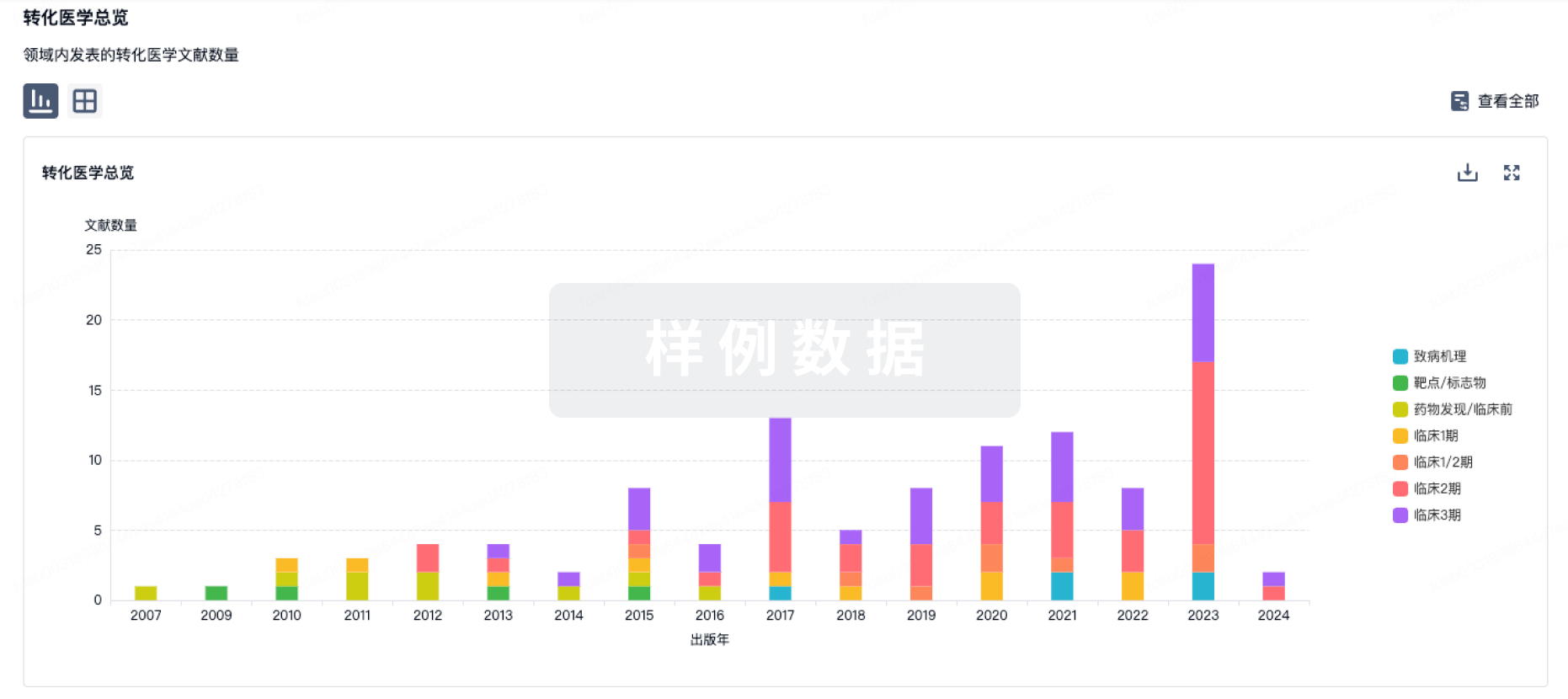
药物交易
使用我们的药物交易数据加速您的研究。
登录
或
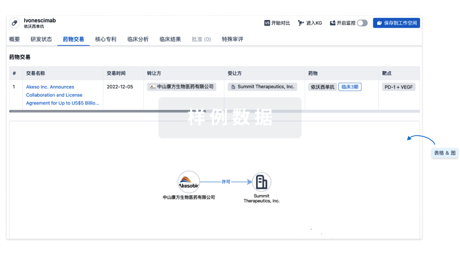
核心专利
使用我们的核心专利数据促进您的研究。
登录
或
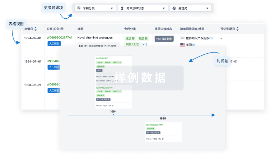
临床分析
紧跟全球注册中心的最新临床试验。
登录
或
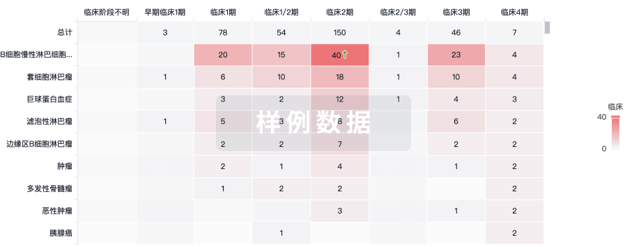
批准
利用最新的监管批准信息加速您的研究。
登录
或
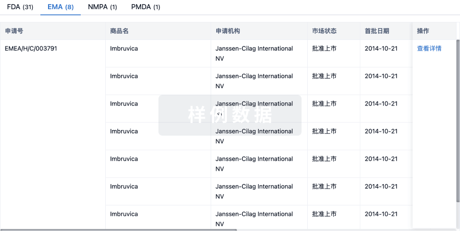
生物类似药
生物类似药在不同国家/地区的竞争态势。请注意临床1/2期并入临床2期,临床2/3期并入临床3期
登录
或
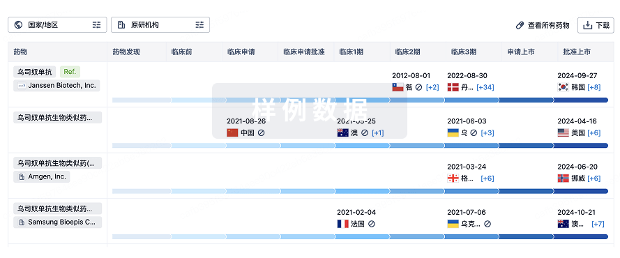
特殊审评
只需点击几下即可了解关键药物信息。
登录
或
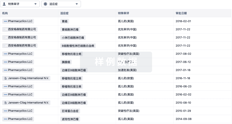
提出任何生物医药研究问题
让每一步生物医药决策都精准制胜
基于十亿级生物数据与全球情报的精准 AI,赋能早期药物发现
立即开始免费试用!
智慧芽新药情报库是智慧芽专为生命科学人士构建的基于AI的创新药情报平台,助您全方位提升您的研发与决策效率。
立即接入生物医药数据包
智慧芽新药库数据也通过智慧芽开放平台,以MCP或API服务形式对外开放。立即使用我们的MCP服务定制您的LLM智能体。
生物序列数据库
生物药研发创新
免费使用
化学结构数据库
小分子化药研发创新
免费使用