预约演示
更新于:2026-03-08

Stanford University School of Medicine
更新于:2026-03-08
概览
标签
肿瘤
血液及淋巴系统疾病
内分泌与代谢疾病
小分子化药
降解型分子胶
诊断用放射药物
疾病领域得分
一眼洞穿机构专注的疾病领域

暂无数据
技术平台
公司药物应用最多的技术
暂无数据
靶点
公司最常开发的靶点
暂无数据
| 排名前五的药物类型 | 数量 |
|---|---|
| 小分子化药 | 19 |
| 降解型分子胶 | 2 |
| 诊断用放射药物 | 2 |
| 分子胶 | 2 |
| CAR-T | 2 |
关联
31
项与 Stanford University School of Medicine 相关的药物


100 项与 Stanford University School of Medicine 相关的临床结果
登录后查看更多信息
0 项与 Stanford University School of Medicine 相关的专利(医药)
登录后查看更多信息
22,455
项与 Stanford University School of Medicine 相关的文献(医药)2025-12-22·JACS Au
Mycobacteriophage Functionalized Magnetic Nanocrystal Clusters for Highly Sensitive and Rapid Detection of
Mycobacterium tuberculosis
Article
作者: Bogyo, Matthew ; Rao, Jianghong ; Wang, Ting ; Dai, Sheng-Yao ; Ibrahim, Jawad ; Fu, Qunfeng ; Xiao, Zhen ; Murugesan, Kanagavel ; Hajfathalian, Maryam ; Yen, Charles ; Banaei, Niaz
Tuberculosis caused by Mycobacterium tuberculosis (Mtb) is one of the most dangerous diseases globally. Mtb poses a heavy death toll, especially in low-resource settings, where inadequate diagnostic capabilities greatly hinder treatment and prevention. Here, we present a rapid and cost-effective bacilli-capturing method that uses magnetic nanoclusters conjugated with mycobacteriophages. The mycobacteriophages provide Mtb recognition functionality, and the binding of the nanoparticles with attenuated Mtb H37Rv and Mycobacterium bovis Bacillus Calmette-Guérin (BCG) was visualized by electron microscopy. The magnetic nanocrystal clusters have an excellent separation efficiency. A nearly 100% capturing efficiency and high specificity toward mycobacteria species were obtained. Magnetically separated mycobacteria were disrupted by ultrasound to facilitate the rapid release of cellular adenosine triphosphate (ATP) for bioluminescent detection. Using portable and inexpensive devices, we achieved rapid detection of Mtb at as low as 1000 bacilli per sample in artificial sputum, urine, and whole porcine blood within 35 min. This method demonstrates excellent potential for point-of-care tuberculosis diagnosis in resource-limited settings.
2025-12-16·ANALYTICAL CHEMISTRY
Polarized Distribution of Lipid Droplets with Long Acyl Chains and Unsaturation are Hallmarks of Human Intestinal Enteroid Differentiation
Article
作者: Liu, Yueming ; Heilshorn, Sarah C. ; Klett, Katarina C. ; Johansson, Patrik K. ; Enejder, Annika
Cell polarization and differentiation require increased energy mobilization and cell membrane synthesis, whereby mitochondria and lipid droplets (LDs) play key roles. However, how these metabolic organelles organize at the subcellular level to efficiently meet energy demands in human intestinal organoids is unclear. To address this, we introduce coherent anti-Stokes Raman scattering (CARS) microscopy multiplexed with confocal fluorescence microscopy to spatially map LDs and mitochondria throughout cell differentiation in human intestinal enteroids. The results show an overall decrease in LDs over time, though less pronounced for cells positive for proliferation or stemness markers. The LD depletion was observed in the apical region, resulting in a polarized distribution to the basal side. A similar mitochondrial polarization pattern was also observed in differentiated enteroids. Spectral CARS further shows that LDs postdifferentiation contain lipids with signatures of longer acyl chains and a higher degree of unsaturation. These observations demonstrate that polarized metabolic and lipid supply infrastructures are formed to support intestinal cell differentiation in organoid cultures.
2025-11-01·Journal of voice : official journal of the Voice Foundation
Auditory-Perceptual and Pupillometric Evaluation of Vocal Roughness and Listening Effort in Tracheoesophageal Speech
Article
作者: Doyle, Philip C ; Parsa, Vijay ; Farahani, Mojgan
OBJECTIVES:
This study evaluated auditory-perceptual judgments of perceived vocal roughness (VR) and listening effort (LE) along with pupillometric responses in response to speech samples produced by tracheoesophageal (TE) talkers.
METHODS:
Twenty normal-hearing, naive young adults (eight men and twelve women) served as listeners. Listeners were divided into two groups: (1) a with-anchor (WA) group (four men and six women) and (2) a no-anchor (NA) group (four men and six women). All were presented with speech samples produced by twenty TE talkers; listeners evaluated two auditory-perceptual dimensions-VR and LE-using visual analog scales. Anchors were provided to the WA group as an external referent for their ratings. In addition, during the auditory-perceptual task, each listener's pupil reactions also were recorded with peak pupil dilation (PPD) measures extracted as a physiologic indicator associated with the listening task.
RESULTS:
High interrater reliability was obtained for both the WA and NA groups. High correlations also were observed between auditory-perceptual ratings of roughness and LE, and between PPD values and ratings of both dimensions for the WA group. The inclusion of an anchor during the auditory-perceptual task improved interrater reliability ratings, but it also imposed an increased demand on listeners.
CONCLUSIONS:
Data obtained offer insights into the relationship between subjective indices of voice quality (ie, auditory-perceptual evaluation) and physiologic responses (PPD) to the abnormal voice quality that characterizes TE talkers. Furthermore, these data provide information on the inclusion/exclusion of audio anchors and potential increases in listener demand in response to abnormal voice quality.
673
项与 Stanford University School of Medicine 相关的新闻(医药)2026-03-05
TORONTO, March 05, 2026 (GLOBE NEWSWIRE) --
Profound Medical Corp.
(NASDAQ:PROF; TSX:PRN) (“Profound” or the “Company”), a commercial-stage medical device company that develops and markets innovative interventional MRI (“iMRI”) procedures, today reported financial results for the fourth quarter and full year ended December 31, 2025. Unless specified otherwise, all amounts in this press release are expressed in U.S. dollars and are presented in accordance with U.S. generally accepted accounting principles (U.S. GAAP).
Business Highlights
Q4-2025 revenue grew 43% year-over-year and 13% sequentially quarter-over-quarter to a record $6.0 million.
Profound’s TULSA-PRO
®
qualified sales pipeline also continues to grow, and currently stands at 110 new systems being classified within one of the “Verify, Negotiate and Contracting” stages.
The Company’s TULSA-PRO installed base
stood at 78 as of December 31, 2025
and, due to its strong capital sales pipeline, Profound currently expects to reach approximately 120 installs by the end of 2026.
Profound continued to see a wide variety of prostate disease patients treated by its TULSA-PRO customers in the fourth quarter of 2025:
67% were treated for prostate cancer only, 17% were hybrid patients suffering from both prostate cancer and benign prostatic hyperplasia (“BPH”), 13% were salvage, and 3% were men with BPH only;
For cancer grade, 5% were GG1, 57% were GG2, 29% were GG3, and 9% were GG4 & GG5;
By intention-to-treat, 44% were whole gland; 24% were sub-total but more than half the gland; 24% were hemi-ablations, and 9% were focal therapy; and
For prostate size, 9% were <20cc; 42% were 20-40cc; 31% were 40-60cc; 17% were 60-100cc; and 2% were over 100cc.
In October 2025, Profound unveiled new, real-world data from the internationally recognized
Busch Center
. The data — marking the center’s milestone of 500 completed TULSA Procedures™ — demonstrated the procedure’s versatility and success in treating a broad spectrum of prostate diseases, severities, and aggressions.
As user interest in Profound’s technologies continues to build, the Company is deploying its own direct sales team in North America, while partnering with select strategic distribution partners to support the business potential and the customer base in other parts of the world. In November 2025, Profound:
Regained exclusive distribution rights for TULSA-PRO in Canada;
Entered into an exclusive distribution and supply agreement for its TULSA-PRO and Sonalleve
®
technologies in Saudi Arabia with
Al Faisaliah Medical Systems Co. (FMS)
, a subsidiary of one of the Kingdom’s most prominent business conglomerates,
Al Faisaliah Group (AFG)
; and
Entered into a strategic distribution agreement with
Getz Healthcare
to introduce TULSA-PRO in Australia and New Zealand.
Also in November, Profound announced that the Hong Center Scottsdale, led by Dr. Y. Mark Hong of
Integrative Urology
in Phoenix, Arizona, achieved a world-first milestone: 200 TULSA Procedures performed independently by a urologist, without radiologist involvement.
In November/December 2025, Profound launched its TULSA-AI
®
Volume Reduction module for optimizing the treatment of patients with BPH at the Radiological Society of North America meeting in Chicago, IL. The use of AI to streamline the workflow and reduce procedure times is a significant advancement that makes using TULSA-PRO for treating enlarged prostate just as efficient as other modern procedures, but with the advanced benefits of precision and customization to any prostate shape or size. The Company believes the reduced procedure times for BPH will increase adoption of the TULSA Procedure and triple Profound’s total available market in prostate disease to about 600,000 patients annually.
In December 2025,
Pejman Ghanouni, MD, PhD
, Associate Professor in the Department of Radiology, Division of Body MRI at Stanford University School of Medicine, received the Cum Laude award for his presentation titled "CAPTAIN Randomized Controlled Trial of MRI-Guided Transurethral Ultrasound Ablation (TULSA) Versus Robotic Radical Prostatectomy" at the 2025 Radiological Society of North America ("RSNA") Annual Meeting.
Also in December, the Company significantly strengthened its balance sheet via the closing of a $36.0 million registered direct offering in the United States and an upsized $6.45 million private placement in Canada. Both the registered direct offering and private placement were structured as straightforward equity investments with no warrant coverage and were led by healthcare-dedicated investors alongside existing shareholders.
In January 2026, Profound announced two U.S. commercial milestones.
The Johns Hopkins Hospital
(Baltimore, MD) treated its first non-clinical-trial prostate cancer patient using the Company’s TULSA-PRO system, marking the official launch of Profound’s technology at one of the world’s most influential centers for prostate cancer innovation and coinciding with the opening of Johns Hopkins Medicine’s new iMRI suite. Soon thereafter, the world-renowned
Mount Sinai Hospital
(New York, NY) successfully treated its first prostate cancer patient with the TULSA-PRO system, becoming the first health system in the New York metropolitan area to offer the TULSA Procedure.
In February 2026,
PRO FAMILIA Specialist Hospital
in Rzeszów, Poland completed its 500th Sonalleve
®
Procedure.
Also in February, the Company received the 2025/2026 Mount Logan Award from
INOVAIT
, the Canadian national network for commercializing breakthroughs in image-guided therapy (IGT) and AI. The award recognizes Profound’s achievement of significant milestones, including treating the 4,000
th
TULSA Procedure patient, securing reimbursement for the TULSA Procedure by the U.S. Centers for Medicare & Medicaid Services (CMS) at Urology APC Level 7, and establishing strategic partnerships to expand patient access to TULSA-PRO globally.
Today, Profound is pleased to announce that Laurence Klotz, M.D., FRCSC, an esteemed urologist and professor of surgery at the University of Toronto and the Sunnybrook Chair of Prostate Cancer Research, will present the first clinical outcomes from the Level 1 post-market CAPTAIN trial comparing the safety and efficacy of the TULSA Procedure with robotic radical prostatectomy in men with localized prostate cancer next week at the 41st Annual European Association of Urology (EAU) Congress in London, UK.
EAU26
is a premier academic urology meeting, and the Company is pleased that the data have been selected for inclusion in the meeting’s Late-Breaking and High-Impact session on Friday, March 13
th
.
“We continued to execute well, delivering record revenues, an expanded TULSA-PRO installed base, and a stronger capital sales pipeline again in the fourth quarter — validating our belief that Q3-2025 marked an important inflection point in our business,” said Arun Menawat, Profound’s CEO and Chairman. “Moving forward, with the strengthening of our balance sheet via the completion of our financing in December, and based on several upcoming potential catalysts, including the clinical outcomes from the Level 1 post-market CAPTAIN study, we believe that we are on the right path toward driving high double digit to low triple digit revenue growth and, ultimately, positive cash flows and net profitability.”
Summary Fourth Quarter 2025 Results
For the quarter ended December 31, 2025, Profound recorded revenue of approximately $6.0 million, with $2.3 million from recurring - non-capital revenue, which consists of the sale of TULSA-PRO one-time use devices service revenue related to extended warranties, and $3.7 million from the one-time sale of capital equipment. Fourth quarter 2025 revenue was up 43% from $4.2 million for the same three-month period a year ago.
Gross margin for the fourth quarter of 2025 was 67%, compared to 71% in the prior year period. The lower than usual fourth quarter 2025 gross margin was primarily due to product mix and new market introductory prices with international distributors in Saudi Arabia and Australia.
Total operating expenses in the fourth quarter of 2025 were approximately $11.4 million, up marginally from $11.3 million in the prior year period. The increase in operating expenses was primarily due to increased headcount, increased sales force, commission payments, and increased travel and infrastructure costs to support the Company’s growth.
Fourth quarter 2025 net loss was approximately $8.2 million, or $0.27 per common share, compared to a net loss of approximately $4.9 million, or $0.20 per common share, in the three months ended December 31, 2024.
Summary Full Year 2025 Results
For the year ended December 31, 2025, Profound recorded revenue of approximately $16.1 million, with $9.7 million from recurring non-capital revenue and $6.4 million from the one-time sale of capital equipment. This compares to revenue of approximately $10.7 million in the year ended December 31, 2024.
Profound’s full year 2025 gross margin was 71%, compared to 66% in the prior year period. Gross margin expansion in 2025 was primarily due to increased selling prices coupled with the growth in the number of capital systems sold.
Total operating expenses in the year ended December 31, 2025 were approximately $52.6 million, compared with $40.1 million in 2024. The increase in operating expenses was primarily due to increased headcount, increased enrolment for the CAPTAIN trial and higher material expenditures and travel associated with the trial, increased testing and design modification, increased sales force and commission payments, and increased travel and infrastructure costs to support the Company’s growth. Partially offsetting these increases was a decrease to insurance due to lower premium rates and a reduction in trade receivable write-offs.
Profound recorded a net loss for the year ended December 31, 2025 of approximately $42.6 million, or $1.41 per common share, compared to approximately $27.8 million, or $1.12 per common share, for the year ended December 31, 2024.
Liquidity and Outstanding Share Capital
As at December 31, 2025, Profound had cash of approximately $59.7 million.
As at March 5, 2026, Profound had 36,293,640 common shares issued and outstanding.
For complete financial results, please see Profound’s filings, which will be made available under Profound’s pro
,
and on Profound’s
website
under “SEC & SEDAR+ Filings.”
Changes to Board of Directors
Profound also announced today that Kris Shah has resigned from its Board of Directors and Frank Baylis has been appointed as Mr. Shah’s successor, effectively immediately.
Mr. Baylis joined Baylis Medical Company in 1989, serving as its President until 2015. During his tenure, the company grew into an internationally recognized developer of medical devices used in cardiac electrophysiology, structural heart disease, and spine procedures. He returned to Baylis Medical Company in 2019 after serving as Liberal Member of Canada’s Parliament for the federal riding of Pierrefonds-Dollard from 2015 to 2019. During his time at Baylis Medical Company, his leadership and vision helped build the company from a small start-up into a global leader, ultimately leading to its acquisition by Boston Scientific in 2022 for $1.75 billion. Mr. Baylis also co-founded the consulting business OME Group in 1991, which was sold to Ernst and Young in 2011. Currently, Mr. Baylis is the Executive Chairman of Baylis Medical Technologies, a leader in the development and commercialization of innovative medical devices in the fields of interventional radiology and spine procedures. Headquartered in Canada, the company also provides contract manufacturing services to some of the world’s leading medical device companies.
Mr. Baylis is an active board member for Epineuron Technologies, a clinical-stage company developing a novel class of bioelectronic therapy that offers a transformative approach to aid in the treatment of injured peripheral nerves. In the past he has served on the boards of MEDEC, Campus de Technologie de Sante (CTS), and the Business Advisory Committee of HTX. His list of accomplishments includes several patents, the Ernst and Young Entrepreneur Award for Healthcare in Quebec (2011) and the University of Waterloo Alumni Achievement Award (2014). Mr. Baylis holds a Bachelor of Science in Electrical Engineering from the University of Waterloo, and while attending, he met Mr. Shah, and together they founded Baylis Medical Technologies.
“On behalf of the Board and staff of Profound, I would like to take this opportunity to thank Kris for his many contributions to the Company and wish him all the best in his future endeavours,” said Dr. Menawat. “I am also very pleased to welcome Kris’ long-time business partner, Frank, to our team. He is not only a successful entrepreneur and experienced executive, but also has a breadth of relevant business and Board experience that should be invaluable as Profound executes the next stages of its growth strategy.”
Conference Call Details
Profound Medical is pleased to invite all interested parties to participate in a conference call today at 4:30 pm ET during which time the results will be discussed.
To participate in the conference call by telephone, please pre-register via this
link
to receive the dial-in number and your unique PIN.
The call will also be broadcast live and archived on Profound's website in the Investors section
here
.
About Profound Medical Corp.
Profound is a commercial-stage medical device company and an innovator in interventional MRI (iMRI) procedures, enabling precise, incision-free therapies that improve clinical confidence, procedural control, and patient outcomes. By leveraging real-time MRI guidance, Profound’s technologies are designed to replace uncertainty with clarity across treatment planning, delivery, and confirmation.
The company’s flagship platform,
TULSA-PRO
®
, enables MRI-guided, incision-free prostate therapy designed for precision and flexibility. The
TULSA Procedure™
allows physicians to see, treat, and confirm therapy in real time, supporting personalized treatment strategies across the continuum of prostate care—from whole-gland to subtotal, hemi, multifocal, and focal treatment. This approach enables individualized care for the full spectrum of prostate disease, including prostate cancer and/or benign prostatic hyperplasia (BPH), while minimizing side effects typically associated with surgery or radiation, such as urinary incontinence and/or erectile dysfunction.
Profound also commercializes
Sonalleve
®
, an MRI-guided therapy that provides a non-surgical treatment option for pain palliation of bone metastases, desmoid tumors, and osteoid osteoma, as well as for common gynecologic conditions including uterine fibroids and adenomyosis. Sonalleve delivers targeted therapy with no incisions, no blood loss during the procedure, no overnight hospital stay, and faster recovery — and, in gynecologic applications, enables uterine-sparing treatment that may help preserve fertility. Profound is also exploring additional clinical applications for Sonalleve, including non-invasive ablation of abdominal cancers and hyperthermia-based cancer therapies.
Profound Medical’s technologies are approved across major global markets. TULSA-PRO is cleared or approved in the United States, Europe, Canada, Saudi Arabia, India, Australia/New Zealand, and the UAE, while Sonalleve is cleared or approved in the United States (HDE), Europe, Canada, China, and Saudi Arabia.
Through real-time MRI guidance and data-driven innovation, Profound is advancing the future of MRI-guided therapy — expanding access to precise, personalized, and incision-free treatment options worldwide.
Forward-Looking Statements
This release includes forward-looking statements regarding Profound and its business which may include, but is not limited to, the expectations regarding the efficacy of Profound’s technology in the treatment of prostate cancer, BPH, uterine fibroids, adenomyosis, palliative pain treatment, desmoid tumors, and osteoid osteoma; the extent and timing of Profound’s completion of TULSA-PRO
®
system sales from its qualified sales pipeline; Profound’s expectations for future revenues; and the success of Profound’s commercialization strategy and activities for TULSA-PRO and Sonalleve. Often, but not always, forward-looking statements can be identified by the use of words such as "plans", "is expected", "expects", "scheduled", "intends", "contemplates", "anticipates", "believes", "proposes" or variations (including negative variations) of such words and phrases, or state that certain actions, events or results "may", "could", "would", "might" or "will" be taken, occur or be achieved. Such statements are based on the current expectations of the management of Profound. The forward-looking events and circumstances discussed in this release, may not occur by certain specified dates or at all and could differ materially as a result of known and unknown risk factors and uncertainties affecting the Company, including risks regarding the medical device industry, regulatory approvals, reimbursement, economic factors, the equity markets generally and risks associated with growth and competition. Although Profound has attempted to identify important factors that could cause actual actions, events or results to differ materially from those described in forward-looking statements, there may be other factors that cause actions, events or results to differ from those anticipated, estimated or intended. No forward-looking statement can be guaranteed. Other factors and risks that may cause actual results to differ materially from those set out in the forward-looking statements are described in Profound's Annual Report on Form 10-K and other filings made with U.S. and Canadian securities regulators, available at
and
. Except as required by applicable securities laws, forward-looking statements speak only as of the date on which they are made and Profound undertakes no obligation to publicly update or revise any forward-looking statement, whether as a result of new information, future events, or otherwise, other than as required by law.
For further information, please contact:
Stephen Kilmer
Investor Relations
skilmer@profoundmedical.com
T: 647.872.4849
Profound Medical Corp.
Consolidated Balance Sheet
As at December 31, 2025 and 2024
In USD (000s)
2025
$
2024
$
Assets
Current assets:
Cash
59,723
54,912
Trade and other receivables, net
7,200
7,045
Inventory
8,238
5,801
Prepaid expenses and deposits
928
1,307
Total current assets
76,089
69,065
Trade and other receivables, net
300
-
Property and equipment, net
698
425
Intangible assets, net
138
261
Right-of-use assets, net
184
396
Deferred tax assets, net
66
87
Total assets
77,475
70,234
Liabilities
Current liabilities:
Accounts payable
1,563
1,317
Accrued expenses and other current liabilities
3,815
2,835
Deferred revenue
445
419
Long-term debt
-
1,737
Lease liabilities
213
257
Income tax payable
39
-
Total current liabilities
6,075
6,565
Deferred revenue
388
49
Long-term debt
4,499
2,924
Lease liabilities
-
203
Other non-current liabilities
79
71
Total liabilities
11,041
9,812
Commitments and contingencies
Shareholders’ equity
Common shares, no par value, unlimited shares authorized, 36,293,640 and 30,039,809 issued and outstanding at December 31, 2025 and 2024, respectively
323,839
281,552
Additional paid-in capital
25,310
21,298
Accumulated other comprehensive income
5,025
2,742
Accumulated deficit
(287,740)
(245,170)
Total shareholders’ equity
66,434
60,422
Total liabilities and shareholders’ equity
77,475
70,234
Profound Medical Corp.
Consolidated Statements of Operations and Comprehensive Loss
In USD (000s)
Three months ended
December 31, 2025
$
Three months ended
December 31, 2024
$
Year ended
December 31, 2025
$
Year ended
December 31, 2024
$
Revenue
Recurring - non-capital
2,302
2,679
9,730
8,240
Capital equipment
3,675
1,498
6,368
2,440
5,977
4,177
16,098
10,680
Cost of sales
1,986
1,214
4,705
3,643
Gross profit
3,991
2,963
11,393
7,037
Operating expenses
Research and development
4,272
4,649
20,596
16,965
Selling, general and administrative
7,088
6,658
32,051
23,134
Total operating expenses
11,360
11,307
52,647
40,099
Operating loss
7,369
8,344
41,254
33,062
Other (income) expenses
Net finance (income) expense
(160)
(332)
(1,070)
(1,436)
Net foreign exchange (gain) loss
940
(2,828)
2,134
(3,808)
Total other (income) expenses
780
(3,160)
1,064
(5,244)
Net loss before income taxes
8,149
5,184
42,318
27,818
Income tax expense
12
(92)
231
144
Deferred tax (recovery) expense
14
(146)
21
(146)
Total income tax (recovery) expense
26
(238)
252
(2)
Net loss attributed to shareholders for the year
8,175
4,946
42,570
27,816
Other comprehensive (income) loss
Item that may be reclassified to (income) loss
Foreign currency translation adjustment
(275)
(1,968)
(2,283)
2,823
Net loss and other comprehensive loss for the year
7,900
6,914
40,287
30,639
Loss per share
Basic and diluted net loss per common share
0.27
0.20
1.41
1.12
Basic and diluted weighted average common shares outstanding
30,726,382
25,770,800
30,232,966
24,765,503
Profound Medical Corp.
Consolidated Statements of Cash Flows
For the years ended December 31, 2025 and 2024
In USD (000s)
2025
$
2024
$
Cash flows from operating activities
Net loss for the year
(42,570)
(27,816)
Adjustments to reconcile net loss to net cash provided by operating activities:
Depreciation of property and equipment
373
707
Amortization of intangible assets
187
229
Non-cash lease expense adjustment
(37)
(38)
Share-based compensation
5,490
2,581
Interest and accretion expense
63
600
Change in amortized cost of trade and other receivables
-
(307)
Changes in operating assets and liabilities:
Trade and other receivables
(172)
186
Inventory
(2,822)
656
Prepaid expenses and deposits
464
31
Accounts payable, accrued expenses and other liabilities
378
815
Deferred revenue
371
(948)
Income taxes payable
42
-
Deferred tax liabilities
-
(58)
Deferred tax assets
26
(91)
Net cash used in operating activities
(38,207)
(23,453)
Cash flows from investing activities
Purchase of property and equipment
(176)
-
Purchase of intangible assets
(66)
-
Net cash used in investing activities
(242)
-
Cash flows from financing activities
Issuance of commons shares
42,436
62,106
Payments of financing costs
(1,016)
(4,895)
Repayments of long-term debt
(290)
(2,560)
Proceeds from the exercise of stock options
8
45
Net cash provided by financing activities
41,138
54,696
Net increase in cash and cash equivalents
2,689
31,243
Effect of exchange rate changes on cash
2,122
(2,544)
Cash, beginning of year
54,912
26,213
Cash, end of year
59,723
54,912
高管变更财报并购
2026-02-27
·出入
(应粉丝留言整理成文,欢迎指正)
在人类浩瀚无垠的基因图谱中,偶尔会出现一些极其微小却足以引发毁灭性系统崩溃的排版错误。当这种错误悄然发生在X染色体上的某一个特定微小区段时,一个罕见而残酷的生命魔盒便被无情地打开了。这便是医学界所称的眼脑肾综合征(Lowe Syndrome,亦简称为Lowe综合征或OCRL综合征)。
这是一种发病率仅为五十万分之一的X连锁隐性遗传极罕见病,这一遗传特性意味着它几乎只将魔爪伸向男童,而女性通常作为无症状或仅有轻微眼部特征的基因携带者 。患病男童自出生起,便面临着生命三大核心防线的全线告急与系统性崩溃:他们的双眼往往在出生时便被先天性白内障或随后的青光眼所蒙蔽,剥夺了认知世界的初见之光;他们的大脑深陷严重的全身性肌张力低下(Hypotonia)与不可逆的智力发育迟缓;而在随后的短短数周至数月内,他们的肾脏将遭遇灾难性的近端肾小管功能障碍,维持生命的宝贵营养物质如流水般从尿液中流失,最终在青春期或成年早期不可避免地走向慢性肾衰竭(CKD) 。绝大多数患者的生命时钟,会在40岁前因肾衰竭、严重感染或癫痫等并发症戛然而止 。
然而,在面对如此令人绝望的疾病图景时,人类的医学探索从未停止。在过去五年(2021年-2026年)里,全球医学界、基础生物学界与罕见病患者倡导组织紧密携手,对这一罕见病的认知与反击达到了前所未有的历史高度。从细胞微观机制的多年盲区被一举照亮,到通过海量筛选实现的“老药新用”正式进入人体临床试验阶段,再到CRISPR碱基编辑技术在实验室中展现出的惊艳破局能力,科研人员正在与死神和时间进行着激烈的赛跑。本报告将全景式、深层次地拆解近五年全球针对Lowe综合征的致病机制突破、小分子靶向药物进展、基因疗法潜能及临床生命支持治疗的最新优化指南,力求客观、严谨、深情地呈现人类与罕见病死磕的最新战况。硬核科普软着陆:OCRL基因密码与“细胞物流大罢工”
要真正理解Lowe综合征那摧枯拉朽般的破坏力,我们必须跨越宏观的器官表象,深入潜入人体细胞的微观运转世界。Lowe综合征的万恶之源,是位于人类X染色体长臂Xq26.1区段的OCRL基因发生了致病性突变 。该基因在正常情况下,负责编码一种至关重要的酶类——OCRL-1酶(即肌醇多磷酸-5-磷酸酶,Inositol polyphosphate 5-phosphatase)。
为了让这一复杂的生化过程易于理解,我们可以将人体内的每一个细胞视作一座高度现代化、运转极度繁忙的微型城市。在这座城市中,存在着名为“高尔基体(Golgi network)”和“内体系统(Endolysosomal system)”的核心设施,它们共同构成了这座城市的“中央物流分拣中心”与“快递运输网络” 。在这个庞大且精密的细胞物流网络中,有一种存在于细胞膜和囊泡上的特异性脂质分子被广泛使用,它被称为PI(4,5)P2(常简称为PIP2,即磷脂酰肌醇-4,5-二磷酸)。在细胞的物流体系中,PIP2完美地扮演着“快递条形码”或“物流集装箱定位标签”的角色。当特定的货物(如维持正常生命活动的受体蛋白质、离子通道或细胞外营养物质)需要被精准运送到细胞膜表面或被吞噬进细胞内部时,必须依赖PIP2这个“条形码”的标记、指引与锚定 。
然而,细胞内的物流法则极其严苛:当包裹着货物的囊泡成功送达目的地后,这些完成使命的PIP2“条形码”必须在第一时间被迅速撕毁、降解并循环回收。如果任由它们堆积,整个物流通道的识别系统就会陷入彻底的混乱,甚至导致物理通道被废弃的集装箱彻底堵死。而OCRL-1酶,正是那个由基因指派的、负责精准降解PIP2的“顶级物流回收员”或“系统清理工”。
当OCRL基因发生先天性突变,导致这位不可或缺的“物流清理工”OCRL-1酶完全缺失或失去酶活性时,一场微观层面的灾难便无可挽回地降临了,这直接导致了细胞功能的全面崩溃: 首先是细胞物流的彻底瘫痪。由于缺乏OCRL-1的降解作用,未被处理的PIP2脂质分子在细胞内体等亚细胞区室中开始疯狂堆积。这种异常的堆积会直接扰乱细胞内部的结构支撑网络——细胞骨架(尤其是肌动蛋白Actin网络),导致其发生异常的过度交联和扭曲变形 。失去正常骨架支撑的细胞,其物质运输、细胞分裂和极性维持等基础生命活动陷入全面停滞。 随之而来的是多器官防线的连环崩盘。在肾脏组织中,高度依赖细胞内吞作用的近端肾小管上皮细胞无法通过“物流吞噬”机制回收初尿中的低分子量蛋白质、氨基酸、重碳酸盐和各种关键矿物质,营养物质直接漏出体外,导致了极其严重的肾性Fanconi综合征(Renal Fanconi Syndrome)。在大脑中,由于物流运输的阻断,神经元发育过程中的信号受体无法正常回收,神经细胞的迁徙网络被无情切断,直接引发了严重的智力低下与异常的行为模式。在眼部,晶状体上皮细胞的发育和角膜结构的维持因细胞极性丧失而陷入紊乱,最终在出生时便表现为双侧先天性白内障与极高比例的婴幼儿青光眼 。
不再需要堆砌晦涩难懂的生化名词,当代医学界如今已清晰且深刻地认知到:Lowe综合征绝对不是孤立的、单一器官的退行性病变,而是一场由最底层的脂质代谢失控所引发的、席卷全身的“细胞物流大罢工”。核心板块(一):科研盲区被逐一照亮——近5年疾病机制的颠覆性与补充性认知
在2021年至2026年这极具活力的五年间,全球顶尖的医学与遗传学期刊(如《Nature Genetics》、《The American Journal of Human Genetics》、《Pediatric Nephrology》及《Frontiers in Cell and Developmental Biology》等)密集发表了一系列极具分量的基础研究成果。这些研究以抽丝剥茧的方式,揭开了Lowe综合征在发病机制上曾长期困扰医学界的诸多谜团。这些机制层面从“知其然”到“知其所以然”的飞跃,为后续的靶向药物开发指明了前所未有的精确方向。1. 破解脑部症状的独立密码:ApoER2/Reelin信号通路的枢纽作用
在过去的几十年中,临床医学界非常清楚OCRL基因突变会直接导致肾脏漏出营养和眼部晶状体浑浊,但对于为何这种基因缺陷会跨越血脑屏障,导致患者出现重度神经发育迟缓、智商通常停留在中重度智力障碍区间(均值IQ在40-54之间),以及引发极度固执、频繁发脾气等特殊行为表型的问题,科学界一直知之甚少 。 直到2024年,一项发表于核心期刊的关键神经生物学研究取得了突破性进展,该研究首次明确揭示了OCRL-1的缺失直接阻断了人类神经元中至关重要的ApoER2/Reelin信号通路的正常运作 。Reelin是一种在大脑早期发育和成熟大脑突触可塑性中起决定性作用的关键蛋白,它就像是神经元迁徙和建立连接的“交通指挥官”。而ApoER2则是神经元表面接收Reelin指令的专属天线受体。该研究通过人体神经元细胞模型证实,缺乏OCRL-1酶会导致ApoER2受体在细胞内部的内体运输中被错误拦截,无法被正常回收到细胞膜表面。这使得神经元由于缺失了“天线”,从而对Reelin发出的发育信号“充耳不闻” 。这一微观通路的发现,首次从分子受体层面完美解释了Lowe综合征患者中枢神经系统异常、智力受损的直接原因,并为未来针对中枢神经系统退行性病变和行为异常的靶向神经药物开发,提供了一个确切且极具潜力的全新干预靶点。2. 基因型与表型的深度关联:为何突变基因相似,生命命运却截然不同?
在临床肾脏病学领域,存在着一种与Lowe综合征在基因起源上完全同源的罕见病——Dent病2型(Dent disease type 2,简称Dent-2)。这两种疾病皆由同一条X染色体上的OCRL基因突变引起,然而患者的临床命运却天差地别:Dent-2患者通常仅表现出较轻的近端肾小管病变(如低分子量蛋白尿),而脑部和眼部的多系统症状极其轻微甚至完全缺如 。这种“同源不同命”的现象,一直是遗传学界的热门悬案。 近五年来,随着基因测序技术的普及与大样本数据的积累,中国学者团队发布了一项涵盖83名患者(Lowe综合征与Dent-2患者)的大型基因型-表型相关性队列研究,彻底阐明了这一现象背后的分子逻辑。研究明确指出,突变发生在基因的特定位置以及突变的具体类型,直接决定了生命防线崩溃的广度与深度。数据表明,在患有严重多系统症状的Lowe综合征患者中,导致蛋白质翻译提前强制终止的**截短突变(Truncating mutations)**占比高达70.8%,且绝大多数(81.3%)集中在OCRL基因后半段的第13至23号外显子(Exon 13-23)区域;相反,症状较轻的Dent-2患者的突变多集中在第2至12号外显子,且截短突变仅占31.4% 。研究人员进一步通过分子三维构象模拟提出,位于OCRL基因后端特定结构域(如5'-磷酸酶结构域或RhoGAP样结构域)的突变,会引起更为广泛的蛋白质折叠灾难,导致细胞内试图弥补OCRL功能的其他同源磷酸酶(如INPP5B)代偿机制彻底失效,从而引发了毁灭性的全系统“构象疾病(Conformational disease)”效应 。这一发现不仅大幅提高了临床基因遗传咨询的准确性,也让学界认识到,针对不同外显子突变的患者,未来可能需要采取高度差异化的精准治疗策略。3. 斑马鱼与果蝇模型的全面介入:填补在体(In Vivo)生理研究的长期空白
长久以来,Lowe综合征的机制研究过度依赖于体外培养的患者成纤维细胞或肾小管上皮细胞系。然而,单个细胞的病变无法完全解释多器官间复杂的相互作用。近五年来,以斑马鱼(Zebrafish)和果蝇(Drosophila)为代表的活体动物模型的广泛构建与应用,极大地加速了机制验证与新药初筛的步伐。 在果蝇模型中,研究人员通过敲除果蝇体内唯一的OCRL直系同源基因(dOCRL),成功再现了幼虫肾细胞(Nephrocyte)在内体-溶酶体系统中的严重结构和功能缺陷,证实了OCRL在肾细胞排泄功能中的细胞自主性作用 。而在更为复杂的斑马鱼模型中,OCRL缺陷不仅完美再现了人类患者的低分子量蛋白尿,还首次在活体层面上直观地展示了肾小管由于内吞作用缺陷而发生的病理性缩短,以及肾小管上皮初级纤毛(Primary cilia)功能的丧失 。这些兼具高效繁殖能力与基因可编辑性的动物模型,如今已成为测试高通量化合物药物筛选与基因拯救实验的最前沿阵地,使得将体外实验转化为活体疗法的周期被大幅缩短。核心板块(二):治疗手段的快速迭代——从小分子“老药新用”到CRISPR基因编辑的黎明
如果说前面详述的机制研究是科学家在黑暗中为攻克疾病绘制的敌军分布图,那么针对PIP2脂质代谢异常和DNA突变序列开展的靶向干预,就是人类真正向Lowe综合征发起的武装反攻。面对罕见病新药研发周期动辄长达十余年且试错成本高达数亿美元的困境,近五年来,医学界采取了最为务实且高效的双轨并行策略:在短期内,通过海量的高通量筛选,在已获监管部门批准的庞大药物库中寻找能够“跨界”纠正OCRL细胞表型的“老药新用(Drug Repurposing)”方案;在长期内,倾注全力研发旨在彻底修正基因代码的“基因疗法(Gene Therapy)”。
目前,这两条战线均取得了堪称里程碑式的突破性进展,部分先锋药物已正式步入人体临床试验阶段。为便于读者直观了解最新战况,以下整理了截至2026年针对Lowe综合征及OCRL相关疾病的核心在研疗法全景数据:在研疗法 / 药物名称药物类型与技术平台核心作用机制与治疗靶点当前研发与临床阶段(截至2026年)主要资助与主导机构阿佩利司 (Alpelisib / Piqray)
小分子靶向药物
(原抗癌药,老药新用)特异性PI3K激酶抑制剂,通过调节激酶活性,间接纠正异常的肌动蛋白骨架分布与内体内吞脂质代谢。
临床二期试验阶段 (Phase 2)
试验代号ALPEDENT,已在罗马Bambino Gesù医院启动患者入组。
剑桥大学Gurdon研究所、Lowe Syndrome Trust、LSA Piclidenoson
小分子靶向药物
(原抗炎药,老药新用)A3腺苷受体高亲和力激动剂,直接靶向恢复受损肾细胞的关键内吞与重吸收功能。
临床二期试验阶段 (Phase II)
由Can-Fite主导,于2026年中期启动,核心关注肾脏功能拯救。
意大利TIGEM研究所、Can-Fite BioPharma、Telethon基金会 雷帕霉素 (Rapamycin) + 他汀类药物联用小分子药物联用组合抑制过度激活的细胞内调节因子,协同降低细胞内的异常信号传导。
临床前深度评估阶段
在实验室患者细胞中实现表型逆转,展现巨大临床转化潜力。
普渡大学生物科学系 (Claudio Aguilar团队) ABE8e-NG (单碱基编辑)新型基因编辑疗法腺嘌呤单碱基编辑器,在不剪断DNA双链的情况下,直接将致病点突变(A-T转换为G-C),精准修复OCRL序列。
临床前细胞实验阶段
在体外成功修复携带R844X突变的患者源性成纤维细胞,恢复OCRL表达。
斯坦福大学医学院、上海同济大学、美国NIH等 CRISPR-CasMINI与AAV递送表观基因调控与编辑疗法采用超微型Cas蛋白实现单病毒AAV“即插即用”递送,通过转录激活代偿性5-磷酸酶或修复基因序列。
临床前动物实验阶段
已在果蝇、小鼠等活体动物模型中展开多项功能拯救实验。
斯坦福大学、丹麦Cure Lowe基金会 1. 老药新用:率先冲入临床阶段的“小分子先锋舰队”
由于彻底修正基因的难度极高,科学家们转而思考:能否利用现有的药物,绕过损坏的OCRL酶,通过其他生化路径强行疏通被堵塞的“细胞物流”?答案是肯定的。
阿佩利司(Alpelisib)的跨界大救援:这本是一款由诺华公司开发,已获FDA和EMA批准用于治疗特定类型乳腺癌的强效PI3K(磷脂酰肌醇-3-激酶)抑制剂。剑桥大学Gurdon研究所的Jennifer Gallop博士团队在探究肌动蛋白网络调节的复杂实验中,通过大规模筛选意外且惊喜地发现,Alpelisib能够极其显著地纠正OCRL1缺失所导致的细胞骨架严重紊乱,成功恢复细胞受损的内吞物流功能 。这一发现在学术界引起了巨大轰动。在国际患者组织的强力推动下,2025年6月,在意大利罗马Bambino Gesù儿童医院顶级肾脏病学专家Francesco Emma教授的主导下,针对Alpelisib的Phase 2期人体临床试验(ALPEDENT项目,EUCT官方编号:2024-514196-23)正式拉开帷幕并开始了首批患者入组 。该试验目前虽主要针对病理机制同源的Dent-2病患者进行安全性与疗效探索,但如果临床数据证实该药物能有效逆转近端肾小管的低分子量蛋白尿等核心指标,将毫无疑问地直接惠及广大的Lowe综合征受损肾脏 。
Piclidenoson发起的肾脏功能保卫战:同样是在意大利,TIGEM研究所的Antonella De Matteis教授团队在深入剖析细胞囊泡运输机制的基础上,对超过1300种已知化合物进行了严苛的高通量筛选,最终锁定了一种原本用于治疗银屑病(牛皮癣)等自身免疫性疾病的A3腺苷受体激动剂——Piclidenoson。该团队在体外细胞和动物模型中均无可辩驳地证实,Piclidenoson能够有效恢复OCRL缺陷造成的极其关键的肾脏排泄与重吸收功能 。基于这一坚实的临床前数据,拥有该药物产权的以色列生物技术公司Can-Fite与Telethon基金会达成战略合作,于2026年正式跟进并开启了针对Lowe综合征的第二期临床试验(ClinicalTrials.gov 编号:NCT07410455),预计将在2027年获得初步完成数据 。客观而言,由于该药物此前缺乏未成年人(儿童群体)的安全数据,根据极其严格的医学伦理与临床规范,首批临床试验(Pilot study)必须率先在18岁及以上的成人Lowe患者中开展。若在成人中证实安全性与实质疗效达标,该项目方能进一步向下延伸,造福更广泛的低龄儿童患者 。
雷帕霉素(Rapamycin)与他汀类药物(Statins)的精妙联用:美国普渡大学的Claudio Aguilar团队走了一条更为艰辛的筛选之路。在完成对惊人的40,000多种化合物的全面排查后,他们发现了一个独特的组合方案:将常用于肾移植术后防排斥的免疫抑制剂雷帕霉素,与常用于降血脂、降胆固醇的他汀类药物结合使用。这一精妙的组合能够有效降低Lowe综合征病态细胞中被异常过度激活的特定细胞内调节因子的水平,从而使患者细胞表现出极其显著的表型逆转,开始像健康细胞一样运作。这一方案极具成本优势和临床转化可行性 。2. 基因疗法:CRISPR与单碱基编辑点亮的终极治愈梦想
如果说小分子药物的作用,犹如在已经被堵塞的城市交通网络中临时开辟出几条紧急疏散通道,那么基因疗法(Gene Therapy)的终极目标,则是直接找到导致交通瘫痪的源头设计图纸,擦除错误代码,彻底重建整个城市的物流控制中枢。虽然截至2026年,全球尚无获批正式上市的Lowe综合征基因特效药,但近期在顶尖实验室中取得的一系列硬核突破,让彻底治愈的曙光初现。
ABE单碱基编辑(Adenine Base Editor)展现“分子手术刀”级精准修复:2024年6月,发表在权威学术期刊《人类分子遗传学》(Human Molecular Genetics)上的一项重磅突破性研究,向世界展示了基因编辑技术的全新维度。由美国斯坦福大学医学院与上海同济大学联合组成的跨国科研团队,利用最新的腺嘌呤单碱基编辑器(ABE8e-NG),在提取自真实Lowe综合征患者体内的成纤维细胞中,实施了一场极其精准的“分子级手术”。该患者携带了OCRL基因中极其常见且致命的无义点突变——c.2530 C>T (R844X) 。与第一代传统的CRISPR-Cas9技术在操作时可能会引发DNA双链断裂、从而带来不可预知基因组毒性风险不同,碱基编辑技术更像是在不剪断基因“录音磁带”的情况下,直接在分子层面将错误的遗传字母擦除并完美重写(将A-T碱基对安全地转换为G-C碱基对)。实验结果令人极度振奋:接受ABE编辑修复后的患者细胞,不仅完美恢复了OCRL基因的mRNA转录水平和蛋白质正常表达,更从根本上彻底消除了PIP2脂质分子的病理性堆积。更为神奇的是,原本因物流瘫痪而严重受损的细胞初级纤毛(Primary cilia)也得以奇迹般地重新生长恢复(研究精细测量了长度小于2μm、2-4μm、以及大于4μm的纤毛比例,证实其恢复至健康水平),细胞内部的微管锚定网络及α-肌动蛋白分布也全部复原 。这标志着针对Lowe综合征点突变的基因修正技术在人体细胞层面已宣告全面胜利,奠定了无可动摇的理论基础。
CRISPR-CasMINI与AAV递送系统的技术攻坚:尽管体外细胞实验硕果累累,但要将这把“分子手术刀”安全、高效地递送到人体内数以亿计的病变器官(特别是难以穿透血脑屏障的大脑和结构复杂的肾脏)中,面临着巨大的载体递送瓶颈。由于传统的Cas9编辑蛋白体积过于庞大,很难被完整装入目前临床上最常用、安全性最高的基因递送货车——腺相关病毒(AAV)载体中。为了攻克这一难题,丹麦的罕见病专病组织Cure Lowe基金会投入重资,与斯坦福学者紧密合作,正在全力推进一种基于超微型Cas蛋白(CasMINI)的革命性策略。这种被大幅缩小的CasMINI蛋白实现了AAV单病毒包装的“即插即用(Plug-and-Play)”交付 。目前,该团队一方面尝试通过无核酸酶活性的dCasMINI进行表观遗传激活,试图上调人体内其他能够降解PIP2的代偿性磷酸酶(以此来绕过报废的OCRL-1);另一方面,直接探索通过CRISPR-Pass策略在体内直接纠正突变基因。目前,相关体外筛选体系和基于果蝇、小鼠等动物模型的体内功能拯救实验正在全速展开 。
【医学界严谨提示与客观呼吁】:作为严肃的医学研报,我们必须在此做出客观且极其严谨的澄清:目前所有针对Lowe综合征的基因编辑疗法,均仍处于早期小鼠模型/人体细胞实验阶段或极早期的动物活体验证期。从实验室走向临床人体,依然横亘着巨大的技术鸿沟。在基因递送的器官靶向效率、外源编辑蛋白的长期免疫排斥反应、潜在的脱靶效应以及突破脑部血脑屏障等诸多极其复杂的技术瓶颈被彻底克服之前,绝不可将这些早期突破过度包装、甚至虚假宣传为“即将上市、药到病除的特效药”。公众与患者家属在抱有希望的同时,需保持理智审慎的乐观,切勿轻信非正规渠道的医疗骗局。核心板块(三):生存质量的绝对捍卫——现有对症支持疗法与管理指南的最新优化
在漫长且充满不确定性的等待终极治愈方案(基因治疗或特效靶向药)落地的岁月里,科学、精细的对症支持治疗,是捍卫每一位Lowe综合征患儿生存尊严、延长生命长度与宽度的最后一道坚固防线。在2021年至2026年的医学演进中,针对Lowe综合征临床综合管理的指南,在以下三个核心症状领域实现了极具临床价值的微调与优化。1. 眼科并发症治疗的新型常规武器:外用洛沙坦(Topical Losartan)奇迹般化解角膜瘢痕
Lowe综合征患儿不仅在出生时伴发严重的先天性白内障,同时由于眼球发育异常,极易罹患难治性婴幼儿青光眼。然而,更为棘手的是,这些患儿在经历多次必要的眼科手术(如白内障摘除术)后,或者仅仅是因为其眼表微环境极度脆弱,往往极易发展出顽固的角膜瘢痕(Corneal scarring)甚至极其严重的角膜瘢痕疙瘩(Corneal keloids),这会严重剥夺患儿仅存的一点微弱视力 。传统上,这类严重的角膜混浊几乎只能依靠角膜移植来解决,但这在排斥率极高的罕见病眼部极其困难。 一项在2024-2025年受到全球眼科界高度瞩目的临床回顾性研究与转化医学报告表明,将原本广泛应用于临床降血压的经典口服药物洛沙坦(Losartan)进行制剂改良,以1 mg/mL的浓度配置成外用滴眼液局部滴眼,竟然能够出乎意料地极其显著抑制并逆转角膜的进行性纤维化 。其核心机制在于:洛沙坦能够强效且特异性地阻断角膜基质中转化生长因子-β (TGF-β) 的信号通路传导,从而迫使那些引起异常瘢痕增殖的“罪魁祸首”——肌成纤维细胞(Myofibroblasts)发生凋亡并退场,同时又极其精妙地不影响正常角膜成纤维细胞(Corneal fibroblasts)的自然伤口愈合过程 。多项包含机器学习预测模型的临床病例系列研究证实,规律外用洛沙坦滴眼液不仅能显著提升混浊角膜的透明度,大幅恢复患者的功能性视觉敏锐度,还能在极大程度上替代目前全球极其稀缺的角膜供体移植手术 。这项具有极高性价比的“跨界”治疗方案,无疑为Lowe综合征患儿的终身视觉保护增加了一把最为安全、便捷的利器。2. 肾性Fanconi综合征的精细化阻击:在营养流失战中步步为营
面对肾小管日复一日如同破孔水管般的“漏水”与“漏营养”,现阶段最新修订的国际罕见病临床指南,依然将防线建立在极度严格的液体补充与营养流失补偿之上。 患儿家属与主治医师必须密切配合,确保患儿每日摄入超量的水分以严格补偿多尿带来的严重脱水风险;同时,必须高频度、大剂量地口服补充碱性盐类(如碳酸氢钠、重碳酸盐)以强力纠正由碳酸氢根大量流失引发的严重肾小管酸中毒,临床红线是必须维持血液碳酸氢根浓度在20 mEq/L以上 。此外,由于钙、磷酸盐、钾等电解质如雪崩般丢失,持续口服磷酸盐和钾补充剂是绝对必需的。最新的临床观察尤为强调,应用柠檬酸钾(Potassium citrate)不仅能补充钾离子,其柠檬酸根还能在尿液中与钙结合,极其有效地预防并发症中极其高发且致命的多发性肾钙质沉着(Nephrocalcinosis)与肾结石 。大量长期的临床追踪数据证实,只要能够极其精细化、雷打不动地执行上述电解质与酸碱平衡的替代管理,就能够极其显著地延缓疾病向终末期慢性肾病(即尿毒症期,CKD Stage 4-5)快速恶化的残酷进程,为患儿争取到等待新药上市的宝贵时间窗口 。3. 神经精神与行为干预:从科学角度理解并化解“异常的暴躁与固执”
在临床实践与对患者家庭长期的跟踪随访中,医生们观察到,除了显著的智力发育迟缓外,高达80%以上的Lowe综合征患儿在成长过程中会表现出极其强烈的适应不良行为(Maladaptive behaviors)。其中最具家庭破坏性的是严重的暴怒发作(Temper tantrums)、难以遏制的刻板重复行为以及令人极其崩溃的不可理喻的固执(Stubbornness)。 最新的行为心理干预医学共识强烈呼吁并强调:这绝对不是因为家长“溺爱”或“教导无方”所带来的单纯“性格缺陷或脾气问题”,而是由于前文所述的中枢神经系统突触传递异常(如ApoER2通路受损),直接导致了患儿大脑在感觉处理上存在严重障碍(Sensory processing disorder)与冲动控制中枢受损 。行为干预的策略必须大幅前置:研究表明,当患儿面临环境刺激突然发生改变、心理预期被打断、强迫其停止偏好的重复活动或面临他们无法理解的日常要求时,极其容易触发灾难性的暴怒 。因此,目前的主流治疗指南推荐,一方面要结合高强度的语言沟通治疗与物理康复训练,为患儿建立极度稳定、可预测的日常心理防线;另一方面,当行为问题严重威胁到患儿自身的物理安全或导致家庭彻底失能时,必须在专业儿童精神科医师的严格指导与严密监控下,极度谨慎地使用氯米帕明(Clomipramine,一种三环类抗抑郁药)、帕罗西汀(Paroxetine)或低剂量的利培酮(Risperidone,非典型抗精神病药)等中枢精神药物。这些药物的介入能够有效缓和脑内异常的神经递质风暴,减轻过度的攻击性与强迫症状,从而在最艰难的岁月里提升患儿的社会适应功能与整个家庭的生活质量 。结语:与罕见病死磕的宏大意义——这是人类破解自身基因密码、理解生命底层逻辑的必经之路
Lowe综合征无疑是一种极其残酷、令人心碎的疾病,它用一种极其荒谬且毫不留情的方式,将处于人生起点的幼小生命连同其背后的整个家庭,一同推向了名为“眼、脑、肾同时衰竭”的绝望深渊。然而,人类面对厄运展现出的韧性与智慧同样令人动容。全球最具影响力的权威罕见病组织 Lowe Syndrome Association(LSA)在综合了全球科研趋势后,已经极其坚定地提出了“2030愿景行动路线图(LSA 2030 Roadmap)”,其核心目标只有一个:凝聚全球一切可用的科研与临床资源,致力于在2030年前,将一种或多种具有实质性改变疾病进程意义的治疗手段(Meaningful treatment),切实推向患者可及的临床一线 。
为了达成这一宏伟目标,一场打破物理国界、跨越学术壁垒与信息孤岛的全球性抗争大协作正在波澜壮阔地上演。在社会与心理层面,通过类似于由剑桥大学Gurdon研究所与LSA深度合作发起的“Lowe Syndrome and Me(Lowe综合征与我)”这样的PPI(患者与公众参与)共创影像项目,诸如Haugen、Jablonski、Mondello以及Otani等无数深受病痛折磨的患者家庭,不再是医学论文中冰冷的统计数字,他们拿起相机,记录下抗争疾病的血泪与欢笑。这使得地理空间上被长期孤立的罕见病家庭与深居冷门实验室的科学家们建立了极其深厚的情感共鸣,让晦涩枯燥的科学研究术语被成功转化为有温度、有血肉的生命叙事,极大地唤醒了公众的认知与社会的科研资助意愿 。在数据与信息技术层面,通过诸如Citizen Health等高度隐私保护(符合严格HIPAA合规,拒绝第三方AI滥用)的医疗数据共享平台,全球分散患者庞杂的临床病历与极其珍贵的基因组突变数据,正以前所未有的速度被安全汇聚进统一的大型国际数据池中。先进的专为罕见病构建的AI分析工具,正夜以继日地在数以万计的病历页中寻找深层的突变规律与并发症的隐秘线索,并自动生成详尽的病史摘要,不仅大幅减轻了家属整理医疗档案应对保险申诉的无形重担,更为科学家们寻找下一个突破口提供了最真实、最海量的弹药 。
在这个功利至上的时代,也许有人会冷酷地追问:耗费极其巨大的社会公共资源、顶尖的科研精力与数以亿计的研发资金,去研究一种发病率仅为五十万分之一、甚至大多数医生终其一生都未曾见过的极罕见病,其意义究竟何在?
答案深邃而宏大:研究极罕见病的意义,绝不仅仅是为了在道义上拯救这极小部分不幸在生命基因彩票中“中签”的个体。科学研究的历史早已证明,每一扇被罕见病强行关上的门背后,都隐藏着通往人类生命大厦底层的密道。正是因为对OCRL基因这数十年如一日的死磕,医学界才得以如此深刻地揭示了人类细胞脂质代谢、囊泡运输途径与内吞作用的最底层运行底色。通过彻底弄清PIP2分子复杂的“物流控制机制”,基础科学界正逐步摸爬滚打,触及那些导致人类衰老、复杂心血管疾病、严重神经退行性病变甚至某些恶性肿瘤发生的核心控制密码 。从这个意义上讲,Lowe综合征患儿,是人类基因进化道路上无声的替罪羊,也是引导人类彻底洞悉自身生命奥秘的先驱者。
对抗罕见病,是整个人类对自身基因先天局限性发起的一场不屈且伟大的抗争。当最前沿的CRISPR碱基编辑手术刀在实验室中试图极其精准地擦除宿命的基因乱码时,当被寄予厚望的重组老药开始奇迹般地逆转病变细胞的凋亡进程时,哪怕每一次最微小的科研突破,都是对这些无比脆弱却又无比坚韧的生命发出的无上礼赞。这场从枯燥实验室蔓延至焦灼病床前的生命接力跑,已经进入了最为关键的冲刺阶段。它迫切需要更广泛的公共社会支持、更长效且充足的科研资金注入,以及每一个普通人哪怕最微小却持续的关注。因为,当我们最终合力破解了这五十万分之一的罕见密码时,最终被彻底点亮并长久守护的,必将是整个人类健康的长明灯。
基因疗法临床研究
2026-02-25
近年来,中风、阿尔茨海默病、帕金森病等神经系统疾病的全球发病率持续攀升,对公共健康构成严峻挑战,成为现代医学亟待攻克的堡垒。传统治疗方法多集中于症状管理,难以实现神经修复与再生,疗效存在瓶颈。而干细胞疗法的兴起,标志着神经疾病治疗范式正从“对症维持”转向“根源修复”,为无数患者带来了全新的希望。
干细胞:神经系统疾病的再生“钥匙”
干细胞之所以被视为神经修复领域的革命性工具,源于其以下几大核心生物学特性与作用机制:
细胞替代与结构重建帕金森病、脑卒中等疾病的核心病理是特定神经元的不可逆性死亡或损伤。干细胞具有强大的多向分化潜能,在移植后,能够通过体内微环境的引导,定向分化为多巴胺能神经元、谷氨酸能神经元或神经胶质细胞等,直接替代已损失的细胞,重建受损的神经网络结构。
营养支持与内源性修复干细胞不仅是“种子细胞”,更是活跃的“生物药厂”。它们能持续分泌脑源性神经营养因子、胶质细胞源性神经营养因子、神经生长因子等多种活性物质。这些因子如同“肥料”,能滋养残存的神经细胞,促进其存活、轴突生长与突触形成,并激活大脑内休眠的内源性神经干细胞,启动机体自身的修复程序。
免疫调节与抗炎神经炎症是贯穿多数神经系统疾病进程的关键推手。干细胞具有独特的免疫调节功能,能够抑制过度活化的T细胞、小胶质细胞,减少促炎因子释放,同时促进抗炎环境形成。这对于多发性硬化症、阿尔茨海默病等与免疫炎症密切相关的疾病,提供了重要的治疗策略。
促进血管新生与改善微环境神经修复需要良好的血液供应与微环境。干细胞还能分泌血管内皮生长因子等,促进受损区域新生血管形成,改善局部血液循环与营养供给,为神经再生创造有利的“土壤”。
抗凋亡与保护作用通过调节相关信号通路,干细胞能够抑制损伤后神经细胞的程序性死亡,在疾病急性期或进展期起到重要的神经保护作用,为后续修复赢得时间。
从实验室到临床:前沿研究进展盘点
干细胞疗法已在多种难治性神经系统疾病中开展了广泛且深入的临床探索,部分领域取得了鼓舞人心的进展:
帕金森病2025年3月,我国科学家成功完成的首例自体诱导多能干细胞衍生神经细胞移植治疗帕金森病的临床试验,具有里程碑意义。患者运动功能显著恢复,生活质量大幅提升,验证了细胞替代路径的可行性。此外,多项采用间充质干细胞的研究也显示,其通过营养支持和免疫调节,能有效改善患者统一帕金森病评分量表评分和日常生活能力,延缓病程。
阿尔茨海默病2025年发表于《自然·医学》的II期临床试验为阿尔茨海默病的治疗带来了新思路。研究证实,间充质干细胞静脉输注安全性良好,且能在认知量表评分、脑萎缩速率(特别是海马区)等关键指标上观察到积极信号。其作用可能主要通过抗炎、促进清除异常蛋白和改善脑内代谢环境来实现,为这一缺乏有效疾病修饰疗法的领域开辟了新方向。
脊髓损伤对于脊髓损伤这类毁灭性疾病,干细胞治疗旨在重建神经连接。研究显示,脐带间充质干细胞移植能显著促进急性完全性脊髓损伤患者的感觉与运动功能恢复,部分患者美国脊髓损伤协会分级得到提升。其机制涉及减轻继发性损伤、促进轴突再生和髓鞘修复。
脑卒中针对缺血性脑卒中,2024年的荟萃分析证实,干细胞移植(尤其是间充质干细胞)可有效降低患者死亡率并改善神经功能预后(如改良Rankin评分)。静脉输注因其便捷和无创性,成为主流给药方式之一。细胞疗法有助于减轻缺血半暗带损伤、促进神经重塑和血管新生。
肌萎缩侧索硬化症对于进展迅速的渐冻症,干细胞治疗主要着眼于神经保护与延缓病程。临床研究显示,骨髓间充质干细胞移植后,患者ALS功能评分和肺功能在一定时期内能保持稳定,疾病进展斜率有所放缓,且治疗过程展现出良好的安全性,为患者提供了新的希望。
脑梗
干细胞改善脑梗后遗症
脑梗患者发病后,很多会留下各种后遗症,如语言障碍、肢体麻痹、情感失调等,这些后遗症会严重降低患者生活质量,使其脱离原本正常的社会生活,对患者本人和其家庭都是沉重的打击。而对于脑梗后遗症的治疗一直是医学界的一大难题,近年来,应用“干细胞”治疗脑梗后遗症的再生医疗技术受到世人瞩目。在日本已广泛临床应用,且效果显著。
脑梗塞是由于脑部血管发生堵塞,导致血液和营养物质难以及时输送至脑组织,从而影响脑细胞正常功能的一种疾病。根据受影响的部位不同,患者可能会出现肢体麻木、活动不便等功能方面的障碍,对日常生活造成相当大的影响。
在以往,脑细胞受损坏死导致功能障碍的情况被认为是不可逆的。但随着再生医学技术的发展,研究发现干细胞可促进受损脑细胞的修复及再生,并由此改善脑梗后遗症。
作用机制
利用干细胞的修潜能,能够促进受损脑组织修复及再生。在临床应用中,多采用骨髓来源的干细胞以及脐脂肪干细胞等“间充质干细胞”进行治疗。首先从患者自身采集分离干细胞,培养增殖后再回输到患者体内。进入体内的干细胞会根据“细胞归巢效应”汇聚到受损脑组织。
这些干细胞汇聚至受影响的脑部区域后,可在体内环境的作用下,参与神经细胞及血管相关细胞的修复过程,从而对脑组织的修复与功能改善起到有益作用。具体如下:
神经细胞再生:
干细胞被移植至受影响的脑组织区域后,在适当的体内环境下,可参与向神经细胞及其他脑细胞方向的分化过程。通过这一作用,干细胞可促进受影响区域的功能修复,有助于受损脑部功能的改善。
血管修复:
干细胞还可参与向血管内皮细胞方向的分化过程。脑梗后,患者部分脑部区域的血液循环可能受到影响,通过干细胞促进新生血管的形成,可改善相关区域的血液循环。
保护神经:
干细胞还可分泌多种有助于神经环境调节的物质,例如各种“生长因子”和“细胞因子”等。这些物质可在体内发挥有益作用,有助于维护现有神经细胞的良好状态并为受影响脑组织的修复过程提供有利条件。
抑制炎症:
发生脑梗后,脑部可能会出现一定程度的炎症反应。若炎症反应过于活跃,则会对脑组织环境产生不利影响。研究显示,干细胞对炎症反应具有调控作用,有助于维持脑组织的稳定状态。
预期效果
干细胞治疗脑梗后遗症的预期效果:
1)改善运动功能障碍;
2)改善失语状态;
3)有助于患者情感状态稳定;
4)改善视觉障碍;
5)改善麻痹、疼痛症状;
干细胞治疗还有助于预防脑梗复发。众所周知,脑梗一旦复发,症状会比上一次更严重,因此预防复发也很重要。此外还要注意的是“不能单纯依靠治疗”,对于功能恢复而言,康复训练是必要的,因此治疗的同时也要结合康复训练。
干细胞作为细胞治疗的重要组成部分,在器官修复和组织再生中发挥重要作用,具有广阔的应用前景。“澳大利亚、新西兰、美国、日本等多国家已对干细胞治疗技术进行了扩展性试验。这种技术所涉及的适应症,已经达到了130种,也就是说,130种既往药物治疗无效或者效果很小的疾病,有望通过干细胞来进行治疗。”
以下是近年来全球科学家在干细胞应用于疾病治疗研究领域,取得的部分新突破。
糖尿病
美国《时代》周刊将干细胞治疗糖尿病纳入改变未来医疗的12大创新发明列表中。糖尿病的传统治疗方法是使用降糖药来辅助胰岛素降糖的功能,或者是直接注射胰岛素,这些都不能从根本上完全修复胰岛细胞,也很难彻底治愈糖尿病。
干细胞在胰腺组织微环境的诱导下分化增殖为胰岛细胞,替代受损的胰岛β细胞,分泌胰岛素,可大幅减少服用降糖药,在根本上治愈糖尿病。
2017年来自美国纽约的1型糖尿病患儿Ivan接受了干细胞治疗。Ivan2岁时被发现患有1型糖尿病,导致体弱乏力不能运动。经过两个疗程的治疗后,Ivan血糖控制平稳,胰岛素用量较之前减少,胰岛功能也得到一定程度恢复,活动耐力明显增加。Ivan从治疗前仅可运动5至10分钟到治疗后可以坚持行走1天,其免疫力也明显增强。
骨关节炎
骨关节炎是一种影响关节的退行性疾病。随着时间的推移,保护关节、防止骨头相互摩擦的软骨会断裂。软骨无法进行自我更新,人出生时体内只有这么多细胞,这意味着当这些细胞死亡之后,没有新的细胞取代它们。因此,关节和骨骼会退化。
这就是干细胞介入的地方,它可以转化成许多不同类型的特殊细胞,所以有能力变成软骨细胞,更新曾经不可更新的软骨。并且干细胞可分泌多种生物活性分子,具有调节免疫和抗炎作用。这些特点可减轻和缓解骨关节炎的临床症状,从根本上改善了关节炎患者的软骨缺陷,提升疗效的同时也降低了患者治疗过程中的痛苦和副作用。
2019年4月,《干细胞转化医学》刊发了一篇题为《骨髓间充质干细胞治疗骨关节炎,患者的疼痛和症状得到整体改善,并减少滑膜炎症》文章。12名年龄在45岁至65岁的中度至重度膝骨关节炎患者接受骨髓间充质干细胞治疗。患者被分为3组,每组接受不同剂量的间充质干细胞注射。在12个月结束时,所有患者的疼痛程度和生活质量都有了显著改善。而且在所有的测试剂量下,骨髓间充质干细胞注射都是安全的,剂量越高,结果越有效。
脑卒中
最近的研究表明,干细胞治疗脑卒中(中风)患者也很有希望。
斯坦福大学的一项令人震惊的研究几乎完全逆转了中风对Sonia Coontz的毁灭性影响,Sonia Coontz在31岁时就遭受了脑部损伤。在经历坐轮椅和语言障碍的两年后,她决定参加一项临床试验,该试验利用来自供体的成体干细胞直接注射到中风部位附近的大脑区域。
干细胞治疗后的几个小时内,她能够移动手臂和腿。而在此之前,她几乎完全无法动弹。她和她的医生也注意到她的语言表达有了快速的改善。同样,仅仅几个小时。该项研究的其他患者也发生了惊人的改善,唯一的副作用是“短暂头痛”。研究人员表示,这只是一次试验,只是一个很小的试验,但他们感到震惊。
发表在《STEM CELLS》上的一项I期扩展试验结果首次证明了早期使用自体骨髓单个核细胞(MNCs)治疗急性缺血性脑卒中患者的安全性和可行性。
心 衰
根据最新发表的《中国心血管病报告2018》报告,我国冠心病患者有1100万,心力衰竭患者450万,且发病人数和死亡人数逐年上升。
2015年7月,在美国《干细胞转化医学》杂志上发表的一篇研究报告表明,为受损心肌提供干细胞的新方法已经在治疗严重心脏衰竭领域取得了初步成效!在这项初步研究中,他们发现将干细胞用于治疗心脏衰竭病人是安全可行的,且在治疗一年后,患者的心脏泵血功能都获得了良好改善。
该研究招募了60名严重心脏衰竭的病人,并随机分配其中48名接受干细胞治疗;另外12名维持传统的标准治疗,给予降低血压、减轻心脏负荷、强心利尿等。一年后,48名接受干细胞治疗的患者心脏的“射血分数”相对另外12名患者都有明显改善!同时试验也证实了干细胞疗法在治疗心脏衰竭领域是安全可行的,并没有对患者产生不利影响。
性功能障碍
男性的性功能会随着年龄增大而逐渐衰退,通过干细胞调理,可以靶向修复生殖系统的衰老、退化,恢复或增强性功能;年轻体壮性功能障碍的人,通过干细胞调理可恢复良好的性能力。
2017年,在英国伦敦举行的一项医学会议上,有科学家报道,干细胞治疗可恢复经历了前列腺手术的男性在正常的性交过程中的勃起功能。该报道基于一项I期临床试验:15名接受过前列腺手术治疗后勃起功能障碍的男性,在接受一次干细胞治疗的6个月后,有8名男性性功能恢复正常。
卵巢早衰
卵巢早衰被认为是导致不孕的“不治之症”,是指女性40岁之前由于多种病因出现卵巢功能衰竭。根据卫生部门的统计,这种妇科疾病的发病率逐年攀升。当前尚无法从根本上修复受损卵巢功能,无法恢复生育功能,但干细胞技术的发展,成功地让一些卵巢早衰女性实现了生育梦。
科学研究发现,干细胞可能通过分化卵母细胞或通过旁分泌抑制卵泡凋亡来修复受损卵巢。《分子治疗》杂志上报告称,动物试验显示,干细胞疗法有望安全地治疗卵巢早衰。最近人类临床试验的成功案例再次证实,干细胞治疗卵巢早衰具有十分广阔的前景。而且,干细胞调理可刺激体内荷尔蒙分泌雌性激素,提高体内的雌性激素水平,让女性更年期出现的失眠、多梦、脾气暴躁等症状得到改善,并推迟更年期的到来。
脊髓损伤
脊髓损伤或切断是人体最严重的创伤之一。取决于受伤发生的部位,患者可能再也不能走路,甚至不能移动手臂。长期以来,这种创伤是完全无法弥补的。虽然近年来的一些神经外科手术让患者恢复了部分功能,但结果仍然常常令人失望。
干细胞为这部分患者的未来带来了巨大的希望。在这其中,干细胞提供了替代受损神经的能力,而不是试图修复受损神经。通过将干细胞注射到损伤部位,脊柱可以自我修复,获取正常运作所需的所有成分。
一名34岁的高位截瘫患者在接受自体干细胞移植手术后,恢复了颈、胸腹部知觉。这名患者因车祸致颈3-颈4椎体骨折脱位并脊髓不完全损伤,传统康复治疗手段对其无明显效果,最终干细胞移植手术给他带来了奇迹。
系统性红斑狼疮
红斑狼疮是一种典型的自身免疫性结缔组织病,多见于15~40岁女性。而系统性红斑狼疮(SLE)是红斑狼疮中最常见,也是最严重的一种亚型,男女患病比例为1:7~9。亚洲的患病率和发病率比欧洲人群高出2至3倍。临床上除皮肤表现外,还有器官受累,临床上出现肾损伤表现者占45%-85%。
有研究表明,系统性红斑狼疮主要发病机制与T、B淋巴细胞异常活化有关。同时,近年来的研究中表明,间充质干细胞(mesenchymal stem cell,MSC)对于T细胞、B细胞、NK细胞及DC细胞均有免疫调节的作用,通过细胞间的相互作用及产生细胞因子抑制各种淋巴细胞的增殖及其免疫反应,从而发挥免疫重建的功能。
从中文期刊数据库和中华期刊数据库中查找干细胞治疗系统性红斑狼疮的资料,发现共计171例接受干细胞移植治疗。总体自体外周血造血干细胞移植治疗系统性红斑狼疮患者,1年内有效率93.3%,复发率2.2%,有4.4%移植相关死亡,1年后复发率7.3%。自体骨髓干细胞移植和异体骨髓间充质干细胞移植治疗患者的近期临床效果好,随访1年无复发,无移植相关死亡。
干细胞在临床上应用范围非常广泛,包括血液系统疾病、神经系统疾病、消化系统疾病、心血管系统疾病、呼吸系统疾病、骨骼系统疾病、内分泌系统疾病、免疫系统疾病、泌尿生殖系统疾病,以及抗衰老、增强免疫力等方面。
随着全球医疗水平的不断提升,以干细胞为代表的再生医疗产品,逐渐成为欧美日韩等国尖端医疗科技的“必争之地”,故而有越来越多的患者选择跨国就医,以寻求更优质的医疗服务。下面对日本干细胞抗衰治疗前准备、治疗流程及治疗后注意事项等内容进行详细介绍,为准备赴日就医的患者提供一份参考。
干细胞疗法的赴日就诊流程
1.咨询
请告知我们您想要改善的症状,并提供与健康状态相关的资料(1年以内的体检报告、3个月以内的血液报告以及MRI检查报告等)我们根据您的健康状态给出相关治疗建议。(相关检查也可在日本进行根据自身情况合理选择)
2.预约
咨询结束后,我们会为您寻找选择合适的医疗机构并确认日程,请您提供护照首页照片,为您安排预约。
3.预约完毕
会和您确认预约完成。(部分诊所预约可能有预约金)
4.初诊★
请填写院内问诊表,医生会向您说明干细胞治疗的细节,基于您提供的资料和目前的健康状态,做最适合您的治疗提案。
5.支付
根据所选方案支付治疗费用。
支付方式可分为现金、信用卡支付、海外汇款及电子支付等。(部分诊所预约可能有预约金)
6.细胞提取
细胞提取:医院会进行血液检查,并从耳后或腹部提取5毫米左右的细胞组织。所需时间为20分钟。
采取的组织可为您做有限期限内的保存。
7.细胞培养
一般的培养时间为4-6周。
8.干细胞回输日★
在干细胞回输前,医生会向您详细说明血液检查的结果。医生当天会与您确认最终的治疗内容及方法,并向您展示培养好的干细胞。
为了保持干细胞的活性,请务必在指定时间来院。
9.注入
干细胞疗法会通过皮下注射、或是静脉点滴的方式,将干细胞投予到所需部位。回输时将需要30~60分钟。注入的时候,医生会为您检查第一次来院时、采取细胞组织的伤口。
10.治疗结束
离院前,医院工作人员会为您解说治疗后的注意事项。
当天就可以回国。
11.后续治疗
若想再次问诊或再次接受治疗,请于来日的前4周与我们联系,之后将开始您的干细胞培养。
250万日元起/2亿自体干细胞抗衰改变自己从这里开始
根据自己需求去选择
01治疗需求:如果本身有想治疗的疾病,比如糖尿病,关节炎,脊髓损伤,半月板损伤,变形性髋关节,慢性炎症, 慢性疼痛,末梢神经损伤治疗等等。
02抗衰需求:想衰老的慢一些,比如改善睡眠,改善内分泌,延缓衰老等
03免疫需求:提高免疫力,比如NK免疫疗法,树突细胞疗法,各种免疫性疾病。
•静脉输注(适用于免疫调节、抗衰老、全身修复)。
•局部注射(适用于关节炎、皮肤抗衰、心脏病修复等)。
•脊髓腔注射(适用于帕金森病、脊髓损伤等)。
•卵巢注射(适用于卵巢早衰、生育力下降)。
干细胞是怎么回输到身体里的?不同人方式不同
很多人会问:“干细胞是打到哪儿?会不会痛?是打一针还是输液?”
实际上,根据你的身体状况与需求不同,医生会采用不同的回输方式。
✅静脉输注(IV):
提取干细胞经过培养后通过静脉点滴将干细胞重新注入体内,这些干细胞会聚集到身体受伤的部位,抑制炎症并修复受伤组织,从而抑制导致疼痛的慢性炎症,修复末梢神经等受损部位,以改善疼痛等症状。
静脉回输适用情况:
· 慢性炎症 · 慢性疼痛 · 末梢神经损伤治 · 抗衰治疗 ·糖尿病治疗 · ED治疗
✅ 局部注射:
提取干细胞经过培养后注射到需要治疗的局部部位,促进软骨的再生,改善变形性关节炎的症状。
局部包括:关节痛,变形性关节炎治疗,运动损伤(下面会详细介绍),脱发治疗等,脊髓损伤,帕金森,卵巢早衰等等……
运动损伤适用情况:· 半月板损伤· 膝关节 · 韧带损伤 · 高尔夫肘,网球肘 · 手腕韧带损伤· TFCC 损伤 · 足底筋膜炎 · 肩关节损伤 · 膝盖肌腱炎(跳跃者膝)· Osgood-Schlatter 病肌肉拉伤(肌肉断裂)· 跟腱炎踝关节韧带损伤 · 腱鞘炎。
✅局部美容皮下注射:用于改善皮肤老化、肤质弹性等需求,较为轻量,注射区域可定制。
所有回输方式都是门诊操作,过程舒适快捷,一般无需麻醉,几乎无恢复期,不影响当天返程或日常活动。
03干细胞的价格是透明的吗?到底贵不贵?
价格是大家最关心的点之一。现实中,日本干细胞治疗并不像外界传说的那么“天价”,但确实属于高端医学服务范畴。价格根据以下几项因素浮动。
1、技术路径:新型微创采脂,是使用高端培养技术(不影响细胞活性)。
最新干细胞采取技术需要5-10分钟,也不会影响日常生活。
2、培养技术:是否使用高端培养技术(影响细胞活性)
不同的医院培养的技术也不同,所以不同的技术培养出来的干细胞质量也是不同的,不同的培养技术所产生的细胞活性质量也不同,技术越是先进,那么价格也会相对高一些。(但是贵不等于最好,选择适合的培养方式即可)
想了解更多内容
关于该治疗的安全性和副作用的可能性
我们始终重视此治疗的安全性,但在细胞回输治疗中,仍有以下潜在并发症和副作用的可能性:
与脂肪采集相关的副作用
脂肪组织采集后,可能会引发术后感染、肥厚性瘢痕(疤痕凸起)、伤口出血、采集部位内出血、内出血后腹部皮肤色素沉着、创口疼痛及肿胀等并发症。如果出现这些症状,请咨询医院的主治医生。
此外,极少数情况下可能会出现以下严重的并发症和副作用。如果出现这些症状,我们会立即与医院或联动的医疗机构紧急处理。
贫血(由出血引起)・腹肌损伤・腹膜炎・过敏性休克反应(※如因急性过敏反应导致的冷汗、恶心、腹痛、呼吸困难、血压下降和休克等)
与细胞回输相关的副作用
在进行干细胞回输时,可能会出现回输部位的疼痛、肿胀、内出血等意外并发症。如果发生这些情况,主治医生会判断是否中断或停止治疗,并采取适当的应对措施。注入后如果出现这些症状,请与医院的主治医生联系。此外,极少数情况下可能会发生以下严重的并发症和副作用。如果出现这些情况,我们将紧急处理。
过敏性休克反应(※急性过敏反应如冷汗、恶心、腹痛、呼吸困难、血压下降和休克等)
肺血管阻塞(※由回输的细胞引起,严重时会导致呼吸困难,(日本)国内曾有死亡病例报告)
日本再生医疗诊所推荐
東京MOTIF診所
▲Motif GINZA Clinic
抗衰首先医疗法人社团MOTIF Motif GINZA Clinic(Motif银座医院)
01
再生医疗
外崎登一
Motif银座医院的理事长・院长外崎登一是日本再生医疗治疗专家,已累积超3000例以上的再生医疗治疗经验。外崎理事长本人曾因颈椎疾病接受干细胞治疗,切身体会到再生医疗的治疗效果显著性,并因此立志将日本再生医疗普及给更多期待改善生活质量的人士。
深耕再生医疗逾十年,外崎院长基于整形/美容外科与干细胞的密切关联性,主张以整形外科、美容外科和关节外科为基础,开展再生医学的诊断和治疗。未来,医疗法人motif将不仅服务日本人士,希望能走向全球,为全世界提供安全安心的日本再生医疗。
▲院内图片 侵权必究
医疗法人社团MOTIF Motif GINZA Clinic(Motif银座医院)坐立于日本东京银座,由日本再生医疗名医外崎登一于2021年独立开院。作为日本高端综合性医疗机构,医疗法人 MOTIF以「安心」、「安全」、「有效」、「宾至如归」为理念,提供再生医疗、 美容整形、私密整形等健康・美的医疗服务。“Motif”一词源自法语,意为艺术作品中的主题,也是动力(Motivation)的词源。医疗法人社团MOTIF为每一位人士提供定制化医疗服务,通过提供专业的医疗与美容技术,帮助客人找到自己人生作品的主题,赋予对健康与美更高的追求动力。
干细胞具有两个显著的特性:
特性①:自我复制能力,即可以通过分裂生成更多的同类型干细胞。这一能力使得我们能在实验室中大量培养干细胞。
▲院内图片 侵权必究
特性②:分化能力,能够转变成身体所需的其他类型细胞,如软骨细胞、皮肤细胞或神经细胞等。
以上就是对“什么是干细胞?” 这一疑问的解释。那么,将干细胞应用于我们的身体将带来哪些好处呢?
干细胞的两个效果:
效果一:是帮助修复老化的组织或受损的部分。具体作用包括以下:
血管新生
促进细胞增殖
维持衰弱细胞的生存
保护神经细胞
抑制肝细胞的纤维化
减少皮肤等的疤痕形成
指导细胞移动到特定位置(归巢效应)
软骨再生
干细胞及其分泌的各种因子(如细胞因子、生长因子和干细胞外泌体等上清液成分)共同作用,不仅能够促进组织修复、延缓衰老,还能提升组织的活力,助力关节和神经的修复。
▲院内图片 侵权必究
效果二:干细胞能调节免疫系统,阻止免疫细胞的过激反应,通过抗炎作用有效对抗自身免疫性疾病。
这些特性的结合,使得干细胞在各种治疗中展现出独特的优势。此外,利用个人的干细胞进行治疗不会引起排斥或过敏反应,确保了治疗的安全性。
1,138项涉及干细胞的临床试验,覆盖外伤(整形外科)、呼吸系统、神经系统、心血管系统、免疫系统、消化系统、妇科、精神医学、眼科、泌尿系统、癌症等广泛医疗领域
▲院内图片 侵权必究
▲院内图片 侵权必究
MOTIF的再生医疗
医疗社团法人MOTIF目前的再生医疗:从患者自身采集脂肪并提取出脂肪间的间充质干细胞,在院内培养增值到最佳状态和适当数量后,通过静脉点滴或局部注射的方式将干细胞回输入体内的干细胞疗法。
该院的再生医疗面向成年人士,治疗范围包括随年龄增长而出现的肌肤衰老、慢性疼痛、变形性膝关节病。
▲院内图片 侵权必究
通常,每次大约回输约1亿个~2亿个左右干细胞,医生会根据接受治疗的患者状态与症状制定。
MOTIF再生医疗的医疗特点:
院内独立CPC细胞培养室及储存设备,干细胞无需经过冷冻运输,最大程度保障细胞生存率与活性度。
拥有3,000例以上丰富治疗经验的院长亲自问诊并采集,以安全与疗效为首要考量,认真与每一位客人用心沟通。
领先微创脂肪采集技术,采集时间更短、痛感更小,恢复更快,让采集过程更轻松舒适。
领先无血清培养技术,使用安全的无血清培养基,无需每次培养前大量采集血液,减少对身体的负担。
从采取到培养、回输,全程在院内实行再生医疗,严格管理品质与流程,明确安全责任,让异国治疗更加安心。
坐标东京银座一丁目,独立VIP楼层提供 1 组客户就诊,最大程度保障客户私密性。
严格遵照日本再生医疗相关法规提供治疗,加入日本再生医疗学会健康被害补偿相关保险体制,是全方位保障客户权益的专业再生医疗机构。
MOTIF再生医疗的治疗目标:
增强患者的功能能力,促进身心健康。
提升患者的生活质量(Quality of Life;QOL)。
最小化副作用。
尽量减轻疼痛,确保生活的舒适度。
依照日本厚生劳动省颁布并实施的《确保再生医疗等的安全性等相关法律》规定,为确保提供再生医疗等的安全性,该院已向日本厚生劳动省提交再生医疗等提供计划,并取得三项第二类计划编号与细胞培养加工特定机构编号。
【获批项目】
MOTIF已取得的再生医疗成果-第二类再生医疗计划日本可使用自体脂肪干细胞治疗慢性疼痛 备案号:PB3220075日本可使用自体脂肪干细胞治疗肌肤加龄老化 备案号:PB3210163日本可使用自体脂肪干细胞治疗关节损伤 备案号:PB3210164
▲院内图片 侵权必究
MOTIF已取得的再生医疗成果-细胞培养加工设施(CPC)登记
日本特定细胞加工制造机构
编号:FC3210129该院用于干细胞治疗的细胞,完全由医院内部的细胞培养加工设施(CPC)培养制造。该院在医院内进行细胞培养加工,必须向日本厚生劳动省关东信越厚生局申请并通过一系列严格的标准审核。该院达到审核标准,并且可以在不将细胞委托外部机构培养的情况下,实现在院内完成从采集、培养到回输的完整治疗流程。根据国内医疗机构生产的规定,提交并获准了“特定细胞处理产品制造通知(格式第27号)”。
▲院内图片 侵权必究
▲院内图片 侵权必究
治疗方式:手臂静脉点滴治疗适用症状:
类风湿性关节炎引起的疼痛
肌腱和肌肉疼痛
颈部疼痛、腰痛
神经障碍性疼痛
全身性疼痛
治疗频率:
在刚开始干细胞疗法的第一年,为了帮助恢复到原有健康状态,建议每3~4个月进行一次点滴治疗。随后,根据个人体质,一般建议每年进行一次。
治疗时间:
点滴治疗时长约为30分钟至1小时,
您可以在舒适的环境中,放松地坐在躺椅上完成治疗。
针对慢性疼痛的常见传统治疗比较:
▲院内图片 侵权必究
医疗法人社团MOTIF向成年患者提供针对慢性疼痛的干细胞点滴治疗,通过静脉点滴将自体的脂肪来源干细胞输至体内,促进组织修复、抑制炎症,以达到减轻疼痛的目的。
虽然其效果有个体差异,但是在日本国内外均有改善疼痛等效果的研究报告。
▲院内图片 侵权必究
治疗方式:膝关节局部注射治疗
适用症状:
关节不适
关节疼痛
退行性骨关节病
治疗频率:
根据膝关节损伤程度不同,可能需要多次治疗。建议患者持长期视角,逐步接受治疗。
就诊过程中,需要结合X光或MRI影像沟通确认治疗方案。可通过该院合作机构接受MRI检查。
治疗时间:
注射治疗时长约为5~10分钟,您可以在手术室或处置室的放松环境中,完成治疗。
针对膝关节损伤的常见传统治疗比较:
▲院内图片 侵权必究
膝关节炎是许多人的困扰,通常会被诊断为变形性膝关节炎。
▲院内图片 侵权必究
在初期到中期,患者可能会接受止痛药、贴膏或透明质酸注射等治疗,并可能结合物理治疗以期改善症状。
然而,这些治疗并非根本性的解决方案,病情仍可能逐渐恶化。在疾病进展到后期,严重时可能需要接受人工关节置换手术,即用人工关节替换损坏的关节。
▲院内图片 侵权必究
以往,在保守治疗和手术治疗之间并没有有效的桥梁。干细胞治疗被认为能够填补这一空缺。
医疗法人社团MOTIF向成年患者提供针对关节痛、退行性骨关节病的干细胞注射治疗,通过将脂肪来源的干细胞注射至关节内,达到软骨再生和抑制炎症的目的。
接受干细胞治疗的同时配合适度的康复训练,治疗效果更佳。为避免个人康复方式影响干细胞疗效,该院医护人员会在就诊时提供专业的康复训练指导。
虽然其效果有个体差异,但是在日本国内外均有通过软骨再生与抑制炎症可起到改善关节疼痛等效果的研究报告。
想减少疼痛
不希望病情加重
止痛药和玻尿酸注射不起作用
不想住院
不想做手术
在寒冷的冬天和夏天的空调房会因为受凉而疼痛
▲院内图片 侵权必究
用自己脂肪由来的干细胞进行关节治疗
Motif银座医院提供的干细胞治疗,只需从你的体内提取数克脂肪,从中提取干细胞进行增殖和培养,然后移植到膝关节。移植的干细胞可以使软骨再生,同时也能抑制炎症,减少膝部的疼痛。
▲院内图片 侵权必究
·治疗方式:面部注射治疗
·适用症状:
·皮肤增龄性衰老
·面部松弛下垂
·细纹与皱纹
·治疗频率:
·在刚开始干细胞疗法的第一年,建议每3~4个月进行一次注射治疗;之后每年一次。
·治疗时间:
·注射治疗时长约为15~30分钟,您可以在手术室或处置室的放松环境中接受面部的注射治疗。
·针对肌肤加龄老化的常见传统治疗比较:
▲院内图片 侵权必究
注:孩童时期的皮肤因真皮层厚实、胶原蛋白丰富及弹性纤维组织较少而显得水润有弹性。但随着年龄增长,胶原蛋白和弹性蛋白逐渐退化,真皮层也会逐渐变薄并向外扩展,从而使皮肤出现薄化和皱纹。干细胞治疗能够有效地改善这些衰老迹象。
医疗法人社团MOTIF向成年患者提供因年龄增长引起的皱纹和松弛等皮肤抗衰老的干细胞治疗,通过将脂肪来源的干细胞注射进皮肤,达到皮肤再生等目的。
虽然其效果有个体差异,但是在日本国内外均有关于改善和预防皱纹、松弛下垂等相关效果的研究报告。
该院的面部干细胞治疗效果案例如图所示,图中右侧的图表分别代表“皱纹”和“肤质”的改善情况。图中下降(红色)表示皮肤状态较差,而上升(蓝色)则表示皮肤状态良好。
▲院内图片 侵权必究
注:*1 AGE:终末糖化产物(Advanced Glycation End Products)是蛋白质和糖经过加热反应后形成的物质,具有强烈的毒性,并且被认为是加速老化的因素之一。
*2 SOD:超氧化物歧化酶,这是一种在细胞内分解活性氧的酵素。过量的活性氧被认为是导致癌症、生活习惯病和老化等多种疾病的原因。
MOTIF干细胞治疗流程
。
MOTIF干细胞治疗方式:
MOTIF干细胞治疗流程:
▲院内图片 侵权必究
MOTIF再生医疗特点:
▲院内图片 侵权必究
一家朴实无华,很接地气的日本再生医疗诊所
以下是院内独立CPC细胞培养室及细胞储存设备,避免冷冻损伤,保障细胞鲜活度。
▲院内独立CPC细胞培养室
再生医疗の特定细胞加工物製造(細胞培養加工施設)
用于干细胞治疗的细胞是在医院的细胞培养加工设施(CPC) 中制造的。为了在医院的细胞培养加工设施进行细胞培养,需要向厚生劳动省关东信越福利局申请,并清除严格的检查项目。
在我们医院,我们已经通过了这些标准,可以始终如一地管理仅在→医院收集的培养物→而不会将细胞释放到外部。在 Motif Ginza Clinic,我们已提交并接受了在日本医疗机构生产时的“特定细胞加工产品制造通知
施設番号:FC3230138
院内点滴室
院内手术室
▲院内图片 侵权必究
注意事项;
第一:血常规和膝盖核磁两项(核磁需要 dicom 格式)以上两项直接国内做就可以国内便宜
核磁上岸后做一个双膝关节的即可没必要去日本做,流程只需要提前提供资料以上(核磁)
血项让医生判断一下。医生判断后来日采取,然后培养回输(需要 1 个月确定时间)
第二,膝盖部分关节,我们大部分客人都到会回输二次。在第二次回输前后再做核磁确认效
果,(二次回输日本医院推荐半年间隔)所以一下不用考虑三四次回输这种还是有一个周期
性的。同时日本免费储存五年给客人,(提取方式耳后或大腿内侧)
第三,可以正常提取赴日,机票酒店自己可以就可以预订,约好时间直接有协会专业医疗翻译跟服务人员陪同就医就可以了,治疗费用医院收取,只需要付一个医疗翻译费用几万日元
(一边一个亿自体干细胞回输双腿 2 亿干细胞回输)以上价格不包含机票,酒店,医疗翻译三项,无隐形消费无其它开支费用,可代为客人安排行程方案
东京Reborn诊所
东京Reborn诊所银座院位于东京银座,拥有舒适私人空间的,提供再生医疗服务的预约制诊所。
东京Reborn诊所
诊所致力于将再生医疗治疗作为一种安全、合理的治疗方法,并努力使更多的患者将其作为自己疾病治疗的选择之一。在诊所提供的再生医疗(干细胞治疗)是利用自身细胞进行治疗,因此安全性高,同时比传统治疗更具效果,是一种具有划时代意义、承担新医疗时代的医疗方法。
东京Reborn诊所
诊所提供广泛的再生医疗服务,包括美容、AGA治疗、抗衰老、脑血管疾病等,为了推动再生医疗的发展,提供贴近患者的医疗服务。
东京Reborn诊所
再生医疗专科医院
东京Reborn诊所
东京Reborn诊所(東京リボーンクリニック)是日本的厚生劳动省认可的再生医疗专科医院,拥有众多的再生医疗许可。在疾病治疗上有卓越的成绩。
图片源自:东京Reborn诊所提供资料
另外,利用干细胞皮肤技术修复,帮助改善皮肤,去除角质,深层滋养,使肌肤光滑细腻。
东京Reborn诊所
患者负担极小的干细胞提取
东京Reborn诊所
别家诊所一般用的是微创,而东京Reborn用的是“超微创”专利脂肪粒超微创提取技术。
东京Reborn诊所
通过已获得专利的细胞提取技术,我们在采集患者的细胞时不需要使用传统的“脂肪抽吸”方法。我们只需在脂肪组织上切开大约5毫米大小的口子,即可完成采集,手术时间约为10分钟,术后几乎没有出血,对患者的身体负担也最小化。目前在日本许多医疗机构中使用的“脂肪抽吸”采集方法对患者来说恢复时间长,手术风险高。
细胞培养方面的绝对优势
东京Reborn诊所
Reborn Clinic与国内顶级CPC(细胞培养加工设施)合作。患者自身的干细胞在严格管理下进行培养,确保高质量。该细胞由高度专业的技术和丰富的经验支持,提供最优质的再生医疗服务。
东京Reborn诊所
Tokyo Rebron的培养技术
东京Reborn诊所
储存上我们有日本唯二的两台大型智能冷藏储存罐,无需人工添加液氮,为细胞储存提供稳定可靠的环境。
图片源自:东京Reborn诊所提供资料
运输上培养完成无需冷冻,客人回输当天用专业的智能温控的细胞运输设备,搭乘飞机在4摄氏度的立体(温度、酸碱度、气压)保鲜状态下空运至医院。市场上大部分供应商由于成本问题采用冷冻运输的方式。到达医院后,由于无需解冻,且细胞处于最优质的状态,可以直接由医生调配成点滴回输,简化操作流程。
图片源自:东京Reborn诊所提供资料
而其他家冷冻状态的细胞需要温水解冻,解冻过程中细胞会大量死亡(20%-60%)同时有可能使细胞被污染,且医院非实验室环境,没有可以剔除死亡细胞和杂质污染物的条件和技术。这种状态下回输不仅因为细胞质量不够不能达到治疗目的,且会会因为输入大量的死亡细胞和潜在污染物造成巨大风险。
治疗预期效果及常见疑问
东京Reborn诊所
干细胞的「归巢效应」
干细胞在血管和淋巴管中循环,接收受损组织发出的“求救”信号,聚集到受损部位并将其修复或再生的性质。
组织修复方法
a:分化
在血管中巡逻,找到受损部位,分化成为受损部位的新细胞
b:旁分泌效应
干细胞会释放各种生长因子来帮助修复附近的受损部位。
c:内分泌效应
吸附到肺部等部位的干细胞会释放生长因子并作用于远处的受伤部位。
点滴疗法有望作用于直接给药无法到达的区域。事实上,有众多医学论文中提到干细胞对脑梗塞、肺部疾病、自身免疫性疾病如多发性硬化症、糖尿病、肝硬化和缺血性心脏病均有效。这表明静脉滴注的干细胞在身体的各个部位发挥作用,例如大脑、肺、肝脏和血管。
东京Reborn诊所
常见疑问:
图片源自:东京Reborn诊所提供资料
东京Reborn诊所
东京REBORN诊所是一家专注于再生医疗的诊所,主要采用脂肪来源的干细胞进行治疗,涵盖糖尿病、脑血管疾病、骨关节炎、脊髓损伤、慢性疼痛、脱发、医美等多个领域。
东京REBORN诊所不仅在法律规定的范围内提供安全的再生医学治疗,还从多个角度研究和验证其有效性。我们的目标是确保治疗的安全性和有效性,并利用临床结果推动再生医学的发展。通过优质细胞管理,我们保证干细胞治疗的安全性和有效性。我们相信,通过团队合作、准确诊断和测量治疗效果,可以为患者提供更有利的治疗方案。
1
「再生医疗」激活自体细胞
利用自体“细胞”的力量,
贡献有“爱”的医疗与福祉事业。
2
日本厚生劳动大臣认可医疗机关
再生医疗是由厚生劳动省认可的特别认证再生医学委员会在严格审查(“谁”,“哪里”,“什么/什么样的医疗”,“如何”等)的整个治疗方案且获得批准后才能提交给厚生劳动省。之后,由厚生劳动省给出许可号码之后才可以开展治疗。
医生介绍
山口 修司
Yamaguchi Shuji
东京REBORN CLINIC
再生医疗统管医生
- 专业领域 -
再生医療
整形外科
総合診療科
浦橋 斉悟
Seigo Urahashi
东京REBORN CLINIC
再生医疗医生
- 专业领域 -
整形外科
脊椎・脊髄
1
治疗流程
2
治疗效果
- 静脉滴注的方式进行回输 -
干细胞具有“归巢效应”,它们在血管和淋巴管中循环,能主动发现受损部位并进行修复或再生。
组织修复方法:
1. 分化:干细胞在血管中巡逻,找到受损部位后,分化成该部位所需的新细胞。
2. 旁分泌效应:干细胞释放各种生长因子,帮助修复附近的受损部位。
3. 内分泌效应:吸附到肺部等部位的干细胞释放生长因子,并在远处的受伤部位发挥作用。
点滴疗法有望作用于直接给药无法到达的区域。多项医学研究表明,干细胞对脑梗塞、肺部疾病、自身免疫性疾病(如多发性硬化症)、糖尿病、肝硬化和缺血性心脏病等疾病均有效。这表明静脉滴注的干细胞能在身体的各个部位发挥作用,如大脑、肺、肝脏和血管
可实现多个领域的治疗
再生医疗东京REBORN医院的治疗范围包括糖尿病、脑血管疾病、骨关节炎、脊髓损伤、慢性疼痛、脱发、美容等。
PART 01
脱发
在再生医疗东京REBORN医院,可以通过脂肪干细胞、PRP(富含血小板血浆)毛发再生疗法等多种手段治疗脱发,目前已经为众多头发稀疏、脱发的患者提供治疗。
脂肪干细胞直接注入头皮,可激活毛包干细胞,恢复正常的毛发生长周期,促进毛发生长。
东京REBORN诊断
PART 02
美容
再生医疗东京REBORN医院通过使用从患者自身脂肪中培养的脂肪来源干细胞进行面部注射,可有效改善面部老化症状和面部萎缩症状。
由于干细胞来源于自身的脂肪,可以大规模安全地扩增,可有效提高改善面部衰老的有效性和安全性。与传统的激光疗法、透明质酸和肉毒杆菌素注射等相比,可以使整体肌肤整体更加年轻化。
PART 03
慢性疼痛
慢性疼痛是指持续时间超过预期治疗时间的疼痛,或者与进行性非癌症疾病相关的疼痛。慢性疼痛可以分为神经障碍性疼痛、中枢性疼痛以及心因性疼痛等,会导致持续的疲劳、睡眠障碍、食欲不振、味觉减退、便秘、性欲减退、体重减轻等问题,长期的疼痛还会导致精神上的问题,如焦虑、抑郁和行动能力下降等,严重影响患者的生活质量。
干细胞具有分化为多种细胞类型的能力,包括神经细胞和肌肉细胞,能够修复因损伤或疾病而受损的组织。同时,干细胞能够分泌抗炎因子,减少慢性炎症;调节免疫系统的反应,减少由免疫介导的炎症引起的疼痛。
PART 04
糖尿病
再生医疗东京REBORN医院通过培养从患者脂肪组织中提取的脂肪来源干细胞,并通过静脉点滴的方式输注。
脂肪来源干细胞的投与能够通过“归巢效应”聚集到身体受损部位,有助于修复胰岛的胰岛素生成细胞β细胞,改善其功能。此外,还能够修复糖尿病受损的血管,改善胰岛素抵抗性,从而改善糖尿病症状。
PART 05
脑血管疾病
脂肪干细胞(ADSCs)具有多向分化潜能,可以分化为多种细胞类型,包括神经细胞和血管内皮细胞。脂肪干细胞还具有抗炎和促进血管生成的特性,有助于修复受损的脑组织和改善血流。
东京REBORN诊所
PART 06
骨关节炎
再生医疗东京REBORN医院通过使用自体脂肪干细胞进行局部注射疗法治疗肘部、膝盖和关节疼痛。
脂肪干细胞(ADSCs)具有分化为软骨细胞和其他类型细胞的潜能,可以用于修复受损的软骨组织。脂肪干细胞还具有分泌抗炎和促进组织修复的生长因子的能力,可以减轻关节炎症和疼痛,提高关节功能和患者的生活质量。
PART 07
脊髓损伤
脂肪干细胞具有分化为神经细胞和其他细胞类型的潜力,可能有助于修复受损的神经组织。脂肪干细胞能够分泌多种生长因子和细胞因子,这些因子可以促进神经再生,减少脊髓损伤后的炎症反应,提高患者的运动和感觉功能,改善生活质量
信赖之选:源自日本东京的SENSHIN CLINIC
了解以上知识后,选择一家合法、合规、拥有资质的医疗机构就成了干细胞治疗的重中之重。在日本,只有取得 「特定认定再生医疗等委员会」认证的医疗机构,才能提供真正安全、安心的干细胞治疗。
SENSHIN CLINIC之所以会成为患者干细胞治疗的信赖之选,就在于它不仅是一家获得日本「特定认定再生医疗等委员会」认证的专业再生医疗机构,而且在细胞培养技术、治疗方案以及医师团队等方面都在行业内处于领先地位。
为什么选择SENSHIN CLINIC?
01
官方认证,信赖之选
持有日本再生医疗等委员会特定认证,从细胞培养到回输,全程合法合规,让您毫无后顾之忧。此外还拥有一支专业的医师团队,可为您量身定制个性化的抗衰治疗方案。
SENSHIN CLINIC医师团队
值得一提的是,SENSHIN CLINIC总院长Dr.Richard凭借其过硬的专业能力,成功通过美国再生医学学会(AARM)极其严格的考核,斩获“美国再生医学学会专科医生”这一国际权威认证,成为日本再生医疗界极少数获此国际殊荣的医生之一。
Dr.Richard总院长的这一成就,不仅代表其个人的顶尖医学水准,更标志着SENSHIN CLINIC的再生医疗水平已跻身世界前沿,获得全球再生医学领域的最高认可。
Richard H. Kaszynski MA, MD, PhD,
ABRM Board Certified in Regenerative Medicine (Diplomate, American Board of Regenerative Medicine)
02
品质细胞,活力保障
SEHSHIN CLINIC拥有日本首创全自动3D细胞培养中心,通过利用尖端3D干细胞培养技术,从源头上保证细胞的安全和品质,确保回输到您体内的每一份细胞都充满活力,在达到真正“有效治疗”的同时,还可缩短细胞治疗周期。
日本SENSHIN东京未来医疗中心医师团队研发的「全自动3D干细胞培养机器人CPC中心」的问世,将彻底改变细胞培养方式,这不仅开启了细胞培养的重大临床革命,更是推动了再生医疗实现技术飞跃。
「全自动3D干细胞培养机器人CPC中心」是由日本国立东京大学医学部特邀讲师、SENSHIN东京未来医疗中心创始人樱宗佐理事长和千叶俊明医学博士联合推动的前沿研究项目。
全自动3D干细胞培养机器人核心研发团队
樱 宗佐
SENSHIN东京未来医疗中心创始人
千叶 俊明
医学博士
SENSHIN「全自动3D干细胞培养机器人CPC中心」,在全面提升SENSHIN再生医疗的安全性与疗效的同时,也标志着SENSHIN在细胞治疗源头质量管控树立了行业新标杆。
#02突破技术壁垒!
全自动3D干细胞培养改变细胞培养方式
SENSHIN「全自动3D干细胞培养机器人CPC中心」究竟可以解决细胞培养的哪些难题呢?
干细胞是再生医疗的“生命芯片”。传统2D细胞培养是在现有细胞处理中心中进行培养,采用的是与体内环境不同的2D平面培养方法。这种方法培养的细胞容易过度肥大,导致干细胞品质下降。
除此之外,传统2D培养还面临着规模化生产瓶颈(成本高、效率低、一致性差) 等难题。
而SENSHIN的「全自动3D干细胞培养机器人CPC中心」,使用的是全自动机器人来3D培养干细胞,通过采用独有的3D高密度无纺布,让细胞能够在纤维内部自由渗透,实现立体化增殖,由此可在接近生物体内环境的条件下,实现更高品质的细胞培养。
可以说,SENSHIN「全自动3D干细胞培养机器人CPC中心」的诞生,正是破解传统2D培养等困局的钥匙:
“全自动”保障安全: 机器人精准操作,最小化人为干预,杜绝操作污染风险,确保无菌环境贯穿始终。
“3D环境”孕育高品质: 利用独家3D高密度无纺布,模拟体内立体微环境,细胞得以自由渗透、立体增殖。这种接近生理状态的条件,是培养出保持年轻特性、高活性、高分泌能力(如外泌体:包含复杂RNA和蛋白质、直径在40-100nm等生理活性的小囊泡)的“高品质”干细胞的核心。
“一体化”解决上清液: 系统可高效、自主地生成符合严格内部标准的高品质上清液,彻底摆脱对外购产品的依赖与潜在安全顾虑,实现从细胞到培养产物的全程可控。
SENSHIN「全自动3D干细胞培养机器人CPC中心」,不仅提升了细胞产能,更从根本上提升了用于治疗的干细胞及其衍生物的质量基准线,为后续再生医疗的安全性和有效性奠定了不可动摇的基石。
#03品质铸就疗效!
全自动3D干细胞培养中心的核心优势
接下来,我们从细胞形态、细胞品质、细胞传代三个方面,来具体看一下SENSHIN「全自动3D干细胞培养机器人CPC中心」采用的尖端3D培养技术有哪些卓越优势?
优势一:原生形态,更强再生潜力
· 传统2D:相邻细胞间的相互作用和移动受到限制,细胞容易扁平变形,其功能与体内自然状态相去甚远,可能影响植入后的存活、归巢与修复效能。
· 尖端3D:细胞在与体内相似的细胞环境中自由增殖,具有更强的自我更新、分化潜能和归巢能力。这直接转化为移植后更高的存活率、更有效的组织修复与更稳定的治疗效果,为抗衰与疾病治疗提供更强大的“种子细胞”。
优势二:6倍外泌体,靶向修复力倍增
· 传统2D:由于细胞移动、相互作用和营养供应受到限制,导致外泌体分泌不足,信号传递和修复能力受限。
· 尖端3D:细胞可以自由移动和全方位的互动与营养供应,外泌体量是普通产品的6倍。这些富含生物活性因子的“细胞信使”,是介导抗炎、组织再生、血管新生等关键修复过程的核心。更高浓度的优质外泌体,意味着更显著、更快速的抗衰效果(如皮肤年轻化)和组织损伤修复能力,疗效看得见。
优势三:高效扩增减少损伤,保障安全
· 传统2D:细胞培养操作繁琐,多次传代(细胞分裂),才能获得1次回输所需的1亿个干细胞,且传代次数越多,细胞越容易变形劣化,效果也会有所下降。
· 尖端3D:采用全自动细胞培养机器人进行培养,仅1次传代即可获得约10亿个干细胞,2次传代则可获得约100亿个高品质干细胞,极大减少了体外操作次数和细胞所受压力,有效防止细胞在培养过程中的老化、基因异常等风险,保证了最终回输细胞的安全性、纯净度和旺盛的治疗活性。 同时,缩短培养周期也意味着患者能更快获得治疗的效果。
由此可见,SENSHIN医师团队研发的「全自动3D干细胞培养机器人CPC中心」,不仅产出数量,更确保质量。通过赋予干细胞更接近天然的状态、更强大的修复信号(外泌体)和更安全的体外旅程(减少传代损伤),为再生医疗的安全性和卓越疗效提供了强大的细胞学保障。
SENSHIN CLINIC的干细胞已跨越传统的2D培养技术,采用最新的尖端3D培养技术,通过机器人自动化精准操作,极大降低了人为操作带来的污染风险,并赋予干细胞更接近天然的状态、更强大的修复信号和更安全的体外旅程,为抗衰老和各种疾病的细胞治疗提供了更优质的“种子选手”,真正做到从源头提升疗效。
03
贴心服务,专业关怀
SENSHIN CLINIC坐落于东京高端区域,环境优雅且设备先进,融合了日式的细致关怀与顶尖的日本再生医疗技术,为追求生命质量的健康人士您提供私密、尊贵的一站式健康管理体验。
SENSHIN CLINIC以再生医疗为核心,提供包括自体干细胞治疗、真皮纤维芽细胞移植、免疫细胞疗法在内的多种前沿项目,旨在用日本最前沿的医疗技术,助您唤醒身体活力,延缓衰老步伐。
04SHENSHIN抗衰老综合医疗中心!
中国高净值人士抗衰保健的首选
再生医疗在抗衰老领域备受瞩目。日本作为再生医疗的先驱国,因其在抗衰领域的技术创新和安全性保障,吸引了大量中国高净值人群前来寻求治疗。
在众多的日本医疗机构中,聚焦抗衰老再生领域的SENSHIN东京未来医疗中心,秉承「先进 · 洗练 · 进化」的经营理念,积极引入AI、生物技术、数字健康等医疗与健康领域的前沿技术,汇聚各领域顶尖专家和具有丰富临床经验的医师,为每位患者精研个性化治疗方案。
目前,SENSHIN东京未来医疗中心可为患者安全提供再生医疗、美容皮肤科和专科门诊等医疗服务。其中,再生医疗核心项目有:干细胞治疗、真皮纤维芽细胞治疗、免疫治疗(NK细胞)和PRP疗法等。
干细胞治疗:利用生长因子和外泌体修复损伤组织。通过运用SENSHIN全自动3D细胞培养机器人CPC中心,提供的高品质自体3D干细胞,强力修复受损组织,改善机能,对抗衰老。源头细胞的卓越品质,是再生医疗项目安全高效的根基。
真皮纤维芽细胞治疗:通过提取患者自身的真皮纤维芽细胞,在SENSHIN全自动3D干细胞培养机器人CPC中心进行培养,再移植到面部及身体的皮肤问题部位,促进胶原蛋白生成,从而让肌肤恢复年轻。
从细胞源头确保细胞品质。SENSHIN东京未来医疗中心的干细胞已跨越传统的2D培养技术,采用最新的尖端3D培养技术,将自体细胞以接近体内细胞的形态的方式,安全、高质量地培养,为患者提供安全、可靠的再生医疗和抗衰保健等医疗服务。
「全自动3D干细胞培养机器人CPC中心」的研发,正是SENSHIN医师团队对「 先进 · 洗练 · 进化 」这一理念的践行,通过利用最先进的医疗技术和组建顶尖医疗团队,保障再生医疗的先进性与可靠性,推动抗衰效果的不断进化,为人类的健康长寿和抗衰保健贡献力量!
SENSHIN全自动3D干细胞培养中心
重塑健康未来
在SENSHIN东京未来医疗中心,抗衰与再生医学的未来已来。日本首个「全自动3D干细胞培养机器人CPC中心」的研发,绝非简单的技术升级,而是一次对细胞治疗“质”的重新定义。
我们深知,疗效的根基在于细胞本身。这套革命性系统,确保了从培养源头诞生的每一份干细胞都具备卓越的活性、纯净度与修复潜能,并通过全流程自动化最大程度保障了安全性与一致性。 这为我们的干细胞治疗与外泌体疗法筑起了坚实可靠的质量长城。
让尖端科技护航生命活力,让高品质细胞重塑健康未来。 SENSHIN东京未来医疗中心将持续引领再生医疗的智能化、标准化与精准化进程,依托「全自动3D干细胞培养机器人CPC中心」这一核心引擎,为追求健康长寿的您,提供更安全、更安心、更有效的个性化抗衰与疾病干预方案,开启充满活力的生命新篇章!
Richard H. KASZYNSKI博士
先端再生医疗领头人
SENSHIN CLINIC旨在成为一所引入前所未有的新一代技术的疗愈型医疗中心,提供妇科、乳腺外科、女性美容、男性美容、内科和最先进的再生医疗服务。
作为SENSHIN CLINIC的最高顾问和再生医疗科室负责人,Richard H. KASZYNSKI博士在医学与科研的领域走在了行业先端。
从日本顶尖医学院——神户大学医学部毕业之后,Richard H. KASZYNSKI博士先后在美国哈佛大学医学院以及麻省总医院麻醉科和重症监护科任职,并在其后回到东京大学大学院医学系研究科进行研究。
Richard H. KASZYNSKI博士在
斯坦福大学进行发表
走在现代医学与再生医学研究国际先端的Richard H. KASZYNSKI博士在国际上受到高度认可,不仅是斯坦福大学医学院Stanford solutions联合创始人,更是直接担任驻日刚果民主共和国大使馆高级医疗顾问。
Richard H. KASZYNSKI博士接受
泰国国营电视台采访
目前Richard H. KASZYNSKI博士作为SENSHIN医疗最高顾问之外,也是日本饱负盛名的江东Medical Tower总院长。
SENSHIN医疗着目于女性的生理烦恼,基于Femtech(女性科技)理念,致力于解决女性在每个人生阶段的生理烦恼,通过新一代技术从医疗和美容两方面帮助解决健康方面的问题。
Richard H. KASZYNSKI博士在再生医疗领域与抗衰老领域的研究,给予了SENSHIN医疗为女性患者解决健康与美丽难题的强劲动力。
在上海举行的2023中日德再生医学发展趋势研讨会上,Richard H. KASZYNSKI博士在《日本先端再生医疗的动向及展望》主题演讲中表示,人类寿命最大的影响因素就是老化。而人体老化主要是由于细胞更新的速度小于衰老速度,新鲜的细胞补充不足,而细胞更新的来源就是干细胞。
也正是因为日本早于1981年便开始积极探索干细胞疗法,累积了20余年的科研与临床经验,为SENSHIN医疗的再生治疗与抗衰老服务提供了扎实的基础。
包含间充质干细胞、骨髓间充质干细胞等在内的再生医疗在器官修复等方面的卓越效果与潜力,被国际公认为是攻克衰老与其附带疾病,延长人类健康寿命与抗衰老医美的关键所在。
此外,SENSHIN医疗更致力于从女性的视角出发,解决女性在每个人生阶段所特有的身体问题,希望了解和贴近每位女性的差异,提供适合“您”的治疗,竭尽全力支援每一位女性。
2022年10月,SENSHIN医疗赞助Miss Single Mother日本大会。
2022年11月,SENSHIN医疗赞助Mrs. Global. Earth日本大会。
2023年7月,Senshin医疗赞助Mrs. Universe日本大会。
......
Richard H. KASZYNSKI博士出席
Mrs. Universe日本大会现场
SENSHIN医疗更是相信,女性的每一个再些微的需求都应该被倾听,应该被解决,只有用心关注,从每一个小小的“不适”着手,才能够构建品质生活、健康与美丽的人生。
因此,SENSHIN医疗Richard H. KASZYNSKI博士主张,即便只是一个小小咨询,只要有可能帮助到您,SENSHIN医疗就会为各位的健康 "幸福"人生提供支援,寻找解决方案,并与他们一起绽放笑容。
关于SENSHIN CLINIC的具体治疗项目。
基于“衰老可医治”的理念,2022年创立了SENSHIN CLINIC,聚焦抗衰老领域的科研与临床转化,通过健康寿命管理、个体化干预及技术创新,探索提升生命品质的可行路径,让更多的人可以享受先端医学与科研成果。
SENSHIN CLINIC诊疗的内容包括:
自体干细胞治疗-(抗衰/关节/糖尿病等)
真皮纤维芽细胞疗法
免疫细胞疗法
PRP治疗
NMN点滴
干细胞培养上清
超早期发现癌症治疗性疼痛
Natural Harmony诊所
日本的法律规定,包括干细胞治疗在内,提供再生医疗等医疗服务的医疗机构必须申报治疗内容(再生医疗等提供计划)。由厚生劳动省认可的“特定认定再生医疗等委员会”对再生医疗等提供计划进行审查,以确保提供的再生医疗安全可靠。本诊所已向厚生劳动省申报获得批准的再生医疗等计划,是取得了第二类和第三类计划编号的医疗机构。
关于日本自体脂肪间充质干细胞:
骨髓和脂肪组织中存在的间充质干细胞是可以变成种子的细胞,可以增加新细胞。修复并再生人体的干细胞会随着年龄的增长而减少。当干细胞减少时,组织修复不能及时进行,并表现为老化现象。通过补充这种减少的干细胞,可以修复受伤或衰老的部分,并使整个身体恢复活力。
脂肪干细胞具有两个主要特征:“自我更新能力”和“分化能力”。具有相同特征的其他成体干细胞包括脐带血,胎盘和骨髓,但是脂肪干细胞尤其引人注目,因为它们易于收集并且可以大量收集。
自体脂肪来源的间充质干细胞一旦被从体内移出并进行培养,就会在生长因子的分泌中发挥作用。当返回人体时,它会分化为骨细胞,软骨细胞,成纤维细胞,骨骼肌细胞,脂肪细胞等,并且分泌的生长因子对其他细胞的生长具有积极作用。
自体脂肪再生医疗的特长:不论性别,均可接受治疗;用自己的干细胞恢复活力;没有严重的副作用;治疗更年期的各种症状。
医院专家教授:
东京自然和谐医院由多名再生医疗权威专家教授坐诊:吉村浩太郎。日本杏林大学整形外科教授,日本东京医科大学整形外科主任。1985年获东京医科大学整形外科医学博士,1994年获东京医科大学哲学博士,并曾在1994-1995年间赴美作为密西根大学整形外科客座研究员。日本整形外科学会(JSPRS)理事ID:90-0675,美国整形外科协会(ASPS)准会员ID:31317,日本医学会抗衰老协会理事,日本男性健康协会理事。在脂肪干细胞研究中,吉村教授创立CAL技术,把从脂肪抽吸物中提取出的富含脂肪来源干细胞的基质成分, 添加入等待移植的颗粒脂肪组织中,提高了移植脂肪中的脂肪来源干细胞的含量,使脂肪移植实现了更加稳定、自然治疗效果。
日本熊本大学医学院名誉教授、日本崇城大学教授:诺贝尔奖候选 前田浩。1962年毕业于东北大学,1964年毕业于加州大学戴维斯分校(费尼教授) ,1968年在东北大学医学研究科完成博士课程(医学博士/理学博士) ,1968年哈佛癌症研究所首席研究教授(悉尼法伯教授) ,1971年熊本大学医学部微生物学副教授,1981年熊本大学医学部微生物学教授,2004年从熊本大学退休,退休教授,2005年东条大学教授,2011年东条大学DDS研究所特聘教授。
表参道白山医院院长,山王医院内科医长:森田重文。毕业于独协医科大学医学博士,东京医科大学消化内科前助理教授,国立癌症中心医院病理科,前庆应义塾大学医院放射科,日本胃肠内镜学会认证讲师/胃肠病学家。由日本胃肠内窥镜学会认证的胃肠病学家,日本抗衰老医学认证抗衰老医学专家,日本内科学会认证医师,日本医学会认证的工业医师/健康运动医师,日本胎盘临床医学会会员,日本妇女医学会会员,临床分子营养医学学会会员,杏林预防医学研究所的学术高级课程,IAOMT ASIA会员医生,NPO高浓度维生素C输液治疗学会会员,医疗和牙科协作协会会员。
▲图片:NHC(侵权必究)
NATURAL HARMONY CLINIC(NHC)是一所通过再生医学来实现美丽和健康的诊所。
该诊所一直在研究再生医学,即安全和自然的开展医学治疗,为希望 "以更健康和更美丽的自己为目标 "的男女人士服务。
该诊所使用 "再生医学 "技术,为面部、头皮和身体提供 "美容和健康护理"的服务。
与传统的美容医疗不同,它是一种 "根本性治疗",它更安全、更自然,而且效果极佳。
NHC将再生医学和培育的医疗技术相结合命名为 "再生美容",从身体内部和外部支持美和健康。
NHC再生医疗介绍
01 纤维芽细胞疗法
▲图片:NHC(侵权必究)
纤维芽细胞是构成结缔组织的细胞之一,它可以产生皮肤真皮层的成分,如胶原蛋白、弹性蛋白和透明质酸,维持肌肤的弹性和活力!
(1)治疗内容
▲图片:NHC(侵权必究)
该治疗是采集患者自身的 "纤维芽细胞 ",进行增殖和培养,然后移植到因老化而引起问题的部位。
它能从皮肤内部改善由老化引起的深层皱纹、下垂和凹陷的皮肤等。
皮肤内的胶原蛋白和成纤维细胞随着年龄的增长,数量开始减少,机能出现降低,引起皮肤皱纹、下垂等老化现象。
▲图片:NHC(侵权必究)
该疗法通过移植成纤维细胞可以促进皮肤细胞的增殖和分化,并产生胶原蛋白、弹性蛋白和透明质酸。使皮肤恢复活力,变得更有弹性、更透亮、更水润。
(2)治疗部位
治疗包括黑眼圈、眼皮、笑纹、嘴角下、木偶纹、太阳穴、眼角、眉间、额头、脸颊、嘴唇、痤疮疤痕、颈部、手等部位。
▲图片:NHC(侵权必究)
02 身体(体内)的再生医疗
▲图片:NHC(侵权必究)
该诊所使用自体脂肪来源的间充质干细胞治疗身体内部与年龄增长有关的身体疾病。不仅仅是药物和营养品的 "补充",还是一种 "激发出自己力量 "的医疗方法。
自体脂肪由来的间充质干细胞
间充质干细胞存在于骨髓和脂肪组织中,是能够增加新细胞数量的种子细胞。可以进行修复和再生身体的干细胞随着年龄的增长而减少。
当干细胞减少时,组织修复不能及时完成,就开始表现为衰老现象。通过补充自己的干细胞,可以修复身体受损和老化的部分,使整个身体重新焕发青春。
脂肪干细胞有两个主要特点:自我复制能力和分化能力。
脂肪干细胞一旦从体内取出并进行培养,自体脂肪由来的间充质干细胞就会积极分泌生长因子。
此外,一旦回到体内,就会分化为骨细胞、软骨细胞、纤维芽细胞、骨骼肌细胞和脂肪细胞,它们分泌的生长因子对其他细胞的生长也有积极影响。①脂肪干细胞:同样具有干细胞的增殖能力和自我复制能力
▲图片:NHC(侵权必究)
通过反复分裂,干细胞可以创造出与自己形状和能力相似的其他细胞。②脂肪干细胞:分化为其他组织细胞,有分化能力
▲图片:NHC(侵权必究)
脂肪干细胞不仅是脂肪细胞,必要时,还能根据需要转化为人体的各种组织细胞。
03 更年期障碍
(1)治疗内容
从脂肪干细胞中提取的干细胞,具有补充因疾病或受伤而损失细胞的特性。
脂肪干细胞倾向于通过血管和淋巴管迁移,自己寻找受损部位并进行修复和再生,这被称为 "归巢效应"。
▲图片:NHC(侵权必究)
(2)适应疾病
肺部:COPD、间质性肺炎
肝脏:肝炎、肝硬化
肾脏:慢性肾机能障碍
脑・脊髓:脑出血、脑梗塞
糖尿病
心脏、心肌梗塞
血管年轻化
皮肤年轻化
关节年轻化
▲图片:NHC(侵权必究)
04 人自体活化NK细胞疗法
(1)治疗内容
NK细胞是在免疫系统最前沿工作的淋巴细胞。
这些淋巴细胞检查身体的各个部位,发现并攻击癌细胞和病毒感染的细胞,很容易受到年龄老化的影响,以及饮食、睡眠和压力的影响。
“人自体活化NK细胞的癌症免疫细胞疗法”是一种将取自被治疗者细胞的NK细胞培养三周,将细胞数量增加约1000倍,然后通过输液将细胞注入体内的疗法。
这种提高免疫力的治疗方法使用的是人体自身的细胞,对身体的负担较小。
▲图片:NHC(侵权必究)
(2)适应症
适用于年龄老化引起的身体不适,更年期障碍。
05 慢性疼痛
(1)慢性疼痛
慢性疼痛困扰着日本10%以上的人口,主要有三个原因。
疼痛性(痛觉)疼痛,包括类风湿性关节炎、糖尿病和癌症引起的疼痛。
神经痛(神经障碍性),这是由带状疱疹等感染或交通事故造成的外伤引起的神经损伤。
由心理影响引起的疼痛(心理性),当阿片样物质(大脑中抑制过度疼痛)由于压力或其他心理因素而失去功能时,就会出现这种情况。
(2)治疗内容
▲图片:NHC(侵权必究)
干细胞的抗炎和伤口愈合能力能够改善慢性疼痛的症状。通过改善疼痛,目的也是为了缓解与慢性疼痛的症状绩伴随症状,如不安、抗抑郁状态、行动动机降低和失眠等。
06 特应性皮炎
(1)特应性皮炎
▲图片:NHC(侵权必究)
特应性皮炎是自身免疫性疾病中的一种过敏性皮肤病。
发病原因不明,多见于有家族史・既往史的支气管哮喘、过敏性鼻炎・结膜炎、特应性皮炎等患者,大多数是带有与过敏有关的IgE抗体,而且患者人数在逐年增加。
症状包括湿疹瘙痒的恶化和减轻反复发作,还要考虑各种恶化因素,如干燥、出汗和压力也被认为与此有关。
▲图片:NHC(侵权必究)
(2)治疗内容
▲图片:NHC(侵权必究)
当干细胞被注射到患部时,脂肪干细胞通过血管和淋巴管迁移,自行寻找受损部位进行修复和再生,发挥"归巢效应 "。
与此同时,干细胞的抗炎和免疫抑制作用有望改善皮肤状态,使皮肤细胞恢复活力。
此外,为了减弱瘙痒感,缓解特应性皮炎引起的睡眠障碍和减少外用药物的剂量值得期待。由于使用的是自己脂肪来源的间充质干细胞,因此没有免疫排斥,副作用减少。
07 变形性膝关节炎
(1)变形性膝关节炎
▲图片:NHC(侵权必究)
▲图片:NHC(侵权必究)
变形性膝关节炎,是膝关节的软骨因磨损出现减少,受炎症影响引起受损、出现疼痛和炎症的一种疾病。
该疾病的主要原因包括由于年龄增长导致的关节液粘度下降,由于肥胖导致的膝关节负荷过大以及肌肉萎缩。
该疾病也可能与其他疾病或伤害有关,如类风湿性关节炎、痛风,或因运动或意外造成的韧带或半月板损伤。
该病程分为三个阶段:早期、中期和晚期,治疗方法根据病情发展程度而不同。它在女性中更常见,男女比例为1:4,而且患者年龄越大,患病率越高。
(2)治疗内容
这是一种将自己脂肪来源的间充质干细胞进行培养、增殖并注射到膝关节的再生医疗方法。
该治疗通过归巢效应,在血管和淋巴管内寻找并修复受损部位的细胞,产生抗炎和止痛的物质,抑制膝关节内的炎症,进而达到缓解疼痛的目的
利用来自脐带的干细胞培养上清液等治疗失智症、阿尔茨海默病、脑活性
现代医学对于多数已经发病的失智症患者都是无法根治的。据悉,现今7个人当中就有1个人患有失智症。因此各方都非常重视对失智症和同阿尔茨海默病的早期发现和早期治疗的研究工作。该诊所采用检查、诊所内治疗、家中治疗结合的方式 ,实现综合性的预 防与症状改善效果 。
Tokyo ReLife Clinic
Tokyo ReLife Clinic再生医疗诊所已于2025年1月在东京银座盛大开业,成为了全球首个集临床诊所、实验室与研究机构于一体的再生医疗中心。
早在上世纪80年代,日本就提出了《长寿社会对策大纲》,并在2017年成立了“人生百年时代构想促进室”,对应社会老龄化问题和再生医学的研究和转化应用早已得到该国最高级别的政策支持。
Tokyo ReLife Clinic立足于前沿的抗衰老医学理念,致力于从健康与美容两方面提升人们的生活质量和幸福感。它的出现标志着再生医学领域一个新的里程碑——通过尖端科技“重启生命(ReLife)”,让衰老的细胞焕发新生,为追求青春活力的高端客群带来前所未有的希望和体验。
Tokyo ReLife Clinic再生医疗诊所由日本知名的再生医疗团队创立,其品牌名称“ReLife”寓意着“重获新生”和“重启生命”。这一名称不仅体现出诊所专注于细胞再生、青春焕复的医疗定位,也承载着情感层面的期许:帮助客人找回失去的活力与健康,仿佛赋予生命第二次机会。
Tokyo ReLife Clinic背后的医疗法人及管理团队拥有深厚背景——他们早在日本高端抗衰领域深耕多年,曾在东京自由之丘运营顶级抗衰老机构,积累了丰富的临床经验和卓越口碑。正因如此,“ReLife”品牌自诞生之初便自带信赖感,其专业形象和情感价值紧密相连:既有严谨先进的再生医学作支撑,又蕴含着对生命质量提升的不懈追求。
从创立理念上看,Tokyo ReLife Clinic诞生于全球“抗衰老革命”的浪潮中。在哈佛教授大卫·辛克莱尔提出“衰老本质上是一种疾病”的震撼观点之后,世界各地对逆转衰老的研究迅速升温。日本作为再生医学的领航者,在2014年政府主导完善了再生医疗相关立法和资金支持,临床研究成果全球领先。
Tokyo ReLife Clinic正是在这样的背景下应运而生。诊所秉承“老化并非不可抗”的信念,融合了学术界最前沿的科研成果与自身的医疗实践,以期为患者带来突破性的抗衰治疗体验。其名称“ReLife”既是品牌标识,更代表了一种崭新的医疗哲学:通过再生医学手段让生命重焕活力,让岁月流逝变得可干预、可逆转。地理位置优越,银座抗衰新地标
Tokyo ReLife Clinic坐落于东京市中心最负盛名的银座区域(中央区银座7丁目),地理位置可谓得天独厚,周围交通极其便利。步行数分钟即可到达东京地铁银座站,并临近多条地铁和JR线路,国内外宾客前往诊所十分便捷。作为东京的奢华地标,银座不仅交通发达,更汇聚了顶级酒店、精品商铺和米其林餐厅等完善的配套设施。对于前来就诊的高端客户而言,这里提供了一个兼具医疗与生活便利的理想环境——在接受治疗之余,客人及其随行家人可尽享购物、美食与文化体验,丰富旅日就医的行程。
银座的繁华之中,Tokyo ReLife Clinic营造出一方私密宁静的医疗空间。诊所所在的大厦内安保完善,出入私密,电梯直达专属楼层,充分保障尊贵客人的隐私。周边环境高雅安静,远离喧嚣闹市的嘈杂,使患者在东京中心地带依然能够享受从容放松的诊疗体验。银座作为国际化区域,也方便海外客户的停留:周边高档酒店林立,提供中文、英语等多语言服务的商家众多,消除了国外客户在东京就医期间的语言和环境障碍。
总体而言,Tokyo ReLife Clinic选择银座开院,不仅体现了品牌高端定位,更为客户带来了地利之便:无论本地居民还是远道而来的国际访客,都能在东京心脏地带轻松开启一段重焕健康的新旅程。
医疗班底与技术背景
Tokyo ReLife Clinic由一支明星医疗团队坐镇,他们正是源自东京自由之丘知名高端抗衰机构的原班人马。核心成员包括拥有“日本微整形抗衰第一人”美誉的古山登隆博士等资深专家,以及多年从事细胞治疗研究的再生医学权威。他们在抗衰老与美容修复领域积累了数十年的宝贵经验,对于如何让患者既恢复健康机能又焕发外在美态有着独到见解。团队不仅医术精湛,更具备敏锐的审美修复理念——在制定再生医疗方案时充分考虑个体的容貌管理与整体气质提升,追求内在健康与外在美感的和谐统一。这种“医学+美学”兼修的理念,正是Tokyo ReLife Clinic的重要特色之一,确保每位客户在收获健康的同时,也能焕发年轻神采。
除了强大的临床阵容,Tokyo ReLife Clinic的技术背景同样堪称顶尖。诊所与日本东京大学等世界一流研究机构深度合作,共建再生医疗联合研究项目。这使其成为全球少数直接与顶尖学府共同攻关细胞老化机理的医疗机构之一,确保所采用的技术始终站在科学前沿。诊所内部还设有自营的细胞培养实验室(CPC),配备多项尖端设备和严苛的无菌净化环境,用于对患者自体细胞的提取、培养和加工。这一实验室符合日本最高标准,由诊所团队统一管理,所有过程内部自洽完成。
这种“一体化、内部化”的运营模式使Tokyo ReLife Clinic能够将前沿科研成果快速转化为临床应用,大幅缩短新疗法从实验室到患者身上的时间。例如,诊所引入了最先进的“表观遗传时钟”检测技术来评估细胞的生物学年龄和衰老速度,通过DNA甲基化分析精准量化抗衰疗效。借助这些硬核科技实力,Tokyo ReLife Clinic在保证安全性的前提下,为患者提供实证支撑的再生医疗方案,真正实现“数据驱动的抗衰革命”。总体而言,强大的医疗班底和技术背景赋予了诊所卓越的实力,使其有能力向客户承诺行业领先的治疗效果和安全保障。
作为一家专注抗衰与再生医学的高端诊所,Tokyo ReLife Clinic提供一系列再生医疗项目,涵盖细胞层面到组织修复的多种疗法,主要聚焦在皮肤年轻化、免疫功能调节、疲劳恢复和慢性炎症的调理等方向。其代表性核心项目包括但不限于:
▍自体细胞再生治疗
利用客户自体细胞来实现组织修复与再生的疗法。例如,通过提取自体皮肤纤维芽细胞进行体外培养增殖,再回注于面部以淡化皱纹、紧致肌肤;或提取客户自身免疫细胞、干细胞等用于特定器官组织的修复。此类疗法直击衰老本质,从细胞层面逆转退化进程,常用于面部年轻化、关节软骨修复等需要重建组织结构的场景。细胞再生疗法的优势在于安全性高(使用自体细胞避免排异反应)且疗效持久,可令客户在数月后逐渐感受到肌肤与身体机能的年轻化改变。
其中,颇受瞩目的自体干细胞疗法,主要适用于希望从整体上延缓衰老、提升免疫力、改善体力与慢性疲劳症状的客户。脂肪来源的间充质干细胞在调节免疫、分泌生长因子、抗氧化等方面表现活跃,有助于中年以后体能状态的综合提升。ReLife提供不同级别的干细胞治疗套餐,如1.3亿个细胞、2.5亿个细胞等基础单位,并支持多次疗程组合使用,适用于全年抗衰规划和阶段性养护目标。
▍自体干细胞培养保存
为了将客户自体的干细胞资源“锁定在更青春状态”,诊所还为以治疗为目的的客户提供自体干细胞的培养与长期冷冻保存服务。客户可在年轻或健康状态时提取干细胞样本,经过GMP规范培养扩增后低温存储,未来可用于疾病干预、免疫衰退修复、术后加速康复等多个方向。这项服务也为健康预备人群提供了前瞻性的“生物资产”储备思路。
▍干细胞培养上清液点滴疗法
Tokyo ReLife Clinic提供干细胞培养上清液注射疗法,包括脂肪来源和脐带来源两类,治疗形式包括静脉点滴、面部注射、头皮注射等方式。
外泌体(Exosome)及活性因子富集:这些无细胞成分包含大量细胞分泌的修复性活性物质,包括细胞因子、外泌体、信号分子等,可激活体内组织的自我修复能力。
适用场景:全身疲劳恢复、免疫调节、肌肤活力提升、神经调理与术后恢复。脂肪来源上清液疗法以温和修复为特点,适合初次尝试的客户;而脐带来源则因其分泌物丰富,适合对抗衰有较高预期的客户,或作为密集疗程的加强型配套使用。
▍端粒检测
评估细胞寿命与衰老速度的核心检测
端粒(Telomere)是存在于染色体末端的“保护帽”,它的长度直接反映细胞的复制能力与寿命。每一次细胞分裂,端粒都会缩短,当端粒变得过短时,细胞就会进入衰老状态或凋亡。因此,端粒被公认为“衡量人体衰老程度的分子钟”。
Tokyo ReLife Clinic引进高精度的端粒检测技术,通过采集少量外周血样本,对白细胞中端粒长度进行定量分析,从而判断个体当前的细胞老化水平、未来健康风险及是否处于“加速衰老”状态。
▍表观遗传时钟检测
用DNA甲基化模式,测出身体“真正几岁”
表观遗传时钟检测(Epigenetic Clock)是一项代表全球抗衰医学最前沿的生物技术。它不看你身份证上的年龄,而是通过检测DNA上的甲基化修饰模式,来评估你当前细胞的真实衰老状态,即“生物学年龄”。
Tokyo ReLife Clinic采用国际认证实验室技术,对唾液或血液样本中数万个基因位点的甲基化程度进行建模分析,输出一个科学化的“身体真实年龄”评分,并提供与寿命、健康风险相关的参考指标。
▍吲哚检测(肠道环境检测)
吲哚是由肠道菌群(尤其是某些产酶杆菌属)在分解蛋白质代谢产物——色氨酸时产生的副产物。在正常量下,吲哚有一定的生理作用(如维持肠道屏障功能、参与神经调节);但当肠道菌群紊乱或蛋白质摄入过多、肝肾排毒功能下降时,吲哚会被过度生成,并进一步在肝脏转化为吲哚硫酸盐(Indoxyl sulfate)等毒素。这些毒素是慢性炎症、早衰、甚至慢性肾病(CKD)进展的已知危险因素。吲哚检测能看出什么?
菌群代谢类型是否偏向“毒素型”(即是否存在蛋白质发酵过度);
色氨酸代谢路径是否健康(影响情绪、褪黑素、免疫);
是否需要调节蛋白质摄入、提升益生菌水平或改善解毒路径;
提示肠漏、低度慢性炎症、早期代谢紊乱等风险。
上述重点项目覆盖了抗衰老、美容修复、慢性病干预等多个方向:既有作用全身的系统性疗法,也有针对局部问题的精细方案;Tokyo ReLife Clinic通过组合运用这些前沿技术,为每位客户量身定制综合性的抗衰计划,力求从细胞到器官全方位地延缓衰老进程、修复岁月痕迹,实现由内而外的整体焕新。
Tokyo ReLife Clinic非常欢迎并重视国际客人,根据大量成功案例,总结出以下几类特别适合利用诊所先进再生医疗服务的典型场景:
▍中年女性抗衰老
针对30岁后逐渐出现的肌肤松弛、皱纹增多以及更年期前后的激素波动问题,ReLife提供综合抗衰方案。诸如干细胞回输结合荷尔蒙调节,可从根源改善皮肤质地、平衡内分泌,让爱美女性由内而外重返青春状态;再配合自体纤维芽细胞和干细胞上清液等疗程,还能进一步淡化面部岁月痕迹,恢复紧致亮泽的容颜。对于追求“逆龄”效果的东方女性而言,这里是延缓衰老脚步、留住青春容貌的理想选择。
RD Clinic
RD Clinic
医院介绍
RD Clinic于2004年成立,是日本首家皮肤再生医疗专业诊所。至2018年,RD诊所在皮肤再生医疗中已累积了多达10,000个以上的丰富病例和经验。
RD Clinic成立初期是以烧伤治疗为主,利用皮肤再生医疗让烧伤失去的皮肤再生。此后,RD Clinic亦将这种培养皮肤的治疗技术应用于医学美容领域,和其他医学美容疗程相比,如激光、玻尿酸、化学换肤等,RD Clinic提供另外一种近乎天然的皮肤再生医学美容方案。
北條 元治
RD Clinic创始人
医学博士
利用再生医疗、细胞医疗技术来为医疗界做贡献是我一直的梦想。为了实现梦想我创立了RD Clinic。作为该治疗技术在日本的开发者,我希望更多的患者可以有机会接受到并非整形,而是靠自身力量变年轻的,安全性高的治疗。
医院创始人的北條医生是肌肤再生医疗领域的先锋。他是日本首位引入培养皮肤再生医疗技术的人,并于2004年创立了“RD Clinic”。
北條医生的专业领域是“真皮成纤维细胞补充疗法”,这是一种利用患者自身细胞进行培养并用于治疗的技术。他最初并非专门从事美容外科,而是从事烧伤治疗,专注于培养皮肤的研究。他曾在东海大学的整形外科工作,期间积累了显微外科和再建手术的经验,为再生医疗的发展做出了重要贡献。
诊所
环境
干细胞治疗
脂肪干细胞主要用于创伤治疗、分化、免疫调控、新血管生成等,已经在糖尿病、脑梗塞、肝功能障碍、过敏性疾病的治疗中得到了尝试性应用。脂肪干细胞可作为理想的种子细胞用于因衰老和病变而引起的组织器官损伤的修复。
什么是脂肪干细胞疗法?
从患者腹部采集脂肪组织,从中分离取得间充质干细胞,并把这些细胞培养至数千万个,再通过静脉点滴回输至提供脂肪的患者本人体内。
主要功效
恢复组织细胞的修复功能,促进细胞的再生,恢复年轻面容的同时,身体机能也得到充分改善,有效改善亚健康、早衰等疾病,由内而外真正的有效抵抗衰老。
膝关节再生治疗
站起来或开始走路时膝盖疼痛... 上下楼梯时膝盖疼痛难忍……这些症状可能是“膝关节骨关节炎”。
膝关节再生医疗是利用自身的少量脂肪组织,从中提取干细胞,在专用培养机构中进行培养增殖,再注射到膝关节中。自体细胞将替代软骨,缓解疼痛。与传统的“对症治疗”不同,这是“根本治疗”,效果更持久。
脂肪由来干细胞(ASC)点滴
什么是脂肪由来干细胞点滴?
脂肪由来干细胞点滴是从患者自身的脂肪组织中提取干细胞,通过点滴注射,利用干细胞的再生能力进行全身的抗衰老护理和慢性疼痛治疗。
这种治疗方法在全世界范围内研究,是目前最受关注的革新医疗之一。
针对慢性疼痛的治疗
► 重复发作,持续数月以上的慢性疼痛
► 受伤或疾病等原因已治愈,但疼痛依旧未改善
► 无法治愈的伤病或慢性疾病引起的疼痛
对症治疗等标准疗法未能获得满意效果或由于某些原因不希望使用标准疗法药物治疗的患者,推荐脂肪由来干细胞点滴。
纤维芽细胞治疗
什么是真皮纤维芽细胞?
人体是由一个受精卵进行无数次细胞分裂后形成的,也就是说有的细胞会最终分裂为指甲,有的会变成内脏,而理所当然也存在着一种细胞是专门会分裂成皮肤的。但医学用语当中并不存在皮肤细胞这个词,正解应该是真皮纤维芽细胞哦。
为什么需要再生真皮纤维芽细胞?
年轻皮肤状态好时,我们的皮肤中存在着大量的真皮纤维芽细胞,它们负责维持皮肤中所含的胶原蛋白,玻尿酸和透明质酸的平衡,让皮肤饱满光滑有弹性,美美的。
然而不幸的是这些细胞并非永无止境会进行无限分裂哒。大约20岁后这些细胞会开始逐渐减少,到了50岁就只剩下年轻时的三分之一左右。皱纹、皮肤松弛、皮肤暗黄等现象也就随之产生了。通过真皮纤维芽细胞注射可以修复老化受损的皮肤组织,让皮肤更具有活力。可治疗的部位如下:
治疗流程
在RD Clinic进行治疗的流程是怎样的?
STEP1 首先进行问诊。这一阶段大家可以向医生畅所欲言,将自己肌肤方面的烦恼都告知医生,会得到亲切的对应哦。如果问诊结果满意,可决定当天进行采血和皮肤组织采取,也可另择日期再单独进行。
STEP2 血液检查。可以直接在诊所进行血液检查或者去别的医疗机构进行。需检查的项目为:
1. TPHA(梅毒)
2. HBs抗原(B型肝炎)
3. HCV(C型肝炎)
4. HIV(艾滋病毒)
5. HTLV-1(人類嗜T淋巴球病毒一型)
STEP3 血液检查合格1周后进行皮肤采取。(如果携带验血报告来院并确认需要检查的项目指标合格的情况下可当日进行)。
采取少量耳后皮肤进行细胞培养。采取时会使用麻醉,基本感觉不到疼痛。另外,也会一起采取细胞培养时需要用到的血液。
★之所以采取耳后是由于平日皮肤与身体其他部位的皮肤相比,受日常紫外线照射较少,细胞更加有活力,修复力更强。
STEP4 将采取的皮片送往细胞培养中心。在此要提别说明:RD Clinic所委托的是(特定细胞加工物制造事业者)株式会社cellbank(日本厚生省认可设施)。在这里专门的技术人员会从采取的皮肤中抽出所需的细胞,并在这个基础上进行约10000倍的增值培养。
从开始接手管理这一刻起到培养完成所需大约5周,专业人员会负责保管。
★ 除了用于移植的培养细胞,多培养的部分可以进行冻结保管。也就是说数年后再使用几年前年轻的时候的自己的细胞注射不是梦!!!
STEP5 大约5周后进行移植。将经过培养后原本10000的细胞注射到在意的部位,如小皱纹、法令纹、下垂部位等。被移植的细胞会对受损老化组织进行修复。
★ 移植后细胞安定所需的时间因人而异,一般情况下是1个月到半年左右。
STEP6 术后检查。医院建议大家在进行注射后的几个月之后来进行检查,以便更好地了解术后效果。
案例分享
Before
After
Before
After
Before
After
Natural Harmony Clinic(意为自然和谐,以下简称NHC诊所),成立于2016年,主要提供各类再生医疗项目,以及美肤抗衰点滴、仪器治疗等,是一家颜值和内涵都超级厉害的高端医美抗衰诊所。
这家医院以“不仅是外在的美,内在也要透出健康美丽”为理念,集合了美容外科医生、美体师、营养管理师等丰富经验的专业团队,积极采用先端的科学技术,来为每一位客户打造个性化的健康美容方案。
医院的装修非常有格调,是由活跃在时尚界的知名人士Daichi Miura操刀设计,每个房间都被精心装扮,想要带给客人如同在度假村一般怡人、惬意的环境。
除了诊所的颜值高大上外,院内提供的诊疗项目也非常高端,除了各种美肤抗衰点滴,常规的医美皮肤管理仪器外,更拥有针对“面部肌肤、毛发、身体”的顶级再生医疗项目。
众所周知,日本的再生医疗项目走在世界前列,国家对开展该类项目机构的审核流程十分严密。日本的法律规定:包括干细胞治疗在内,提供再生医疗等服务的医疗机构必须申报治疗内容(再生医疗等提供计划)。由厚生劳动省认可的“特定认定再生医疗等委员会”对再生医疗等提供计划进行审查,以确保提供的再生医疗安全可靠。
NHC诊所严格遵守《确保再生医疗等安全性相关法律》,向厚生劳动省申报的再生医疗第二类和再生医疗第三类计划书均已得到受理并获得批准。会针对每个人不同的症状,结合再生医疗的治疗范畴,提供最佳诊疗方案。
而且NHC诊所的细胞再生培养和保管工作,委托给了同样获得厚生劳动省认定的特定细胞加工物制造许可(执照)的Biotherapy Institute of Japan(设施番号:FA315009、FA3160002)和CellBank(设施番号:FA3150017),合规性和安全性得到了全面的保证。
作为很多明星知名人士御用的“冻龄”秘密基地,NHC诊所始终走在再生医疗的最前沿。提供的再生医疗不仅在日本国内,也是海外许多考虑接受治疗的人士的最佳选择。众多名人都曾到院打过卡哦~
国际摄影师 著名电影导演 蜷川実花
日本著名少女组合成员 小嶋陽菜
日本知名演员&模特,ins人气博主 纱荣子
院内专家团队
NHC诊所包含院长及总括院长在内,合计有10多名医生在职,医生团队在抗衰老、形成外科以及再生医疗等领域都有深入研究,这也是NHC诊所在再生医疗和抗衰老方面如此专业的原因之一。医生数量比较多,小宜妈不一一说明,挑选几位医生代表进行简单的介绍~
理事长 大贺勇人
1999年 佐贺医科大学医学部 毕业
1999年 福冈大学医院形成外科医局 在职
2004年 某大牌美容外科 院长
2010年 某大牌美容皮肤科 院长
2012年 创立东京J's Clinic
所属学会:
日本再生医疗学会
日本美容外科学会 会员
日本形成外科学会 会员
热玛吉 认证医生
艾尔建公司・乔雅登VISTA 认证医生
医师 黑木知明
1997年 千叶大学医学部形成外科 助手
2000年 成田红十字医院形成外科 部长
2011年 昭和大学医学部形成外科 讲师・医局长
2016年 昭和大学藤丘医院形成外科 讲师
2017年 昭和大学横滨市北部医院形成外科 讲师
2018年 昭和大学医院形成外科 副教授
医生简介:黑木医生在入职NHC前,曾在千叶大学医学部、成田红十字病院、昭和大学医学部、昭和大学藤之丘病院、昭和大学横滨市北部病院等知名医院的形成外科担任过讲师,亦是昭和大学病院形成外科的准教授。学术涉及再生医疗,形成外科,烧伤,口盖裂,创伤外科,乳房再造等领域,是一位非常全面的医生。
院内特色项目
与其它美容外科诊所不同,NHC是以干细胞再生医疗、纤维芽细胞面部抗衰、毛发再生、抗衰美肤为主打项目的医院,而且施术项目都与再生医疗有着密不可分的联系。
医院介绍
疫情期间经济停滞,再加上日元走弱,使得口罩之后很多医疗机构陷入了经营困难的境地,再生医疗领域也不能幸免,因此疫情前表现很好的一些再生医疗机构现在被迫使用更有性价比的培养方式,或者更加便宜的培养机构,特别是依赖海外患者的机构更为明显。比如疫情前强调自己用无血清培养基的诊所,开始用患者的血清做培养,单次回输干细胞的数量从疫情前的 2 亿调整到 1-1.5 亿之类的。
但是总有一些机构是不依赖外国患者的,因此他们还沿承之前的高品质宗旨。细胞质量好,价格不高。位于关西的一家大型CPF 是这一类细胞培养加工设施的代表。设施规模大,但是干细胞的出货量并不大。一个原因是采用人工培养的方式,效率低,出货量受限制,另外一个原因日本患者基数本身也小。2025 年这家 CPF 在东京银座新设了一家诊所,很值得推荐。
诊所叫GINZA NOA CLINIC。如果说干细胞是奔驰车,那么 NOA 是 AMG 专卖店,如果干细胞是爱马仕包,NOA 是 Birkin 专柜。NOA 做的是更加精细的再生技术。在拥有大规模培养设施的同时,NOA 院内也设有 CPC。拥有日本最顶级的技术团队,最先进的培养设备,最豪的成本投入,每个环节都做到满级投入。
GINZA NOA的干细胞
日本传统的干细胞胜在安全性高。首先日本是干细胞立法最早的国家,有非常严格细致的再生医疗法律监管,所以在日本的安心之处在于盲选一家也不会踩坑,至少都达到国标,有足够的安全性。另外,相比世界范围也在使用的异体干细胞来说,日本的自体干细胞不存在异体缺陷基因注入导致重大疾病等风险。NOA除了日本传统干细胞的安全保证,还有很多新的技术突破,它是一家科研能力很强的大型CPF。
NOA的干细胞优势,在几个核心指标上均有体现:
细胞年轻:NOA有独特技术对种子细胞进行年轻化复苏,再进行增殖培养,相当于回输的是自己年轻时候的干细胞。
细胞数量高:单次最高可回输2.5-3亿个高活性干细胞。日本绝大部分医院单次回输数量仍然在1-2亿个。
细胞质量突出:拥有大型CPF,追求高品质培养,不惜高成本,高人力,高科研投入。显微镜下对比下可见,NOA的干细胞形状更加规则,形态饱满。
细胞新鲜(活性):拥有移动培养技术,同时拥有院内CPC。日本唯一既在大型CPF完成核心培养,又可以实现院内CPC内剥离即回输的机构。
除此之外,NOA在干细胞治疗的诸多痛点问题上都有独家技术方案:
干细胞回输到相对差的人体内环境后,如何持续保持高活性?
干细胞是活细胞,那么如何在标准化/工厂化的培养中,遵循每个个体独特的生长特性更好养育?
干细胞是全能细胞,不具备靶向性,如何能够靶向性地解决紧急问题?
NOA针对以上问题都有相应的专利方案,鸟哥对此进行了详细解读。篇幅较大,有兴趣的朋友可以点击以下链接(专业性强,较枯燥,适合业内人士阅读)。
GINZA NOA技术详细解读
GINZA NOA的免疫细胞
NOA免疫细胞的技术壁垒也较高。
简单的说包含两个方面,其一是细胞数量:日本目前绝大多数培养中心,对NK的培养单次只能做到5-15亿细胞。NOA单次可以做到90-100亿细胞。
不管是癌症治疗/预防,还是结节/囊肿的控制,一个关键点是免疫细胞的杀伤力能不能跑赢坏细胞的增长力,因此“回输的剂量”自然是重中之重。
第二个方面是免疫细胞的攻击力,即相同数量的免疫细胞,它们的集体杀伤力如何。NOA线粒体激活技术,是指在培养过程中对免疫细胞做年轻化处理的同时,激活细胞内的线粒体(免疫细胞的供能中心,电池),以提高免疫细胞杀伤力,目前可以实现单位细胞杀伤力翻倍。与此同时,更为有力的迭代技术已经在开发中。
GINZA NOA的院内实时CPC
有院内CPC并非新鲜事,不过院内CPC 本质上是“小作坊”。干细胞的培养是一项高科技工作,单打独斗的小型培养室不具备科研能力,一两个雇佣培养士,按部就班的用业界已经公开的技术进行细胞复制,所以技术上的落后,管理的不严格是它最大的问题。
然而,院内CPC有它独特的优势,它可以提供新鲜的干细胞。据此,GINZA NOA率先在院内设置了实时CPC。即核心培养环节在自营的大型CPF进行,两地运输采用“移动的培养室”,即干细胞在运输过程中也在生长增殖。给患者回输前,再从培养基中进行剥离和清洗,实现了「0小时」种子细胞处理,「0小时」新鲜培养回输,这在日本是独一的。不仅全程不冷冻,更不会在运输过程中损失活性。
日本权威机构资质
获得厚生省认可的
再生医疗资质
在日本,再生医疗需由日本厚生劳动省认可的「再生医疗特别认证委员会」进行严格的审查。通过后,才允许将治疗计划书提交给厚生劳动省。之后,由厚生劳动省授予“认可号码”后方可以进行治疗。
厚生省审批的资质也分不同的层级,值得一提的是,「GINZA NOA」获得的是其中最优级资质,该资质甚至很多超大型的培养CPC都无法取得。
NOA的脂肪采集
GINZA NOA的脂肪采集选用独特手法,几乎没有痛感且侵袭性低,在不给身体造成负担的情况下采集脂肪,手术只需要15分钟即可完成。
通常来说,出于培养的需要,要满足一定的细胞采集量,吸脂手术的方法需要采集约 100 毫升左右脂肪。而GINZA NOA只需采集约 20 个米粒大小的脂肪颗粒,并且单次采脂获取的优质种子干细胞数量可供培养20 次。
100 项与 Stanford University School of Medicine 相关的药物交易
登录后查看更多信息
100 项与 Stanford University School of Medicine 相关的转化医学
登录后查看更多信息
组织架构
使用我们的机构树数据加速您的研究。
登录
或
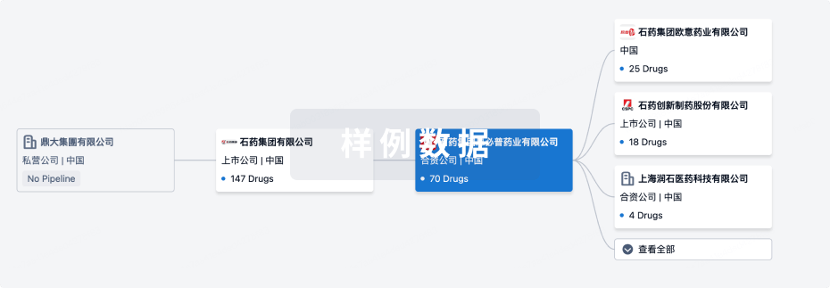
管线布局
2026年07月28日管线快照
管线布局中药物为当前组织机构及其子机构作为药物机构进行统计,早期临床1期并入临床1期,临床1/2期并入临床2期,临床2/3期并入临床3期
药物发现
4
27
临床前
其他
10
登录后查看更多信息
当前项目
| 药物(靶点) | 适应症 | 全球最高研发状态 |
|---|---|---|
GW604714X ( MPC1 x MPC2 ) | 糖尿病 更多 | 临床前 |
UK-5099 ( MPC1 x MPC2 ) | 糖尿病 更多 | 临床前 |
AKOS005153046 ( MPC1 x MPC2 ) | 糖尿病 更多 | 临床前 |
TAS-0612 ( Akt x p70S6K x p90 RSK ) | 急性淋巴细胞白血病 更多 | 临床前 |
PMD-026 ( p90 RSK ) | 复发性儿童急性淋巴细胞白血病 更多 | 临床前 |
登录后查看更多信息
药物交易
使用我们的药物交易数据加速您的研究。
登录
或
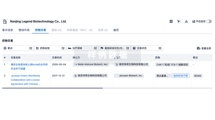
转化医学
使用我们的转化医学数据加速您的研究。
登录
或
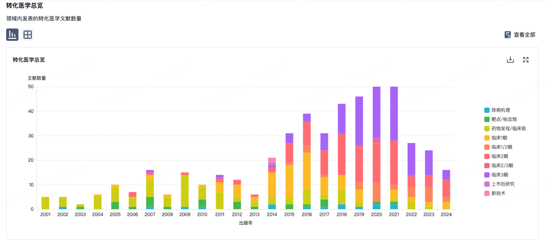
营收
使用 Synapse 探索超过 36 万个组织的财务状况。
登录
或
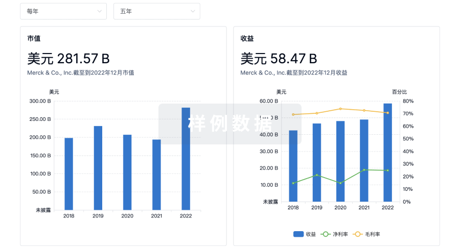
科研基金(NIH)
访问超过 200 万项资助和基金信息,以提升您的研究之旅。
登录
或
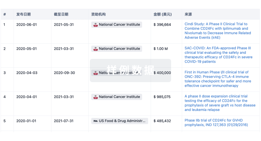
投资
深入了解从初创企业到成熟企业的最新公司投资动态。
登录
或
融资
发掘融资趋势以验证和推进您的投资机会。
登录
或
芽仔
全新生物医药AI Agent 覆盖科研全链路,让突破性发现快人一步
立即开始免费试用!
智慧芽新药情报库是智慧芽专为生命科学人士构建的基于AI的创新药情报平台,助您全方位提升您的研发与决策效率。
立即开始数据试用!
智慧芽新药库数据也通过智慧芽数据服务平台,以API或者数据包形式对外开放,助您更加充分利用智慧芽新药情报信息。
生物序列数据库
生物药研发创新
免费使用
化学结构数据库
小分子化药研发创新
免费使用