预约演示
更新于:2026-06-15
Chongqing Medical University
更新于:2026-06-15
概览
标签
肿瘤
其他疾病
神经系统疾病
小分子化药
单克隆抗体
蛋白水解靶向嵌合体(PROTAC)
疾病领域得分
一眼洞穿机构专注的疾病领域

技术平台
公司药物应用最多的技术
靶点
公司最常开发的靶点
关联
靶点 |
作用机制 |
在研机构 |
原研机构 |
在研适应症 |
非在研适应症 |
最高研发阶段 |
首次获批国家/地区 |
首次获批日期 |

靶点 |
作用机制 |
在研机构 |
原研机构 |
在研适应症 |
非在研适应症 |
最高研发阶段 |
首次获批国家/地区 |
首次获批日期 |

靶点 |
作用机制 |
在研机构 |
原研机构 |
在研适应症 |
非在研适应症 |
最高研发阶段 |
首次获批国家/地区 |
首次获批日期 |
NCT07625020
Ultrasound-Guided Nerve Hydrodissection for Diabetic Lower Limb Entrapment Neuropathy: A Randomized, Single-Blind, Sham-Controlled Trial
NCT07625007
Shortwave Intervention for Diabetic Peripheral Neuropathy: A Randomized, Single-Blind, Sham-Controlled Trial(SIDPN)
NCT07298889
Effect of High Versus Low Positive End-Expiratory Pressure on Intubation-Free Survival in Patients With Pneumonia or ARDS Receiving Noninvasive Ventilation: A Multicenter Randomized Controlled Trial
100 项与 重庆医科大学 相关的临床结果
登录后查看更多信息
登录后查看更多信息
2026-12-31INTERNATIONAL JOURNAL OF HYPERTHERMIA
Metabolic characteristic changes in local tissues after high-intensity focused ultrasound treatment for uterine fibroids
Article
作者: Mu, Yan ; He, Kun ; Shi, Qiuling ; Yang, Xi ; Huang, Guohua ; Ming, Xue ; Zhu, Tong ; Liu, Xiaofang
PURPOSE:
To investigate the changes in the metabolome of fibroid tissue after high-intensity focused ultrasound (HIFU) treatment, providing insights into its potential mechanisms of action.
MATERIALS AND METHODS:
Thirty patients with uterine fibroids who scheduled for hysterectomy were included. Among them, 15 patients had HIFU previously (HIFU group), and 15 patients did not receive HIFU (Control group). The tissues of treated fibroid, myometrium, endometrium, and new-emerging fibroids were collected. Gas chromatography-mass spectrometry (GC-MS) was applied for metabolomic analysis.
RESULTS:
GC-MS metabolomics analysis identified 94 metabolites. There was no significant difference in metabolite profile between the newly emerged fibroid tissue in the HIFU group and fibroid tissue in the Control group, and also no significant difference in the myometrial tissue and endometrial tissue between the two groups. In contrast, a significant difference was observed in metabolites between the treated fibroid tissue and the myometrial tissue in HIFU group. The levels of malus pumila acid, dimethyl fumarate, fumaric acid, methylphenylacetic acid, benzoic acid, and phosphoenolpyruvic acid were significantly higher (p < 0.05), and acetophenone, hexanoic acid, and idketoglutaric acid monoamide exhibited significantly decreased levels in the HIFU group (p < 0.05). Biomarker analysis revealed that malus pumila acid and nicotinic acid were elevated in the HIFU group, whereas erketoglutaric acid monoamide, hexanoic acid, and acetophenone were significantly reduced (p < 0.05).
CONCLUSION:
HIFU ablation may have a reprogramming effect on energy metabolism in the treated fibroids. The findings of this study provide a new direction for exploring the future development of biomarkers for combined metabolic regulation therapy.
2026-12-31JOURNAL OF DERMATOLOGICAL TREATMENT
Physical activity as a potential nonpharmacologic strategy to prevent seborrheic dermatitis: a cohort study in the UK biobank
Article
作者: Mao, Rui ; Liu, Jiachen ; Yang, Ziye ; Yang, Li ; Zhang, Tong ; Zhang, Tongtong
BACKGROUND:
Seborrheic dermatitis (SD) is a chronic inflammatory skin disease with limited long-term treatment options. The association between physical activity and incident SD, as well as the potential mediating role of depressive symptoms, remains unclear.
OBJECTIVE:
To investigate the association between physical activity and incident SD and to assess whether depressive symptoms mediate this relationship.
METHODS:
Data from 396,903 participants in the UK Biobank were analyzed. Cox proportional hazards models were used to examine the association between physical activity and incident SD. Regression-based mediation analyses were performed to quantify the extent to which depressive symptoms mediated this association.
RESULTS:
Physical activity above the recommended minimum (≥600 MET-min/week) was associated with a lower risk of incident SD. The greatest risk reduction was observed at 3-5 times the recommended level (1,800-3,000 MET-min/week; HR 0.80, 95% CI 0.71-0.90). Participants meeting criteria for healthy physical activity had a 28% lower risk of incident SD (HR 0.72, 95% CI 0.64-0.81). Mediation analyses showed that 17% of the protective association was explained by reduced depressive symptoms (indirect HR 0.83, 95% CI 0.77-0.88), while 16% was attributable to the direct effect of physical activity (direct HR 0.84, 95% CI 0.77-0.93).
CONCLUSIONS:
Physical activity may serve as a non-pharmacologic strategy for the prevention of SD, partly through alleviating depressive symptoms. The maximal benefit was observed at 3-5 times the recommended minimum activity level.
2026-12-31INTERNATIONAL JOURNAL OF HYPERTHERMIA
Effect of high-intensity focused ultrasound ablation on lesion stiffness and symptoms of adenomyosis
Article
作者: Shi, Qiuling ; Chen, Wenzhi ; Tian, Xin ; Zi, Dan ; Shen, Xuan ; Ren, Xiaoyu
OBJECTIVES:
To investigate the relationship between lesion stiffness and symptoms of patients and to evaluate the effectiveness of high-intensity focused ultrasound ablation (HIFUa) against lesion stiffness and symptoms of patients with adenomyosis.
METHODS:
A total of 124 patients with adenomyosis treated with HIFUa were included. Baseline characteristics and adenomyotic lesion stiffness were collected to identify factors associated with stiffness. Data on the changes of uterine lesion stiffness (E value), the menstrual volume score (PBAC), and the dysmenorrhea numeric rating scale (NRS) score before and 1, 3, and 6 months after HIFUa treatment in 42 patients were collected to explore the effects of HIFUa on stiffness and symptom changes.
RESULTS:
Before HIFUa, adenomyotic lesion stiffness was significantly greater than that of the normal myometrium. Adenomyotic lesion stiffness was significantly positively correlated with moderate to severe dysmenorrhea (r = 0.731), however, there was no significant linear correlation with hypermenorrhea (r = 0.153). Compared with that at pretreatment, stiffness at 3 and 6 months significantly decreased (that is, the elasticity increased) (p < 0.01). The NRS and PBAC scores at 1, 3, and 6 months were significantly lower than those at before HIFUa (p < 0.01), and the decrease in the NRS score was related to the decrease in stiffness.
CONCLUSION:
For adenomyosis patients with moderate to severe dysmenorrhea, lesion stiffness has been proven to be an objective reflection of dysmenorrhea severity. Lesion stiffness can be used as an imaging index to evaluate dysmenorrhea severity. Dysmenorrhea relief after HIFUa is related to decreased lesion stiffness.
2026-06-14
2026-06-12
·药圈头条
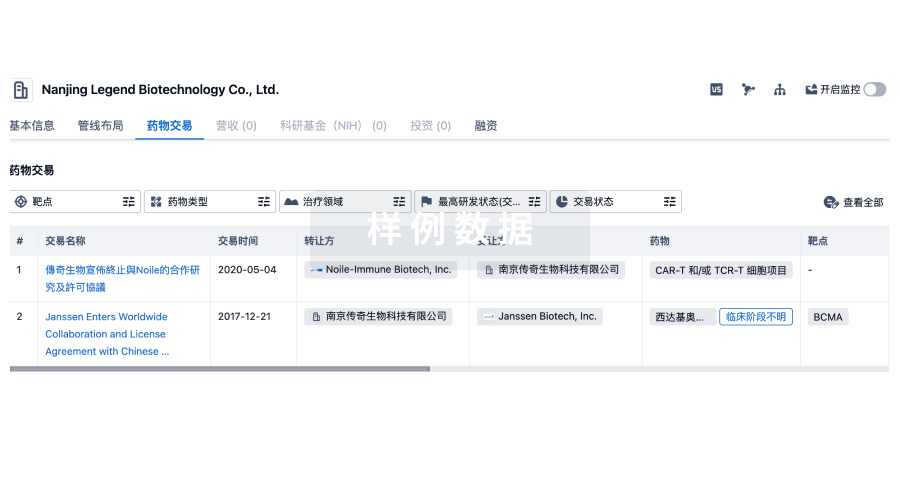
100 项与 重庆医科大学 相关的药物交易
登录后查看更多信息
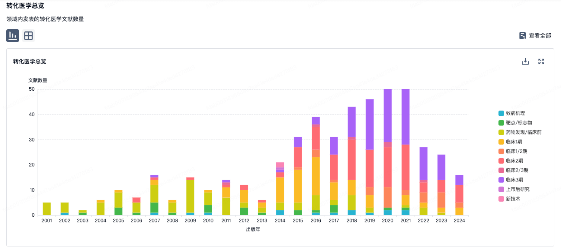
100 项与 重庆医科大学 相关的转化医学
登录后查看更多信息
组织架构
使用我们的机构树数据加速您的研究。
登录
或
管线布局
2026年07月21日管线快照
管线布局中药物为当前组织机构及其子机构作为药物机构进行统计,早期临床1期并入临床1期,临床1/2期并入临床2期,临床2/3期并入临床3期
药物发现
13
42
临床前
其他
1
登录后查看更多信息
当前项目
登录后查看更多信息
药物交易
使用我们的药物交易数据加速您的研究。
登录
或
转化医学
使用我们的转化医学数据加速您的研究。
登录
或
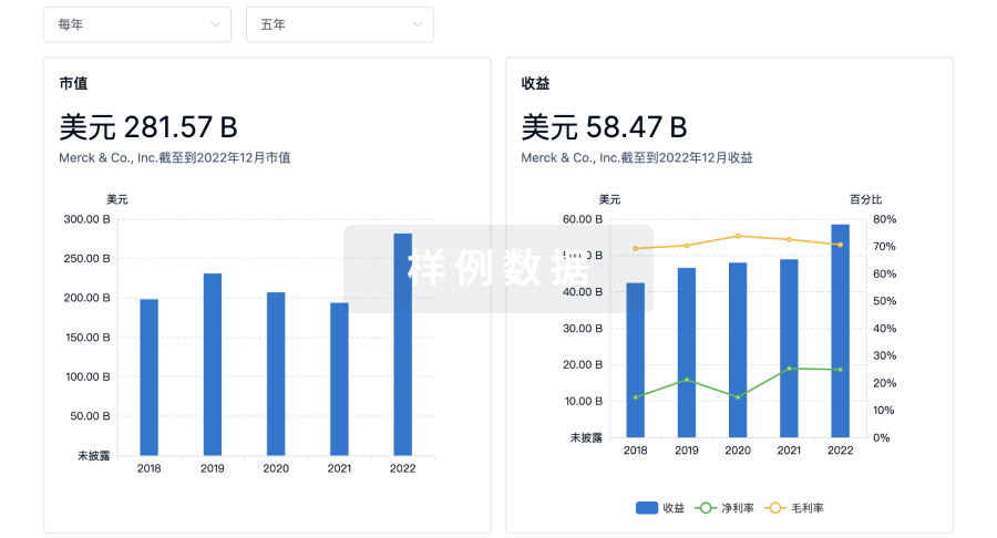
营收
使用 Synapse 探索超过 36 万个组织的财务状况。
登录
或
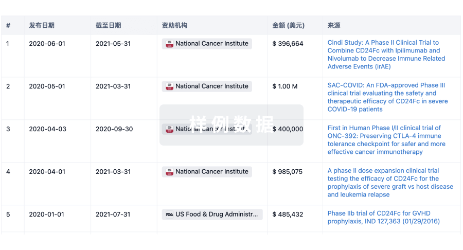
科研基金(NIH)
访问超过 200 万项资助和基金信息,以提升您的研究之旅。
登录
或
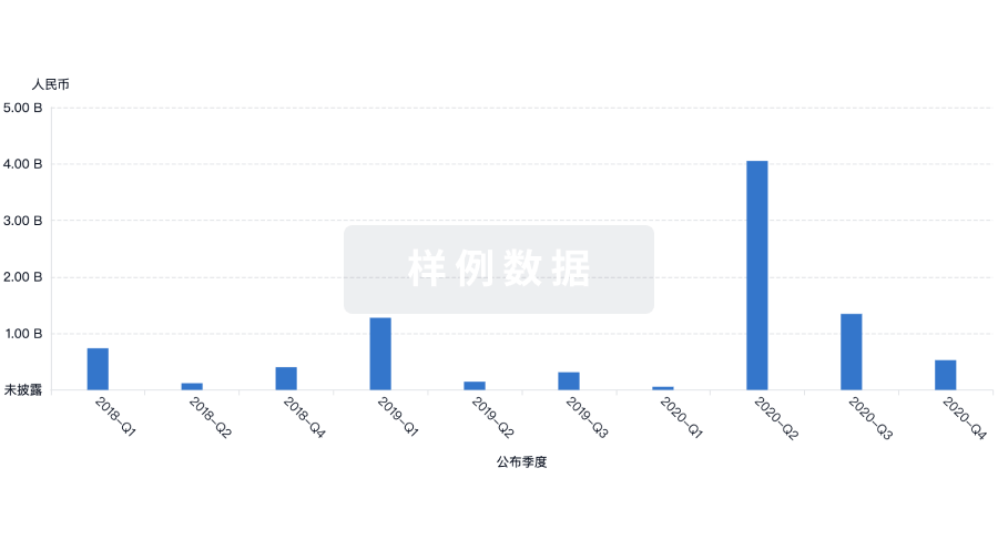
投资
深入了解从初创企业到成熟企业的最新公司投资动态。
登录
或
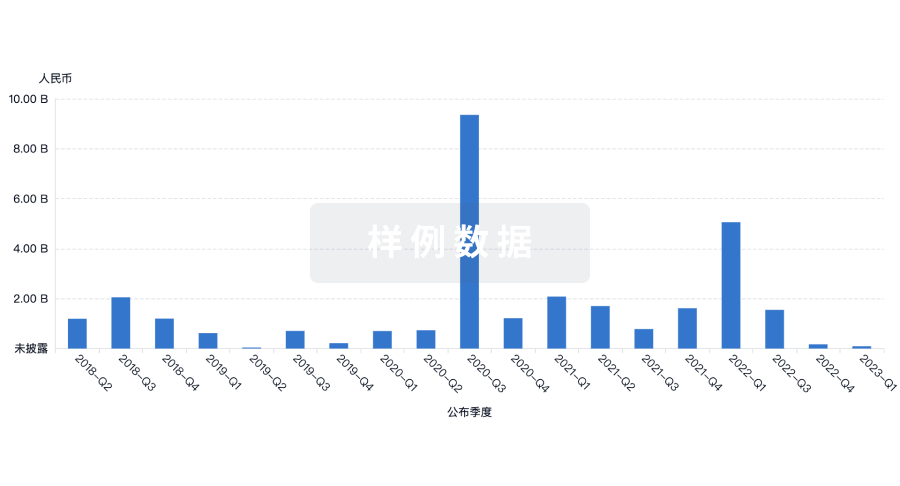
融资
发掘融资趋势以验证和推进您的投资机会。
登录
或
芽仔
全新生物医药AI Agent 覆盖科研全链路,让突破性发现快人一步
立即开始免费试用!
智慧芽新药情报库是智慧芽专为生命科学人士构建的基于AI的创新药情报平台,助您全方位提升您的研发与决策效率。
立即开始数据试用!
智慧芽新药库数据也通过智慧芽数据服务平台,以API或者数据包形式对外开放,助您更加充分利用智慧芽新药情报信息。
生物序列数据库
生物药研发创新
免费使用
化学结构数据库
小分子化药研发创新
免费使用