预约演示
更新于:2026-01-30

Revvity, Inc.
更新于:2026-01-30
概览
标签
肿瘤
神经系统疾病
其他疾病
蛋白水解靶向嵌合体(PROTAC)
基因疗法
疾病领域得分
一眼洞穿机构专注的疾病领域

技术平台
公司药物应用最多的技术
靶点
公司最常开发的靶点
关联
靶点 |
作用机制 |
在研机构 |
原研机构 |
在研适应症 |
非在研适应症 |
最高研发阶段 |
首次获批国家/地区 |
首次获批日期 |


靶点 |
作用机制 |
在研机构 |
原研机构 |
在研适应症 |
非在研适应症 |
最高研发阶段 |
首次获批国家/地区 |
首次获批日期 |

靶点 |
作用机制 |
在研机构 |
原研机构 |
在研适应症 |
非在研适应症 |
最高研发阶段 |
首次获批国家/地区 |
首次获批日期 |

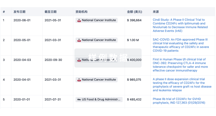
DRKS00032367
Exchange of human material for diagnostics development and production
NCT04370119
Screening for SARS-CoV-2-Infections and Monitoring of Serological Responses to SARS-CoV-2 in Healthcare Workers
NCT04141982
Clinical Concordance Evaluation of the T-SPOT®.TB Assay Performance Using Positive Selection With the T-Cell SelectTM Kit and Density Gradient Cell Isolation Methods

100 项与 Revvity, Inc. 相关的临床结果
登录后查看更多信息
登录后查看更多信息
2026-02-01INTERNATIONAL JOURNAL OF ANTIMICROBIAL AGENTS
EnvZ mutation–driven downregulation of catecholate siderophore receptors and concurrent TonB complex repression confer cefiderocol resistance in a KPC-producing ST11-KL64 hypervirulent Klebsiella pneumoniae
Article
作者: Zhuo, Xianxia ; Cao, Bin ; Song, Rongrong ; Wang, Chunhui ; Zhao, Jiankang ; Pu, Danni
OBJECTIVES:
Cefiderocol is an ultimate antibiotic option for Carbapenem-resistant hypervirulent Klebsiella pneumoniae (CR-hvKP). While resistance often involves metallo-β-lactamases, mechanisms in KPC-producing strains are unclear. This study aimed to elucidate novel cefiderocol resistance mechanisms in a clinical KPC-producing ST11-KL64 CR-hvKP isolate.
METHODS:
Cefiderocol-resistant mutants were generated through in vitro experimental evolution. Resistance-associated mutations were identified by whole-genome sequencing. Transcriptomic and proteomic analyses were performed to characterize global regulatory changes and were validated by qRT-PCR and targeted genetic manipulation. Additional tests examined siderophore production, intracellular iron levels, bacterial fitness, oxidative stress tolerance, and macrophage survival.
RESULTS:
All high-level cefiderocol-resistant mutants acquired a gain-of-function mutation in the sensor kinase EnvZ (V145G). Integrated transcriptomic and proteomic analyses showed that the envZ mutation drove marked downregulation of catecholate siderophore receptors (cirA and fepA), impairing cefiderocol uptake. In parallel, the TonB-ExbB-ExbD energy transduction complex was independently and stably downregulated, synergistically contributing to resistance. Notably, envZ mutation-associated repression of the enterobactin biosynthesis gene entB paradoxically increased cefiderocol susceptibility, indicating a regulatory trade-off. Resistant mutants exhibited reduced siderophore production, impaired intracellular iron accumulation, and significant fitness costs, including attenuated growth, reduced oxidative stress tolerance, and decreased survival within macrophages.
CONCLUSIONS:
In conclusion, this work uncovers a novel cefiderocol resistance mechanism in KPC-producing ST11-KL64 CR-hvKp, initiated by the envZ mutation, which causes the downregulation of catecholate siderophore receptors. This receptor repression, combined with the stable downregulation of the TonB-ExbB-ExbD energy complex, severely impairs cefiderocol's "Trojan horse" active uptake. This resistance mechanism is accompanied by a fitness trade-off, providing critical insights into the evolution of these superbugs.
2025-12-16ACS Omega
A Bifunctional Antibody Conjugate for Marking the Location of DNA Binding Proteins on DNA Fibers
Article
作者: Shaik, Althaf ; Seidman, Michael M. ; Gali, Himabindu ; Bellani, Marina A. ; Pokharel, Durga ; Ling, Chen
The cellular response to DNA damage can be marked by the appearance of immunofluorescent foci of DNA Damage Response (DDR) proteins. These serve as surrogates for DNA damage and are frequently interpreted as denoting specific lesions. For example, double-strand breaks (DSBs) are potent inducers of the DDR, whose best-known factor is the phosphorylated histone variant H2AX (γ-H2AX). The association with DSBs is so well established that the reverse interpretation that γ-H2AX invariably implies DSBs is routine. However, this conclusion is inferential and has been challenged. The resolution of this question has been hampered by the lack of methods for distinguishing the location of DDR proteins relative to DSBs caused by sequence indifferent agents. Here, we describe an approach for marking the location of DDR factors in relation to DSBs on DNA fibers, illustrated by γ-H2AX. We synthesized a two-arm "Y" conjugate containing biotin and trimethylpsoralen (TMP) coupled to a secondary antibody. After exposure to ionizing radiation, which introduces multiple lesions, including DSBs, cells were fixed, permeabilized, and incubated with a primary antibody against γ-H2AX followed by binding of the conjugated secondary antibody to the primary antibody. Exposure to long-wave UV light covalently linked psoralen to the DNA. DNA fibers were spread, and the immunofluorescence of the biotin tag indicated the location of the target protein. This technique denotes the location of γ-H2AX along DNA fibers following ionizing irradiation of the cells and the relationship between DSBs and γ-H2AX. Our results demonstrate that, after radiation, most γ-H2AX signals detected on DNA fibers are located at internal sites rather than at ends.
2025-10-01APPLIED RADIATION AND ISOTOPES
Aromatic ring tritiation and tritium decay catastrophe
Article
作者: Filer, Crist N
During her development of the radioimmunoassay (RIA) in the late 1950's, Rosalyn Yalow also discovered a curious property involving the 125I peptides used in her work. She found that many 125I peptides suffered irrevocable damage as they individually decayed. As a result of their altered structures, these non-radioactive fragmented peptides could no longer bind to the biological target and interfere with the non-decayed 125I peptide binding. This useful outcome acted to preserve the potency and molar activity of the remaining 125I peptides. Yalow termed this phenomenon "decay catastrophe." Recently, the first documented instance of tritium decay catastrophe for a fully tritiated methyl group (-C3H3) was reported. In this current work, an example is provided which also extends tritium decay catastrophe to a high molar activity radioligand labelled with tritium in several aromatic ring positions.
100 项与 Revvity, Inc. 相关的药物交易
登录后查看更多信息
100 项与 Revvity, Inc. 相关的转化医学
登录后查看更多信息
组织架构
使用我们的机构树数据加速您的研究。
登录
或
管线布局
2026年02月08日管线快照
管线布局中药物为当前组织机构及其子机构作为药物机构进行统计,早期临床1期并入临床1期,临床1/2期并入临床2期,临床2/3期并入临床3期
药物发现
2
13
临床前
其他
12
登录后查看更多信息
当前项目
登录后查看更多信息
药物交易
使用我们的药物交易数据加速您的研究。
登录
或
转化医学
使用我们的转化医学数据加速您的研究。
登录
或
营收
使用 Synapse 探索超过 36 万个组织的财务状况。
登录
或
科研基金(NIH)
访问超过 200 万项资助和基金信息,以提升您的研究之旅。
登录
或
投资
深入了解从初创企业到成熟企业的最新公司投资动态。
登录
或
融资
发掘融资趋势以验证和推进您的投资机会。
登录
或
生物医药百科问答
全新生物医药AI Agent 覆盖科研全链路,让突破性发现快人一步
立即开始免费试用!
智慧芽新药情报库是智慧芽专为生命科学人士构建的基于AI的创新药情报平台,助您全方位提升您的研发与决策效率。
立即开始数据试用!
智慧芽新药库数据也通过智慧芽数据服务平台,以API或者数据包形式对外开放,助您更加充分利用智慧芽新药情报信息。
生物序列数据库
生物药研发创新
免费使用
化学结构数据库
小分子化药研发创新
免费使用