预约演示
更新于:2026-05-12

Radius Health, Inc.
更新于:2026-05-12
概览
标签
皮肤和肌肉骨骼疾病
内分泌与代谢疾病
肿瘤
小分子化药
合成多肽
疾病领域得分
一眼洞穿机构专注的疾病领域

技术平台
公司药物应用最多的技术
靶点
公司最常开发的靶点
关联
靶点 |
作用机制 |
在研机构 |
原研机构 |
在研适应症 |
非在研适应症 |
最高研发阶段 |
首次获批国家/地区 |
首次获批日期 |


靶点 |
作用机制 |
在研机构 |
原研机构 |
在研适应症 |
非在研适应症 |
最高研发阶段 |
首次获批国家/地区 |
首次获批日期 |

靶点 |
作用机制 |
在研机构 |
原研机构 |
在研适应症 |
非在研适应症 |
最高研发阶段 |
首次获批国家/地区 |
首次获批日期 |

NCT04626141
Time to Healing, Loss of Fixation, and Loss of Alignment in Supracondylar Distal Femur Fractures Among a Geriatric Population Treated With Abaloparatide: A Double-Blind Placebo Controlled Study
NCT05387798
A Phase 3, Open-label Extension Study to Assess the Safety, Tolerability, and Efficacy of RAD011 (Cannabidiol Oral Solution) in Patients With Prader-Willi Syndrome
NCT05098509
A Phase 2/3, Randomized, Double-Blind, Placebo-Controlled Study of RAD011 (Cannabidiol Oral Solution) for the Treatment of Patients With Prader- Willi Syndrome
100 项与 Radius Health, Inc. 相关的临床结果
登录后查看更多信息
登录后查看更多信息
2025-09-01Bone Reports
Abaloparatide effects on BMD in acetabular regions corresponding to DeLee and Charnley zones in women with postmenopausal osteoporosis
Article
作者: Pearman, Leny ; Winzenrieth, Renaud ; Torkelson, Jared ; Wang, Yamei ; Bostrom, Mathias P ; Humbert, Ludovic ; Krohn, Kelly ; Sheth, Neil P ; Boxberger, John I
Background:
Acetabular bone loss in patients with osteoporosis is associated with increased risk of acetabular fragility fractures, significant morbidity, and can increase risk of complications in patients undergoing total hip arthroplasty. The anabolic osteoporosis treatment abaloparatide increases total hip areal bone mineral density (BMD), but its effect on acetabular BMD is unknown.
Methods:
Anatomical landmarks were identified in DXA scans from a random subgroup of postmenopausal women with osteoporosis (PMO) treated with abaloparatide 80 μg/d or placebo (n = 250/group) from the phase 3 ACTIVE trial to virtually place a hemispherical shell model of an acetabular cup and define regions of interest corresponding to DeLee and Charnley zones 1 (R1), 2 (R2), and 3 (R3). Changes in BMD from baseline at 6 and 18 months were calculated. Statistical significance was tested using a mixed model with repeated measures. Local mean changes in BMD were depicted by alignment of DXA scans via intensity-based registration onto a reference scan.
Results:
Abaloparatide significantly increased acetabular areal BMD in all three DeLee and Charnley zones at 6 and 18 months versus placebo. Mean BMD increases with abaloparatide were 8.38 % (R1), 7.25 % (R2), and 9.73 % (R3) at 18 months. BMD increases were homogenously distributed throughout the regions. With placebo, localized losses in BMD were noted after 18 months.
Conclusions:
Abaloparatide treatment rapidly and progressively increases BMD in acetabular zones in PMO.
Clinical trial number:
NCT01343004.
2025-07-05JBMR Plus
The effect of abaloparatide on the proximal femur in men with osteoporosis assessed by three-dimensional dual-energy X-ray absorptiometry
Article
作者: Boxberger, John ; Wang, Yamei ; Mitlak, Bruce H ; Binkley, Neil ; Humbert, Ludovic ; Dhaliwal, Ruban
Abstract:
Abaloparatide treatment significantly increased BMD at the LS, TH, and FN compared with placebo in men with osteoporosis in the phase 3 ATOM trial. The current study used 3D-DXA modeling to evaluate the effects of abaloparatide on cortical and trabecular compartments of the proximal femur in ATOM study participants. Proximal femur DXA images were retrospectively analyzed using 3D-DXA (3D-Shaper software v2.12.0, 3D-Shaper Medical, Barcelona, Spain) to evaluate changes in bone parameters from baseline at months 6 and 12 in all randomized men from the ATOM trial. Between-group comparisons were made for percent change from baseline data based on a mixed-effect repeated-measure model with treatment, visit, treatment-by-visit interaction, and type of DXA scanner as fixed effects. Other covariates include BMI, age, and baseline values of bone parameters. Abaloparatide treatment significantly increased integral volumetric BMD (vBMD) (3.7%), trabecular vBMD (7.0%), cortical thickness (1.1%), and cortical surface BMD (1.7%) at 12 mo compared to baseline (p < .0001). Changes were greater for abaloparatide compared to placebo for all 4 parameters (p < .01). Significant increases from baseline compared to placebo in integral vBMD (2.7% vs −0.1%, p < .0001) and trabecular vBMD (6.1% vs −0.6%, p < .0001) were also observed at 6 mo. In conclusion, in men with osteoporosis, abaloparatide improved proximal femur 3D-DXA parameters broadly consistent with results in postmenopausal women in the ACTIVE study, adding to the growing data on abaloparatide bone structure effects at the hip.
2025-03-15JOURNAL OF BONE AND MINERAL RESEARCH
Bone turnover markers predict changes in bone mineral density in men treated with abaloparatide: results from the abaloparatide for the treatment of men with osteoporosis (ATOM) study
Article
作者: Brown, Jacques P ; Binkley, Neil ; Wang, Yamei ; Eastell, Richard ; Adler, Robert A ; Kendler, David ; Lewiecki, E Michael ; Orwoll, Eric S ; Mitlak, Bruce H
Abstract:
Early increases in bone turnover markers (BTMs) in response to anabolic therapy correlate with 18-mo BMD increases in postmenopausal women with osteoporosis; however, this relationship has not been assessed in men. In this analysis, the correlation between changes from baseline in fasting intact serum procollagen type I N propeptide (PINP) and serum CTX at 1, 3, 6, and 12 mo and percent increase from baseline in BMD at 12 mo in men from the randomized phase 3 ATOM study (NCT03512262) were evaluated using Pearson’s correlation coefficients. The uncoupling index (UI), a measure of the balance between markers of bone formation (PINP) and bone resorption (CTX), with positive UI favoring bone formation, was calculated. Results in men were compared to 12-mo results for women from the ACTIVE study using the z score test after Fisher’s Z transformation. In abaloparatide-treated men, PINP increases at 1 mo (r = 0.485), 3 mo (r = 0.614), 6 mo (r = 0.632), and 12 mo (r = 0.521) were highly correlated (p < .0001) with 12-mo LS BMD increases. The mean UI for abaloparatide-treated men was greater than placebo as early as 1 mo (2.26 vs −0.25). At month 3, the mean UI for men was greater (1.32) than for women (0.88) (p < .001). There was a significant correlation between 3-mo UI and LS BMD at 12 mo in both men (r = 0.453; p < .001) and women (r = 0.252; p < .01). UI at months 6 and 12 were also significantly correlated with 12-mo LS BMD in men and women, but the correlation was stronger in men than women. These data support that early changes in BTMs in men treated with abaloparatide are associated with subsequent changes in BMD similar to what has been reported in women.
2026-04-24

2026-04-17

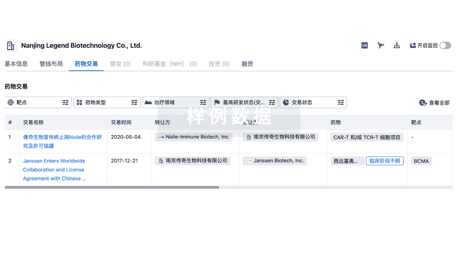
100 项与 Radius Health, Inc. 相关的药物交易
登录后查看更多信息
100 项与 Radius Health, Inc. 相关的转化医学
登录后查看更多信息
组织架构
使用我们的机构树数据加速您的研究。
登录
或
管线布局
2026年07月21日管线快照
管线布局中药物为当前组织机构及其子机构作为药物机构进行统计,早期临床1期并入临床1期,临床1/2期并入临床2期,临床2/3期并入临床3期
临床前
1
1
临床1期
申请上市
1
2
批准上市
其他
4
登录后查看更多信息
当前项目
登录后查看更多信息
药物交易
使用我们的药物交易数据加速您的研究。
登录
或
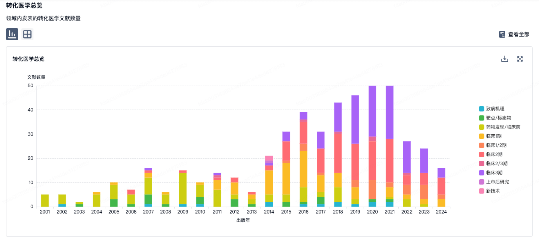
转化医学
使用我们的转化医学数据加速您的研究。
登录
或
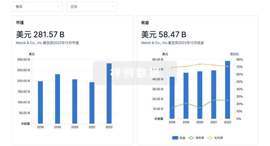
营收
使用 Synapse 探索超过 36 万个组织的财务状况。
登录
或
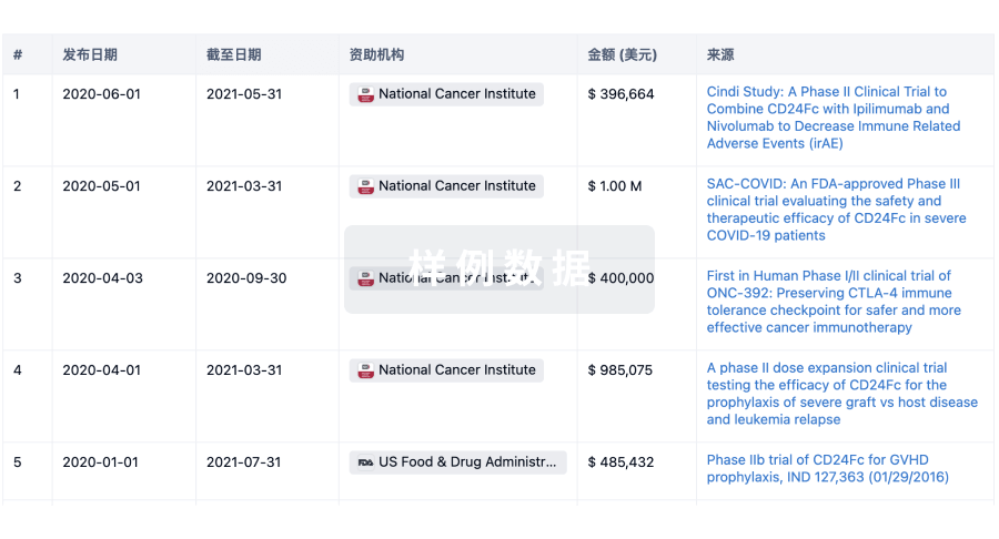
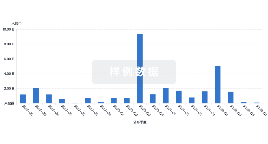
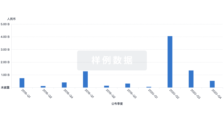
科研基金(NIH)
访问超过 200 万项资助和基金信息,以提升您的研究之旅。
登录
或
投资
深入了解从初创企业到成熟企业的最新公司投资动态。
登录
或
融资
发掘融资趋势以验证和推进您的投资机会。
登录
或
芽仔
全新生物医药AI Agent 覆盖科研全链路,让突破性发现快人一步
立即开始免费试用!
智慧芽新药情报库是智慧芽专为生命科学人士构建的基于AI的创新药情报平台,助您全方位提升您的研发与决策效率。
立即开始数据试用!
智慧芽新药库数据也通过智慧芽数据服务平台,以API或者数据包形式对外开放,助您更加充分利用智慧芽新药情报信息。
生物序列数据库
生物药研发创新
免费使用
化学结构数据库
小分子化药研发创新
免费使用