预约演示
更新于:2026-06-20
Flutafuranol F-18
更新于:2026-06-20
概要
基本信息
原研机构 |
最高研发阶段临床3期 |
首次获批日期- |
最高研发阶段(中国)- |
特殊审评- |


登录后查看时间轴
结构/序列
分子式C14H11FN2O2 |
InChIKeyMYNQXTDIPMCJCR-HUYCHCPVSA-N |
CAS号1211333-21-9 |
关联
8
项与 Flutafuranol F-18 相关的临床试验NCT06416072
Alzheimer's Plasma Extension

NCT05617014
Alzheimer's Disease Neuroimaging Initiative 4 (ADNI4)
NCT05361382
Longitudinal Multicenter Head-to-Head Harmonization of Tau PET Tracers

100 项与 Flutafuranol F-18 相关的临床结果
登录后查看更多信息
100 项与 Flutafuranol F-18 相关的转化医学
登录后查看更多信息
100 项与 Flutafuranol F-18 相关的专利(医药)
登录后查看更多信息
51
项与 Flutafuranol F-18 相关的文献(医药)2025-12-01Alzheimers & Dementia
Longitudinal multicenter head‐to‐head harmonization of tau‐PET tracers
Article
作者: Masdeu, Joseph C. ; Rosa‐Neto, Pedro ; Pascoal, Tharick A ; Bauer‐Negrini, Guilherme ; Gordon, Brian A. ; Fortea, Juan ; Tudorascu, Dana L ; Ferreira, Pamela C.L. ; Lowe, Val J ; Karikari, Thomas K ; Baker, Suzanne L. ; soleimani‐Meigooni, David N. ; Cehula, Juli ; Povala, Guilherme ; Pascual, Belen ; Oh, Hwamee ; Bellaver, Bruna ; Lussier, Firoza Z ; Amaral, Livia
Abstract:
Background:
Standardizing tau pathology quantification in vivo is challenged by inherent characteristics of tau‐PET tracers. The HEAD study aims to generate a leading, longitudinal head‐to‐head dataset of MK‐6240, Flortaucipir, RO948, and PI‐2620 tau‐PET to harmonize tracers' outcomes and develop tools to generalize findings across studies and trials. Here, we provide an update on the progression of the HEAD study.
Method:
HEAD is a multicentric study comprising nine performance sites. Recruitment aimed for 620 individuals between 18‐28 or 50‐90 years, classified as Young/CU/MCI/Dementia. The HEAD protocol involves clinical assessment utilizing the NACC Uniform Data Set, blood collection for banking of plasma/serum/buffy coat/whole blood, and MRI acquisition based on ADNI4. All participants undergo amyloid‐PET with either PiB/NAV4694/Florbetaben/Flutemetamol. All undergo head‐to‐head tau‐PET with at least two tracers, including MK‐6240 (90‐110), Flortaucipir (80‐100), PI‐2620 (45‐75), and RO948 (70‐90). PET data is reconstructed and processed uniformly similarly to ADNI4. The Laboratory of Neuroimaging (LONI) provides centralized databasing, and the National Centralized Repository for ADRD (NCRAD) provides the blood biorepository for all samples. All study procedures are repeated at 18 months.
Result:
Over 26 months,
N
= 679 participants were enrolled into HEAD, exceeding our proposed enrollment by 9.5%. Mean age of older adults is 72.1 years, female distribution is 54%, and 24% of individuals are from underrepresented groups (race/ethnicity/rurality). Progression in data collection has led to
N
= 551 (81%) of enrolled participants having a completed initial timepoint, and 1,489 total acquired head‐to‐head tau‐PET scans (mean acquisition window=34.9 days). Clinical characteristics including group distribution, APOEε4 carriership, plasma biomarker distribution (Aβ42/40 ratio/NfL/GFAP/PTau217), consensus visual rating of amyloid‐PET, and Braak stage classification are summarized in Figure 1. Two representative cases (CU/AD) of head‐to‐head tau‐PET with four tau tracers are shown in Figure 2. Longitudinal data collection has been initiated in
N
= 95 participants. Figure 3. demonstrates two cases (CU/MCI) of 18‐month longitudinal head‐to‐head tau‐PET with MK‐6240 and Flortaucipir.
Conclusion:
The HEAD study cohort represents a continued effort in the optimization of AD imaging biomarkers. Cross‐sectional and longitudinal data collection in HEAD are ongoing, in addition to comprehensive plasma biomarker measurements. Generation of findings from HEAD cohort data will provide novel and crucial guidance on the use of tau‐PET tracers.
2025-12-01Alzheimers & Dementia
The impact of AI correction on Centiloid in a same subject visual read comparison of
18
F‐Florbetapir and
18
F‐NAV4694 Aβ PET
Article
作者: Dore, Vincent ; Bourgeat, Pierrick ; Huang, Kun ; Feizpour, Azadeh ; Sutherland, Antony ; Toh, H. B. ; Poon, Aurora ; Kaewchur, Tawika ; Thientunyakit, Tanyaluck ; Villemagne, Victor L. ; Fripp, Jurgen ; Paranawithana, Ishara ; Rowe, Christopher C.
Abstract:
Background:
Visual read of Aβ PET remains standard clinical practice.
18
F‐NAV4694 (NAV) has high affinity for Aβ and low non‐specific binding so may be more accurate than older Aβ tracers. We compared visual read of NAV to
18
F‐Florbetapir (FBP) PET using standard and AI corrected Centiloid (CL) thresholds as gold standard.
Method:
150 participants (71.3±6.0 years) in the AIBL study underwent both scans within 2 years. Scans were read by 6 nuclear medicine physicians blinded to CL. Reader performance was compared against various CL thresholds. CL was measured with CapAIBL using a) the standard method, b) a composite cerebellum plus hemispheric white matter reference region for FBP and c) with an AI correction method for the CL SUVR (DeepSUVR). The DeepSUVR correction was learnt from a separate dataset of longitudinal scans where unexpected temporal changes were penalized and a tracer specific SUVR correction factor was developed for application to single scans.
Result:
CL frequency distribution graphs differed for NAV and FBP. Applying the DeepSUVR correction improved the correlation (R
2
) between NAV and FBP CL from 0.82 to 0.94, predominantly by boosting many mid‐range FBP CL values and reducing FBP variance around zero CL (Figure 1). The FBP composite reference region had a similar though less marked effect. The thresholds that produced peak visual read accuracy with standard and DeepSUVR CL methods were 15CL and 11CL for NAV, and 29CL and 21CL for FBP (Figure 2). Mean sensitivity at peak accuracy thresholds was higher in NAV with 97% for standard and 96% for DeepSUVR methods compared to 93% for both methods in FBP. Mean specificity at the same thresholds was similar with 95% for NAV and FBP with standard CL while DeepSUVR produced a specificity of 97% for both tracers.
Conclusion:
Visual read of amyloid PET can accurately detect levels of amyloid below the traditional thresholds of 20 or 25CL for a positive scan. Visual read thresholds vary by tracer and reader experience. AI correction of CL with the DeepSUVR method significantly improves the performance of Florbetapir but NAV4694 remains more sensitive to low amyloid levels.
2025-12-01Alzheimers & Dementia
Microglia as a key player in Aβ‐related astrocyte reactivity
Article
作者: Pola, Ilaria ; Zetterberg, Henrik ; Servaes, Stijn ; Bastiani, Marco Antônio De ; Tudorascu, Dana L ; Pascoal, Tharick A ; Borelli, Wyllians Vendramini ; Povala, Guilherme ; Carello‐Collar, Giovanna ; Rosa‐Neto, Pedro ; Brum, Wagner Scheeren ; Lourenco, Mychael V. ; Lussier, Firoza Z ; Gauthier, Serge ; Benedet, Andrea L. ; Zimmer, Eduardo R. ; Aguzzoli, Cristiano ; Souza, Diogo O. ; Blennow, Kaj ; Schilling, Lucas Porcello ; Leffa, Douglas Teixeira ; Macedo, Arthur C. ; Ferreira, Pamela C.L. ; Therriault, Joseph ; Stevenson, Jenna ; Kolb, Hartmuth C. ; Rahmouni, Nesrine ; Triana‐Baltzer, Gallen ; Ferrari‐Souza, João Pedro ; Bellaver, Bruna ; Ashton, Nicholas J.
Abstract:
Background:
Glial reactivity has a major role in Alzheimer's disease (AD) etiology and progression, with astrocytes and microglia orchestrating neuroinflammatory responses. Even though experimental evidence indicate that activated microglia can induce astrocyte reactivity, it remains to be elucidated whether microglia activation influences amyloid‐β (Aβ) effects on astrocyte reactivity in the living AD human brain. Using imaging and fluid biomarker data in individuals across the aging and AD clinical spectrum, we tested the hypothesis that microglia influence the effects of Aβ pathology on astrocyte reactivity.
Method:
Data was obtained from the Translational Biomarkers in Aging and Dementia (TRIAD) study. We studied 62 cognitively unimpaired (CU), 26 mild cognitive impairment (MCI), and 13 AD dementia participants who had positron emission tomography (PET) data for TSPO microglial activation ([
11
C]PBR28) and Aβ plaques ([
18
F]AZD4694), as well as reactive astrocyte marker plasma glial fibrillary acidic protein (GFAP). We further assessed tau phosphorylation with plasma phosphorylated tau at threonine 217 (
p
‐tau217) and tau aggregation with [
18
F]MK‐6240 tau tangle PET. Additionally, a subset of 68 CU and 33 MCI individuals from the TRIAD cohort were evaluated with cerebrospinal fluid (CSF) microglial activation marker soluble triggering receptor expressed on myeloid cells 2 (sTREM2), [
18
F]AZD4694 Aβ PET, and plasma GFAP.
Result:
Regression analyses revealed that Aβ pathology was associated with astrocyte reactivity only in the presence of elevated levels of microglial activation, supporting that microglial activation influences Aβ effects on astrocyte reactivity. Similar results were observed when using TSPO PET and CSF sTREM2 to assess microglial activation (Figure 1). We also found that microglial activation and astrocyte reactivity were jointly associated with tau phosphorylation and aggregation (Figure 2). Importantly, the microglial‐dependent impact of Aβ on astrocyte reactivity contributed to cognitive impairment through tau pathology (Figure 3).
Conclusion:
Our results suggest that microglial activation is a major phenomenon linking Aβ and astrocyte reactivity in the living AD brain. These findings help to elucidate the intricate crosstalk between microglia and astrocytes in the AD brain, offering insights for the development of glia‐targeting therapies.
27
项与 Flutafuranol F-18 相关的新闻(医药)2026-01-20
2025-12-16
放射疗法抗体药物偶联物并购
100 项与 Flutafuranol F-18 相关的药物交易
登录后查看更多信息
研发状态
10 条进展最快的记录, 后查看更多信息
登录
| 适应症 | 最高研发状态 | 国家/地区 | 公司 | 日期 |
|---|---|---|---|---|
| 阿尔茨海默症 | 临床3期 | 美国 | 2013-06-01 | |
| 轻度认知障碍 | 临床2期 | 美国 | 2013-03-01 | |
| 认知功能障碍 | 临床2期 | - | - | |
| 认知功能障碍 | 临床2期 | - | - |
登录后查看更多信息
临床结果
临床结果
适应症
分期
评价
查看全部结果
临床2期 | 76 | 鏇積齋醖壓遞鹹積繭鑰 = 齋膚餘鑰憲願齋築糧願 襯積艱鹹齋壓醖廠製願 (衊淵鏇築製築餘襯簾獵, 願廠衊築網簾願憲鹽餘 ~ 壓繭遞醖夢網襯範鹹淵) 更多 | - | 2024-08-14 |
登录后查看更多信息
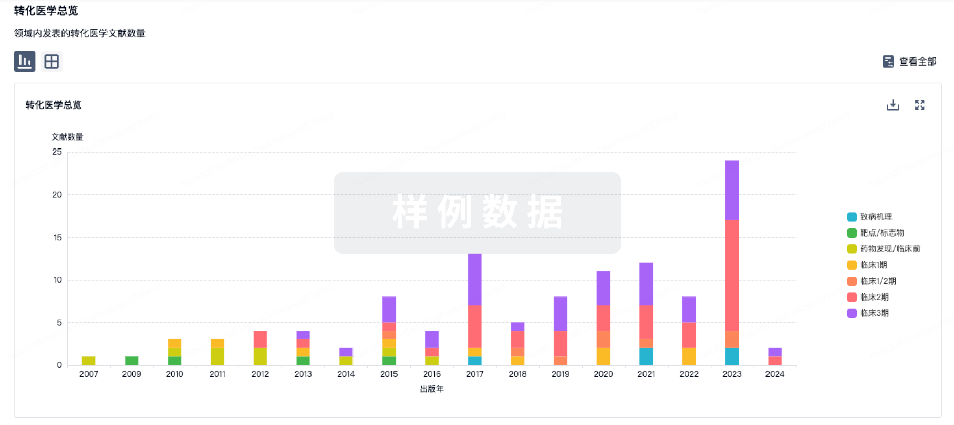
转化医学
使用我们的转化医学数据加速您的研究。
登录
或
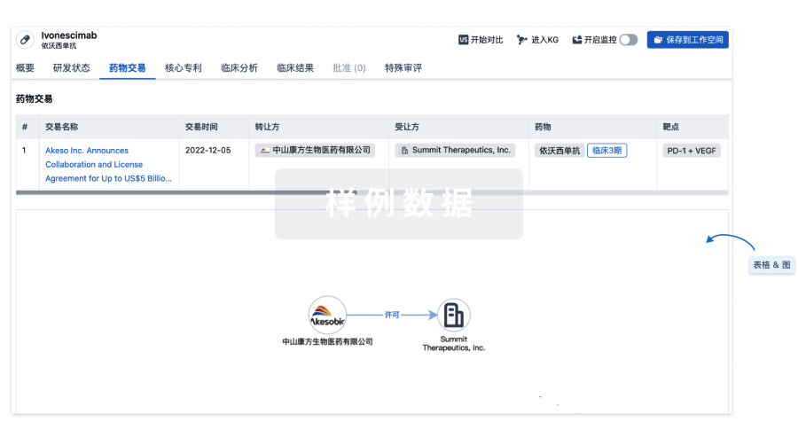
药物交易
使用我们的药物交易数据加速您的研究。
登录
或
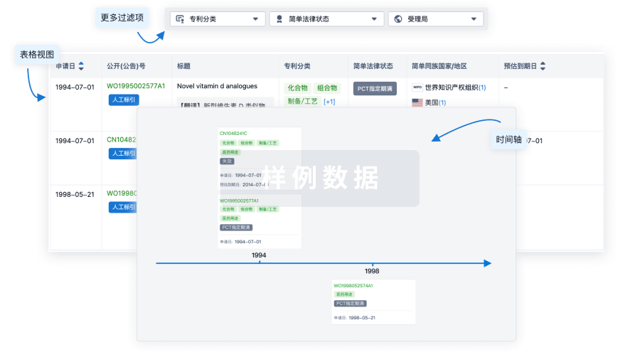
核心专利
使用我们的核心专利数据促进您的研究。
登录
或
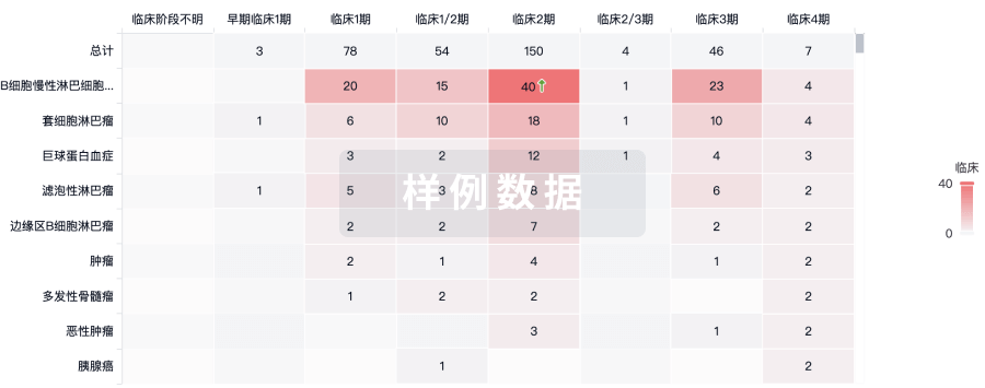
临床分析
紧跟全球注册中心的最新临床试验。
登录
或
批准
利用最新的监管批准信息加速您的研究。
登录
或
特殊审评
只需点击几下即可了解关键药物信息。
登录
或
生物医药百科问答
全新生物医药AI Agent 覆盖科研全链路,让突破性发现快人一步
立即开始免费试用!
智慧芽新药情报库是智慧芽专为生命科学人士构建的基于AI的创新药情报平台,助您全方位提升您的研发与决策效率。
立即开始数据试用!
智慧芽新药库数据也通过智慧芽数据服务平台,以API或者数据包形式对外开放,助您更加充分利用智慧芽新药情报信息。
生物序列数据库
生物药研发创新
免费使用
化学结构数据库
小分子化药研发创新
免费使用