预约演示
更新于:2026-05-01
Artenimol
双氢青蒿素
更新于:2026-05-01
概要
基本信息
原研机构 |
在研机构 |
非在研机构- |
最高研发阶段批准上市 |
首次获批日期 中国 (1997-01-01), |
最高研发阶段(中国)批准上市 |
特殊审评特殊审批 (中国) |

登录后查看时间轴
结构/序列
分子式C15H24O5 |
InChIKeyBJDCWCLMFKKGEE-ISOSDAIHSA-N |
CAS号71939-50-9 |
关联
25
项与 双氢青蒿素 相关的临床试验NCT07557927
A Multicentre, Randomised, Double-blind, Positive-control Clinical Trial Evaluating Dihydroartemisinin Tablets for the Treatment of Discoid Lupus Erythematosus
This study is a multicentre, randomised, double-blind, double-dummy, phase II clinical trial with a positive-control group, designed to evaluate the efficacy and safety of dihydroartemisinin tablets in the treatment of discoid lupus erythematosus (DLE).
开始日期2026-04-30 |
申办/合作机构 |
ChiCTR2500102437
Preoperative Oral Dihydroartemisinin on Tumor Neoantigens and the Tumor Immune Microenvironment in Triple-Negative Breast Cancer: A Single-Center, Open-Label, Single-Arm Pilot Study
开始日期2025-10-15 |
申办/合作机构 |

ChiCTR2500103939
A prospective phase II clinical study of radiotherapy combined with dihydroartemisinin (DHA) in the treatment of brain metastases from breast cancer
开始日期2025-07-01 |
申办/合作机构 |

100 项与 双氢青蒿素 相关的临床结果
登录后查看更多信息
100 项与 双氢青蒿素 相关的转化医学
登录后查看更多信息
100 项与 双氢青蒿素 相关的专利(医药)
登录后查看更多信息
2,580
项与 双氢青蒿素 相关的文献(医药)2026-06-01·TOXICOLOGY AND APPLIED PHARMACOLOGY
MiR-29a/DNMT3A axis participates in Dihydroartemisinin's suppression on lung adenocarcinoma: Implications for overcoming acquired resistance to EGFR-TKIs
Article
作者: Tang, Jing ; Qiu, Ping ; Chen, Liwen ; Liu, Yu ; Chen, Renjie ; Zhu, Yayu ; Ling, Huijuan ; Liu, Yaqing
Dihydroartemisinin (DHA), the traditional antimalarial agent, has attracted significant interest as potential anticancer drug, yet its underlying mechanisms are incompletely understood. Herein, we showed that DNA methyltransferase 3 alpha (DNMT3A), a de novo DNA methyltransferase was substantially inhibited by DHA in both in vitro and in vivo lung adenocarcinoma settings. Overexpression of DNMT3A effectively counteracted DHA's inhibition on the proliferation of HCC827 and A549 cells. Intriguingly, DNMT3A was observed to be up-regulated in Gefitinib-resistant HCC827 (HCC827/GR) cells as compared with parental Gefitinib-hypersensitive HCC827 cells. DNMT3A overexpression effectively counteracted the inhibition of Gefitinib on HCC827 cells. Conversely, si-RNA mediated DNMT3A knockdown generated considerable inhibitory effects on HCC827/GR cells, and further increased Gefitinib sensitivity. Furthermore, combination of DHA and Gefitinib generated significant synergistic effects on the suppression of cell viability and induction of apoptosis of both HCC827 and HCC827/GR cells. Finally, our results showed that DHA effectively up-regulated the expression of miR-29a. Transfection with miR-29a mimics substantially suppressed DNMT3A expression whereas with miR-29a inhibitor effectively up-regulated DNMT3A in HCC827 and A549 cells. Furthermore, miR-29a inhibitor effectively restored DHA's suppression on DNMT3A expression in both cell lines. Overall, these data demonstrate that DHA has therapeutic effects on lung adenocarcinoma by down-regulating DNMT3A through miR-29a. Understanding of this mechanism will have implications for overcoming acquired resistance in epidermal growth factor receptor (EGFR) - targeted therapy by using tyrosine kinase inhibitors (TKIs).
2026-05-01·BIOCHIMICA ET BIOPHYSICA ACTA-GENERAL SUBJECTS
Protective effects of dihydroartemisinin against sterile inflammation and oxidative stress in alveolar macrophages
Article
作者: Yeh, Wei-Lan ; Chang, Chen-Ni ; Chen, Yen-Chang ; Wang, Yu-Wen ; Lu, Dah-Yuu ; Lin, Wei-Ting ; Huang, Jun-Ting ; Chen, Jia-Hong ; Lin, Jia-Yu ; Tsai, Cheng-Fang
BACKGROUND:
Pulmonary alveoli are highly vulnerable to oxidative stress due to high oxygen tension, with alveolar macrophages serving as the primary defense. Dihydroartemisinin (dha), an active derivative of Artemisia annua L., possesses potent biological activities. We investigated the efficacy and mechanisms of dha against acute lung injury (ALI).
METHODS:
Inflammation and oxidative stress were induced in MH-S alveolar macrophages using PMA and TNFα. In vivo, an ALI mouse model was established via intratracheal TNFα administration, followed by oral dha treatment (3 days). We assessed inflammasome activation, reactive oxygen species (ROS) levels, and lung pathology.
RESULTS:
In MH-S cells, dha significantly inhibited PMA- and TNFα-induced inflammasome activation by downregulating NLRP3, ASC, and cleaved-caspase-1. Dha also reduced total and mitochondrial ROS production, likely through SOD2 upregulation. In mice, oral dha effectively alleviated TNFα-induced immune cell infiltration, cytokine secretion, pulmonary edema, and early-stage fibrotic remodeling. These protective effects were associated with attenuated pulmonary inflammasome activation and IL-1β expression.
CONCLUSIONS:
Dha exerts robust anti-inflammatory and anti-oxidative effects by modulating the NLRP3/ASC axis and mitochondrial ROS production, thereby mitigating pulmonary structural damage.
GENERAL SIGNIFICANCE:
These findings highlight dha as a promising therapeutic candidate for acute inflammatory lung diseases, providing a mechanistic basis for its clinical potential.
2026-05-01·HUMAN IMMUNOLOGY
The therapeutic potential of Dihydroartemisinin for autoimmune disease: Effects on the Th17/Treg balance
Review
作者: Mao, Yuwen ; Zhao, Yanni ; Zhou, Juncheng ; Yimin, Reyihanguli ; Gao, Yudong
Autoimmune diseases (AID) are chronic inflammatory conditions resulting from aberrant recognition of self-antigens by the immune system. Appropriate ratios of the subsets of CD4+ T cells, T helper cells 17 (Th17) and regulatory T cells (Treg), are necessary for immune homeostasis with imbalances associated with immune dysregulation and AID. These observations imply that restoration of Th17/Treg balance may represent a therapeutic approach for AID. Dihydroartemisinin (DHA) is a metabolite of artemisinin which has been shown to suppress excessive Th17 cell differentiation, restoring Th17/Treg balance and ameliorating AID. The current review summarizes recent progress on DHA and adjustment of Th17/Treg balance in autoimmune diseases such as systemic lupus erythematosus, rheumatoid arthritis and inflammatory bowel disease. Reference material was largely from that indexed in PubMed, Web of Science, China National Knowledge Infrastructure, Google Scholar and Wanfang Database between 2010 and 2025 with a few studies from 2008 and a focus on publications from the last five years.
63
项与 双氢青蒿素 相关的新闻(医药)2026-04-30
本期内容聚焦于2026年4月21日至4月28日的膀胱癌研究动态,精心筛选并汇总了多项具有代表性的前沿研究进展。为了方便读者更好地把握研究方向,我们将这些文献进行分类整理,力求呈现一个系统、全面的研究概览。 本文汇总了膀胱癌领域的最新高影响力研究,涵盖药物开发、治疗策略、诊断技术及基础机制。
发表在《Cancer Discovery》(IF 33.3)的一项研究揭示,野火烟雾暴露不仅危害呼吸健康,还与膀胱癌等多种癌症风险显著增加相关,提示环境因素在癌症预防中的重要性。
《European Urology》(IF 25.2)发表的回复文章强调,免疫检查点抑制剂联合放化疗在肌层浸润性膀胱癌中展现出保膀胱治疗的良好潜力,为患者提供了新的治疗选择,推动了肿瘤免疫治疗与传统疗法的融合。
《Biosensors and Bioelectronics》(IF 10.5)报道了一种创新的DNA纳米结构结合CRISPR/Cas12a信号放大技术,实现了膀胱癌尿液细胞的高灵敏无创检测,极大提升了诊断的准确性和实用性。
《Nano Letters》(IF 9.1)提出基于尿素酶驱动纳米马达的膀胱内化疗新策略,显著提高药物在肿瘤中的分布和疗效,实现肿瘤显著缩小和早期复发抑制,展现出广阔的临床应用前景。
《Advanced Science》(IF 14.1)的一项研究揭示,膀胱癌中O-GlcNAc化修饰通过破坏抗原呈递机制促进肿瘤免疫逃逸,指出了免疫治疗的新靶点,助力提升膀胱癌免疫治疗效果。
整体研究为膀胱癌的个性化治疗和精准管理提供了重要理论基础和临床指导。
药物类:
1.Mol Cancer Ther(IF:5.5):靶向整合素β6的抗体-药物结合物优化用于膀胱内给药治疗非肌层浸润性膀胱癌;
2.Bioorg Chem(IF:4.7):新型二氢青蒿素-布洛芬杂合物的设计、合成及抗膀胱癌活性研究;
3.Sci Rep(IF:3.9):恩格列净增强顺铂在化疗耐药EJ138膀胱癌细胞中的活性:抗糖尿病药物在癌症治疗中的重要性;
治疗类
1.Eur Urol(IF:25.2):伊匹单抗和纳武利尤单抗联合化放疗作为肌层浸润性膀胱癌保膀胱治疗的二期试验回顾;
2.Nano Lett(IF:9.1):纳米马达辅助膀胱内化疗用于膀胱肿瘤缩小及早期肿瘤再生抑制;
3.Eur J Cancer(IF:7.1):高危膀胱癌检查点抑制剂与辅助BCG试验中的删失模式及结果不一致性分析;
4.Int Immunopharmacol(IF:4.7):STING激活型纳米药物在癌症免疫治疗中的机制、设计与治疗结局;
5.Minerva Urol Nephrol(IF:4.2):基于ROBUUST 2.0注册研究的上尿路尿路上皮癌国际指南管理的多中心真实世界依从性分析;
6.Minerva Urol Nephrol(IF:4.2):经尿道肿瘤切除术后延迟或不完整BCG方案对非肌层浸润性膀胱癌患者的不良影响:系统综述;
7.Curr Oncol(IF:3.4):高危非肌层浸润性膀胱癌的放射治疗:现有证据与未来方向;
手术类:
1.Eur Urol Oncol(IF:9.3):肌层浸润性膀胱癌的深度系统反应:根治性膀胱切除术仍然有必要吗?;
2.Eur Urol Open Sci(IF:4.5):根治性膀胱切除术后的生活质量:多种评估工具下新膀胱与回肠膀胱造口结局的Meta分析;
3.Minerva Urol Nephrol(IF:4.2):高容量三甲机器人中心20年机器人辅助根治性膀胱切除术生存结局的演变;
诊断类:
1.Biosens Bioelectron(IF:10.5):双功能DNA多价结构结合稳定捕获尿路上皮癌细胞与CRISPR/Cas12a信号放大用于膀胱癌检测;
2.Eur Urol Open Sci(IF:4.5):基于放射组学的可解释模型结合白光膀胱镜图像预测膀胱癌分级的新方法;
3.Ann Surg Oncol(IF:3.5):ASO作者反思:成像肿瘤异质性揭示膀胱癌隐匿性淋巴结风险;
4.Front Oncol(IF:3.3):循环肿瘤DNA作为尿路上皮癌的预后与监测生物标志物;
其他类:
1.Cancer Discov(IF:33.3):野火烟雾暴露与癌症风险增加相关;
2.Nat Rev Urol(IF:14.6):伯明翰的膀胱癌研究正在蓬勃发展;
3.Nat Rev Urol(IF:14.6):女性膀胱癌结局较男性更差——偏见、生物学还是两者兼有?;
4.Adv Sci (Weinh)(IF:14.1):O-GlcNAc修饰的TAP1损害抗原递呈并促进膀胱癌免疫逃逸;
5.Med Image Anal(IF:11.8):FedSemiDG:域泛化联邦半监督医学图像分割;
6.Eur Urol Oncol(IF:9.3):关于Jurczok N等人“多区域免疫谱分析揭示膀胱癌的预后模式”的评论;
7.Chem Biol Interact(IF:5.4):多维转录组分析及体外实验剖析2',2',4',4'-四溴二苯醚在膀胱癌进展中的作用;
8.Cancer Immunol Immunother(IF:5.1):CD39/CD73介导的免疫抑制与膀胱癌肿瘤侵袭性的相关性;
9.Int J Cancer(IF:4.7):癌症护理质量认证与多种癌症类型患者生存率的相关性:台湾基于人群的队列研究;
10.Immunotargets Ther(IF:4.4):多组学分析揭示m7G甲基化相关基因可能参与TGF-β信号介导的膀胱癌抗PD-L1反应;
11.BJU Int(IF:4.4):不同风险组非肌层浸润性膀胱癌的监测模式:真实世界分析;
12.Hum Mutat(IF:3.7):HSPB6:一种通过PI3K/Akt信号通路抑制上皮-间质转化(EMT)过程的潜在预后生物标志物——基于机器学习与实验验证;
13.Curr Oncol(IF:3.4):日本HER2扩增的肿瘤无关谱系:真实世界流行率及其HER2靶向治疗的意义;
药物类:
1. 一种针对整合素β6的抗体药物偶联物,优化用于膀胱内给药治疗非肌层浸润性膀胱癌
期刊名称:Molecular Cancer Therapeutics
影响因子:5.5
JCR分区:Q1
作者:Christopher M Carosino(一作),Matthew R Levengood(通讯)
单位:Pfizer (United States) Bothell United States
DOI:https://doi.org/10.1158/1535-7163.MCT-26-0077
摘要:非肌层浸润性膀胱癌(NMIBC)占所有新诊断膀胱癌的75%。尽管有旨在替代40余年标准治疗卡介苗(BCG)的新疗法,NMIBC患者仍面临高失败率和复发率。整合素β6(IB6)是一种细胞表面受体,因其在正常成人上皮组织中表达受限且在实体瘤中表达升高而成为临床相关的靶点。IB6靶向抗体药物偶联物(ADC)sigvotatug vedotin已在肺癌中开展研究。本文介绍了针对IB6的ADC PF-08052667,该药物针对NMIBC通过膀胱内给药优化,提高了ADC的肿瘤内摄取效率。PF-08052667载有平均8个单甲基奥柔素E(MMAE)分子,通过葡萄糖苷酸酯连接子连接至IB6特异性的人源化IgG1抗体,体外显示增强活性。膀胱预洗涤进一步改善了药物输送,增强了体内疗效,同时保持极低的全身暴露和无系统毒性。该药物作为首个优化膀胱内给药的IB6靶向ADC,正在一项针对BCG无反应及BCG暴露NMIBC患者的I期临床试验中评估安全性和疗效(NCT07206225)。
总结:本文报道了一种新型抗体药物偶联物PF-08052667,针对非肌层浸润性膀胱癌中高表达的整合素β6设计,利用膀胱内给药方式提高药物局部浓度和疗效,降低全身毒性。该ADC连接了高效的细胞毒素MMAE,通过膀胱预洗提高药物组织渗透,显示出良好体内抗肿瘤活性和安全性,支持其作为首创膀胱内用ADC进行临床试验,尤其针对BCG治疗失败的患者,具有潜在临床价值。
2. 新型双氢青蒿素-布洛芬杂合物的设计、合成及抗膀胱癌活性
期刊名称:Bioorganic Chemistry
影响因子:4.7
JCR分区:Q1
作者:Hui Yu(一作),Qingjie Zhao(通讯)
单位:国家中药功能成分发现与利用重点实验室,上海中医药大学中医药化学生物学上海前沿科学中心,中医药创新研究院,上海市蔡伦路1200号,上海201203,中国
DOI:https://doi.org/10.1016/j.bioorg.2026.109893
摘要:膀胱癌对全球社会经济负担沉重,尽管现有治疗带来一定临床益处,但因复发率高导致死亡率仍然较高。为克服现有治疗局限,设计合成了新型双氢青蒿素-布洛芬杂合物(2a、3a、3b)。其中2a对T24和5637膀胱癌细胞表现出最强抗癌活性,IC₅₀分别为0.44±0.08 μM和0.49±0.07 μM,显著优于双氢青蒿素,并具有良好选择性指数。机制研究显示,2a通过抑制CDK4和CyclinD1表达诱导细胞G0/G1期阻滞,分子对接验证其与CDK4的稳定选择性结合。大鼠药代动力学表明,2a口服生物利用度达16.4%,吸收迅速(Tmax 1.0小时),优于计算预测。综上,2a为具有潜力的膀胱癌CDK4靶向治疗候选药物,体现了基于双氢青蒿素的合理药物设计策略。
总结:本研究设计并合成了新型双氢青蒿素-布洛芬杂合物,发现其中化合物2a对膀胱癌细胞展现出显著的抑制作用,优于原始双氢青蒿素。2a通过阻断细胞周期关键蛋白CDK4及CyclinD1,诱导细胞周期停滞,并且在动物中显示了良好的口服吸收和生物利用度,体现了其作为潜在抗膀胱癌药物的应用前景。该研究为CDK4靶向抗癌药物提供了新的设计思路和候选分子。
3. 恩格列净增强顺铂在耐药EJ138膀胱癌细胞中的活性:抗糖尿病药物在癌症治疗中的重要性
期刊名称:Scientific Reports (Sci Rep)
影响因子:3.9
JCR分区:Q1
作者:Saeedeh Shariati(一作),Dian Dayer(通讯)
单位:Cellular and Molecular Research Center, Medical Basic Sciences Research Institute, Ahvaz Jundishapur University of Medical Sciences, Ahvaz, Iran
DOI:https://doi.org/10.1038/s41598-026-49196-6
摘要:本研究探讨了恩格列净(empagliflozin)对顺铂耐药EJ138膀胱癌细胞的影响。结果显示,恩格列净能显著增强顺铂的抗癌活性,表明抗糖尿病药物在克服化疗耐药性方面具有潜在价值。该发现强调了抗糖尿病药物在癌症治疗中的重要作用,尤其是在提高传统化疗药物效果的可能性,为耐药癌症的新治疗策略提供了理论依据。
总结:该研究发现恩格列净作为一种抗糖尿病药物,能增强顺铂对耐药膀胱癌细胞的治疗效果,提示抗糖尿病药物在癌症化疗中可能发挥辅助作用。这一发现为解决化疗耐药难题提供了新思路,强调了糖尿病药物在癌症综合治疗中的潜在应用。
治疗类
1. 伊匹单抗和纳武利尤单抗联合放化疗作为肌层浸润性膀胱癌的保膀胱治疗:一项二期临床试验的回复
期刊名称:Eur Urol
影响因子:25.2
JCR分区:Q1
作者:Wenlong Lu(一作),Lin Chen(通讯)
单位:上海交通大学附属第六人民医院南院泌尿外科
DOI:https://doi.org/10.1016/j.eururo.2026.04.002
摘要:该文章为关于伊匹单抗和纳武利尤单抗联合放化疗作为肌层浸润性膀胱癌保膀胱治疗的二期临床试验的回复。文章讨论了免疫检查点抑制剂与传统放化疗结合的潜力,重点阐述该治疗方案在保留膀胱功能的同时可能带来的疗效改善。作者回应了之前研究中提出的问题,强调了该方案的安全性和有效性,为未来膀胱癌综合治疗提供了新的思路。
总结:本回复强调了免疫检查点抑制剂(伊匹单抗和纳武利尤单抗)与放化疗联合应用于肌层浸润性膀胱癌的潜力,显示出该方案在保膀胱治疗中具备良好的安全性和疗效,代表了膀胱癌治疗领域的创新方向。此研究为肿瘤免疫治疗与传统治疗的整合提供了临床依据,推动了膀胱癌患者的个体化治疗策略发展。
2. 纳米马达辅助的膀胱内化疗用于膀胱肿瘤缩小及早期肿瘤再生抑制
期刊名称:Nano Lett
影响因子:9.1
JCR分区:Q1
作者:Kristin Fichna(一作),Samuel Sanchez(通讯)
单位:Institute for Bioengineering of Catalonia (IBEC), The Barcelona Institute for Science and Technology (BIST), Baldiri i Reixac 10-12, 08028 Barcelona, Spain
DOI:https://doi.org/10.1021/acs.nanolett.5c05411
摘要:纳米颗粒广泛应用于纳米医学中以实现受控药物递送和提高生物利用度,但其效果常受限于被动扩散,尤其是在膀胱等充满流体的封闭环境中,药物易被快速清除且分布不均,降低治疗效果,导致膀胱癌复发率高。为解决这一问题,本文提出基于介孔二氧化硅纳米颗粒的尿素酶驱动纳米马达(NM),将非肌层浸润性膀胱癌标准化疗药物丝裂霉素C(MMC)装载其中,利用尿液中的尿素驱动纳米马达运动并促进药物扩散。体外实验显示,纳米马达在小鼠膀胱癌细胞中的摄取量是被动纳米颗粒的2.3倍,且以远低于游离MMC的剂量(30 μg/mL对比577.5 μg/mL)实现同等疗效。体内单次膀胱内给药后,肿瘤体积缩小83%,并阻止了早期肿瘤再生,展示了纳米马达辅助递送系统在膀胱癌治疗中的潜力。
总结:该研究创新性地利用尿素酶驱动的纳米马达解决了传统纳米药物传递中被动扩散导致的药物分布不均和快速清除问题,显著提升了药物在膀胱癌细胞中的摄取效率和治疗效果,且有效降低了所需药物剂量,减少副作用风险。此策略通过机械运动增强药物的扩散和分布,首次实现膀胱内化疗的显著肿瘤缩小和早期复发抑制,具有重要的临床转化前景。
3. 免疫检查点抑制剂联合膀胱内卡介苗治疗高危膀胱癌试验中的删失模式及结果不一致性分析
期刊名称:Eur J Cancer
影响因子:7.1
JCR分区:Q1
作者:Dries Develtere(一作),Timothée Olivier(通讯)
单位:Department of Oncology, Geneva University Hospital, Geneva, Switzerland
DOI:https://doi.org/10.1016/j.ejca.2026.116740
摘要:膀胱内卡介苗(BCG)是高危非肌层浸润性膀胱癌(HR-NMIBC)的标准治疗,但对于BCG无反应患者,进一步使用BCG效果有限。为了克服这一问题,最近三项随机对照试验(CREST、POTOMAC和ALBAN)分别研究了将免疫检查点抑制剂(ICI)(sasanlimab、durvalumab和atezolizumab)联合BCG的效果。CREST和POTOMAC显示事件/疾病无进展生存期(EFS/DFS)改善,而ALBAN结果为阴性。文章提出删失(censoring)可能是导致结果不一致的重要原因。试验中因毒性导致的治疗中断可能优先排除体质较差患者,导致剩余患者群体健康状况较好,从而偏倚EFS或DFS结果。对试验删失模式进行分析和基于重建的Kaplan-Meier曲线的敏感性分析显示,少量患者的删失调整即可削弱EFS/DFS的获益。免疫治疗增加毒性,也支持了毒性相关信息删失可能不同程度影响了三项试验。该研究强调在免疫治疗早期疾病应用及开放标签设计中,需系统关注删失机制以确保临床结局的准确解读。
总结:本研究创新性地指出,免疫检查点抑制剂联合BCG治疗高危膀胱癌时,毒性导致的选择性删失(informative censoring)可能是不同试验结果出现不一致的重要原因。通过敏感性分析揭示少数患者的删失调整对生存获益的显著影响,提示临床试验中需严格评估删失机制,特别是在免疫治疗副作用较高的情况下。这一发现为未来设计和解读类似免疫联合治疗试验提供了重要方法学指导,强调了删失偏倚对时间事件终点准确性的潜在威胁。
4. STING激活纳米药物在癌症免疫治疗中的机制、设计及治疗效果
期刊名称:Int Immunopharmacol
影响因子:4.7
JCR分区:Q1
作者:Harshita Singhai(一作),Prashant Kesharwani(通讯)
单位:Next-Generation Translational Nanomedicine Laboratory, Department of Pharmaceutical Sciences, Dr. Harisingh Gour Vishwavidyalaya (A Central University), Sagar, Madhya Pradesh 470003, India
DOI:https://doi.org/10.1016/j.intimp.2026.116692
摘要:环状GMP-AMP合酶(cGAS)-STING通路的激活剂作为癌症治疗的新兴策略,通过调节免疫抑制性肿瘤微环境(TME),利用其检测细胞质双链DNA的天然免疫感应功能,诱导I型干扰素的表达和分泌,从而启动强效的抗肿瘤免疫反应。尽管传统STING激动剂具有治疗潜力,但存在生物利用度有限、非特异性及副作用等问题。基于纳米颗粒的递送系统通过表面修饰和多功能设计,实现靶向传递和可控释放,成为革新方案。本文综述了STING激活纳米药物的最新进展,这些平台整合了化疗与免疫疗法,增强肿瘤抑制效果,诱导长期免疫记忆,并增强免疫检查点抑制剂的疗效。总体来看,这些创新在乳腺癌、卵巢癌、胰腺癌、结直肠癌、膀胱癌、黑色素瘤及肝细胞癌等多种恶性肿瘤中,通过重塑免疫抑制性肿瘤微环境展现了显著的治疗潜力。
总结:该综述系统总结了STING激活纳米药物在癌症免疫治疗中的机制与设计创新,重点突出纳米递送系统在克服传统STING激动剂局限性方面的优势,诸如靶向性和控制释放能力,使得免疫疗法与化疗的协同作用更加有效。通过改善肿瘤微环境,这些纳米药物不仅提升了肿瘤抑制效果,还促进了免疫记忆的形成和免疫检查点阻断疗法的效能,显示出广泛的临床应用潜力。创新点在于通过纳米技术实现的精准递送和多功能整合,为多种恶性肿瘤治疗提供了新策略,推动癌症免疫治疗发展。
5. 上尿路尿路上皮癌管理中多中心真实世界对国际指南依从性研究:ROBUUST 2.0注册研究见解
期刊名称:Minerva Urol Nephrol
影响因子:4.2
JCR分区:Q1
作者:Mariaconsiglia Ferriero(一作),Mariaconsiglia Ferriero(通讯)
单位:Department of Urology, IRCCS Regina Elena National Cancer Institute, Rome, Italy
DOI:https://doi.org/10.23736/S2724-6051.26.06684-X
摘要:
【背景】欧洲泌尿学会(EAU)、美国泌尿学会(AUA)和国家综合癌症网络(NCCN)指南代表了临床实践的重要国际标准。本研究评估了采用肾输尿管切除术治疗的上尿路尿路上皮癌(UTUC)患者在多中心真实世界队列中对EAU-AUA-NCCN指南的依从性。
【方法】基于ROBUUST注册库,进行多中心回顾性分析,评估围手术期治疗的地区依从率及其对肿瘤学结局的影响,采用Kaplan-Meier方法分析。
【结果】2307例患者中,膀胱袖口管理多采用切除术,全球采用率较高(美国88.6%,欧洲90.5%,亚洲89.8%)。术后膀胱内灌注化疗仅实施28.4%,且未显著改善膀胱复发无病生存率(P=0.45)。淋巴清扫在高风险局部晚期(cT3-4)及cN+患者中使用不足,且未显示对癌症特异生存的显著益处。辅助化疗在pT2-T4患者中使用率为27.8%,亚洲采用率显著更高(P=0.03);在pN+患者中辅助化疗率为30.2%,未改善癌症特异生存(P=0.58)。本研究回顾性设计为主要限制。
【结论】真实世界数据显示,围手术期膀胱内化疗、淋巴清扫及辅助化疗等关键质量指标的指南依从性较低,提示需加强这些指南驱动治疗策略的推广和实施。
总结:
本研究通过多中心真实世界数据揭示了上尿路尿路上皮癌围手术期治疗中对国际权威指南的依从性不足,尤其是术后膀胱内化疗、淋巴清扫及辅助化疗的应用均未达到理想水平,且淋巴清扫和辅助化疗未显示明显的生存获益。研究强调了当前临床实践与指南推荐存在差距,提示未来需加强指南的推广和执行,优化患者管理策略,提升治疗质量和预后。该研究创新之处在于基于大规模国际多中心数据,全面评估现实临床环境下的指南依从性及其对肿瘤学结局的影响,具有重要的临床指导和质量控制价值。
6. 经尿道肿瘤切除术后延迟或不完整BCG方案对非肌层浸润性膀胱癌患者的不良影响:系统综述
期刊名称:Minerva Urol Nephrol
影响因子:4.2
JCR分区:Q1
作者:Ettore DI Trapani(一作),Ettore DI Trapani(通讯)
单位:Department of Urology, IEO European Institute of Oncology, IRCCS, Milan, Italy
DOI:https://doi.org/10.23736/S2724-6051.26.06594-8
摘要:
[引言] 卡介苗(BCG)免疫治疗仍是高危非肌层浸润性膀胱癌(NMIBC)的标准治疗。欧洲泌尿学会(EAU)指南建议在经尿道肿瘤切除术(TURBT)后4-6周内开始BCG治疗,但因病理评估、患者因素、医疗系统限制及药物短缺等原因,BCG给药延迟较为常见。本系统综述旨在评估BCG治疗延迟或非传统方案对肿瘤学结局的影响,探寻最佳治疗方案。
[证据获取] 通过PubMed、Scopus和Web of Science数据库检索2010年至今的相关文献,筛选出14篇关于不同BCG方案的研究,其中仅两篇专门讨论高危NMIBC治疗延迟问题。
[证据综合] 研究显示,BCG治疗延迟超过6周与复发无病生存期(RFS)、进展无病生存期(PFS)及癌症特异性生存期(CSS)下降相关,但对于肌层浸润膀胱癌(MIBC)或转移性疾病进展的影响证据尚不充分。即使在延迟情况下,减量或缩短的BCG方案仍能在一定程度上防止疾病进展。
[结论] 本综述强调严格遵守标准BCG治疗时间表的重要性,以降低复发风险;遇不可避免的延迟时,应加强内镜随访并在复发时提供最佳治疗。未来需更多前瞻性研究以明确延迟治疗的长期影响。
总结:
该系统综述首次系统汇总了BCG治疗延迟对高危非肌层浸润性膀胱癌患者预后的影响,明确指出超过6周的延迟与较差的复发和生存指标相关,提示临床应尽量避免治疗延迟。尽管部分研究显示延迟对肿瘤进展的影响尚不确定,但即便是减量或缩短的BCG方案也能提供一定保护,提示在药物短缺或特殊情况下可考虑调整方案。同时,强调了延迟期间严密内镜监测的重要性。该综述为临床实践提供了重要指导,促进对BCG治疗时机和方案灵活性的合理评估,具有较高的临床应用价值和创新意义。
7. 高风险非肌层浸润性膀胱癌的放射治疗:现有证据与未来方向
期刊名称:Curr Oncol
影响因子:3.4
JCR分区:Q2
作者:Lucas Resende Salgado(一作),Anum Aamir(通讯)
单位:Department of Urology and Radiology, Mount Sinai, New York, NY 10029, USA
DOI:https://doi.org/10.3390/curroncol33040225
摘要:
介绍:非肌层浸润性膀胱癌(NMIBC)约占新诊断膀胱癌的78%,其特点是高复发率及不同程度的进展风险。经尿道膀胱肿瘤切除术(TURBT)结合膀胱内治疗为标准治疗,但高风险及卡介苗(BCG)无反应病例的最佳治疗仍有争议。放射治疗(RT),尤其是联合化疗,作为保膀胱替代方案被探索。
材料与方法:本文对高风险NMIBC中放射治疗的作用进行了叙述性综述,重点关注T1期疾病,评估回顾性系列、前瞻性试验、荟萃分析及指南建议。提取了手术范围、同步化疗使用及类型、放疗技术、治疗体积及剂量方案等数据。
结果:早期单用放疗显示完全缓解率有限。近期结合最大范围TURBT与同步放化疗的方案展现出改善的疗效,完全缓解率约80-88%,五年总生存率与手术系列相当。NRG/RTOG 0926二期试验针对复发高风险T1疾病显示81%完全缓解率及良好膀胱保留。荟萃分析数据提示五年无复发生存率约54%,总生存率约70%,但证据有限且多为非随机研究。图像引导及高分割率放疗技术进步或将进一步提升疗效并降低毒性。
结论:确定性放化疗对特定患者具有潜力,但仍属试验性,仅适用于无法或拒绝根治性膀胱切除的患者。需更多前瞻性随机研究明确其在现代治疗中的地位。
总结:
本文综述了放射治疗在高风险非肌层浸润性膀胱癌中的应用现状,尤其针对T1期患者。创新点在于结合最大范围的经尿道切除与同步化疗,显著提高了完全缓解率及膀胱保留率,使其成为根治性膀胱切除术的潜在保守替代方案。尽管当前数据多为非随机、回顾性研究,且证据尚不充分,但近期的临床试验如NRG/RTOG 0926显示出积极结果。未来,现代放疗技术如图像引导及高分割治疗有望进一步提升疗效并减轻副作用。该领域仍需更多高质量随机对照试验以确立放化疗的标准治疗地位,特别是在拒绝或不适合手术的患者中。
手术类:
1. 肌浸润性膀胱癌的深层系统性反应:根治性膀胱切除术是否仍然必要?
期刊名称:Eur Urol Oncol
影响因子:9.3
JCR分区:Q1
作者:Xu-Shan Sun(一作),Xu-Shan Sun(通讯)
单位:Department of Radiation Oncology, CHRU Besancon, Besancon, France; Hopital Nord Franche-Comte, Montbeliard, France
DOI:https://doi.org/10.1016/j.euo.2026.04.003
摘要:肌浸润性膀胱癌(MIBC)患者在接受新型系统性治疗后可能出现深层的肿瘤反应,这引发了对传统根治性膀胱切除术必要性的质疑。本文综述了当前治疗策略,包括新兴的免疫治疗和靶向治疗在内的系统疗法,及其对临床结果的影响。作者探讨了术前治疗对肿瘤负荷的显著降低及其可能改变手术指征的潜力,同时分析了如何精准评估患者的治疗反应以优化个体化治疗方案。文章强调了未来研究需要聚焦于识别哪些患者能够避免根治性手术,从而减轻治疗相关的发病率和提升生活质量。
总结:本文围绕肌浸润性膀胱癌患者在接受深层系统性治疗后,根治性膀胱切除术是否仍为必需进行了探讨。文章指出,随着免疫治疗和靶向治疗的进步,部分患者肿瘤负荷显著减少,可能无需传统手术。通过精准评估术前治疗反应,未来可实现更个体化的治疗策略,减少手术相关风险,提高患者生活质量。
2. 根治性膀胱切除术后生活质量:经尿道新膀胱与回肠导管多种评估工具的荟萃分析
期刊名称:Eur Urol Open Sci
影响因子:4.5
JCR分区:Q1
作者:Ervita Mediana(一作),Hassan Abol-Enein(通讯)
单位:Urology and Nephrology Center, Mansoura University, Egypt
DOI:https://doi.org/10.1016/j.euros.2026.03.005
摘要:[背景与目的] 根治性膀胱切除术(RC)需要尿路改道,常见方式为经尿道新膀胱(ONB)或回肠导管(IC)。ONB保留自然排尿功能,而IC技术相对简单。本研究旨在比较ONB与IC在术后12个月以上的长期生活质量(QoL),以辅助术前共享决策。
[方法] 按PRISMA指南,检索PubMed、Cochrane和Google Scholar至2025年9月15日,纳入成人并随访超过12个月的ONB与IC对比研究。采用新城-渥太华量表评估偏倚,使用Review Manager 5.4分析,异质性通过元回归探讨。
[主要发现与局限] 共19项研究,2379名患者。所有工具(EORTC QLQ-C30、FACT-BL、SF-36和膀胱癌指数[BCI])中,分数越高代表越好生活质量或功能。荟萃结果显示ONB在整体健康状态(EORTC QLQ-C30,MD=-9.42,p=0.009)和功能性健康(FACT-BL,MD=-2.60,p=0.010)上得分较高,而IC组在尿路相关评分(BCI尿路,MD=22.81,p=0.02)上更好。研究间异质性中等至高,地理位置和肿瘤特征对异质性有影响。局限在于观察性研究设计及潜在选择偏倚。
[结论与临床意义] ONB与更高的整体生活质量相关,而IC在尿路功能上表现更好,体现了临床权衡而非单纯优劣。手术选择应个体化,平衡患者对身体形象的偏好与功能管理的挑战。
总结:本研究通过19项涉及2379名患者的研究荟萃分析,比较了根治性膀胱切除术后两种尿路改道方式的长期生活质量。经尿道新膀胱(ONB)在整体健康和功能状态上优于回肠导管(IC),但IC在尿路功能评分上更好,反映出两者在生活质量和功能方面的权衡。异质性受地理和肿瘤因素影响,提示选择术式时应根据患者具体情况和偏好进行个体化决策。
3. 机器人辅助根治性膀胱切除术20年经验的生存结局演变研究
期刊名称:Minerva Urol Nephrol
影响因子:4.2
JCR分区:Q1
作者:Stefano Resca(一作),Simone Morra(通讯)
单位:Department of Urology, AZORG Hospital, Aalst, Belgium
DOI:https://doi.org/10.23736/S2724-6051.26.06722-4
摘要:[背景] 根治性膀胱切除术(RC)是肌层浸润及高危复发性非肌层浸润膀胱癌的金标准。机器人辅助根治性膀胱切除术(RARC)逐渐被采用。尽管技术进步,早期与近期患者的生存差异可能依然存在。本研究对比了比利时AZORG医院高水平机器人中心2003-2016年与2017-2024年接受RARC患者的总体生存(OS)变化。
[方法] 纳入358例患者,3名资深外科医生完成所有手术。比较基线特征,绘制Kaplan-Meier生存曲线,采用Cox回归模型分析整体死亡率(OM),并用18个月标志点分析减少生存偏倚。
[结果] 3年OS:历史队列85%,当代队列93%(P=0.001)。多变量分析显示当代患者OM风险显著降低(HR 0.52,P=0.006),标志点后进一步降低(HR 0.42,P=0.03)。病理T分期(pT3-4)与OM显著相关,Clavien-Dindo 3-4级并发症与早期OM相关。
[结论] 2017-2024年患者整体死亡风险降低约50%,手术技术及围手术期管理的进步是关键因素。病理T分期仍为强预后指标,提示需加强早期诊断和疾病管理。
总结:该研究比较了机器人辅助根治性膀胱切除术在2003-2016年与2017-2024年两时期患者的生存差异,发现近年来患者的总体生存率显著提升,死亡风险降低约一半,显示手术技术和围手术期护理的进步有效改善了预后。然而,病理严重程度依然是影响患者生存的关键因素,强调了早期发现和治疗的重要性。
诊断类:
1. 双功能DNA多价结构整合稳定捕获尿路上皮癌细胞与CRISPR/Cas12a信号放大用于膀胱癌检测
期刊名称:Biosensors and Bioelectronics
影响因子:10.5
JCR分区:Q1
作者:Xiaofei Sun(一作),Haozhi Lei(通讯)
单位:上海交通大学医学院附属仁济医院分子医学研究所(IMM)、泌尿外科、核酸化学与纳米医学上海重点实验室,上海,200127,中国
DOI:https://doi.org/10.1016/j.bios.2026.118710
摘要:由于现有基于尿液的检测方法在实际条件下分析性能有限,膀胱癌的无创检测仍具挑战性。本文报道了一种线性可编程DNA纳米结构,该结构通过多价适配体识别与CRISPR/Cas12a信号转导相结合,实现对肿瘤来源尿液脱落细胞的检测。该多价支架通过杂交链反应组装,增强了配体-细胞结合的稳定性,特别是在机械干扰的检测过程中,结合亲和力较单价适配体提升约14倍。优化后的架构MAP12在模型系统中检测下限为1.1细胞/mL,临床尿液样本诊断性能优异(敏感度92%,特异性88%,曲线下面积AUC=0.9424),支持荧光和侧流设备双信号读取。该研究建立了一个用于可靠、快速且无创癌细胞检测的DNA纳米结构策略,适合实际操作环境。
总结:本研究开发了一种结合多价适配体和CRISPR/Cas12a信号放大机制的DNA纳米结构,用于膀胱癌尿液细胞的高灵敏度检测。通过杂交链反应组装的多价结构显著提高了细胞捕获的稳定性和亲和力,突破了现有尿液检测的性能限制,实现了机械扰动下的稳定识别。该方法在临床样本中表现出高敏感性和特异性,且支持多种信号读取方式,展示了其在无创癌症诊断中的潜力和实用价值。
2. 一种基于新型放射组学的白光膀胱镜图像膀胱癌分级预测可解释模型
期刊名称:Eur Urol Open Sci
影响因子:4.5
JCR分区:Q1
作者:Yewon Choi(一作),Kwang Suk Lee(通讯)
单位:Department of Urology, Urological Science Institute, Gangnam Severance Hospital, Yonsei University College of Medicine, Seoul, South Korea
DOI:https://doi.org/10.1016/j.euros.2026.03.018
摘要:【背景与目的】白光膀胱镜(WLC)是膀胱癌的标准诊断方法,但术前分级准确性仍不足。我们开发了一种多通道放射组学模型,利用WLC图像预测肿瘤等级(低级别[LG]与高级别[HG]),并识别影像生物标志物。
【方法】回顾性收集两中心共423例患者的WLC图像,分割了2624个肿瘤区域用于训练,内部和外部验证集分别包含584和358个区域。从灰度及红绿蓝通道提取放射组学特征,采用系数阈值法与最小绝对收缩选择算子进行特征筛选,训练五种机器学习分类器,评估模型的判别力、校准性和决策曲线分析(DCA),并利用SHapley Additive exPlanations(SHAP)及特征可视化评估模型可解释性。
【主要发现及局限】支持向量机模型表现优异,内部验证AUC为0.87(95%CI=0.84-0.89),外部验证AUC为0.79(95%CI=0.73-0.85)。SHAP分析揭示LG与HG肿瘤存在明显放射组学模式差异。研究局限包括回顾性设计、手动分割及外部验证样本量小且不均衡,外部验证效果反映初步迁移性,非稳健性或普适性。虽校准性良好且阈值≥0.30时净收益明显,但外部数据限制需谨慎解读。
【结论与临床意义】该多通道放射组学模型支持从WLC图像预测肿瘤等级,识别出绿色通道的重要性,为开发实时滤镜工具实现术中风险分层提供基础。
总结:本研究建立了一个结合多通道图像特征的放射组学模型,通过支持向量机有效区分膀胱癌的低级别与高级别肿瘤,模型在内部和外部验证中均表现出较高的准确性和可解释性。该方法利用白光膀胱镜图像,挖掘绿色通道特征,可能为术中实时风险评估提供技术支持,尽管存在回顾性设计和样本限制,研究为膀胱癌术前分级提供了新的计算辅助诊断思路。
3. 影像学揭示膀胱癌隐匿性淋巴结转移风险的肿瘤异质性
期刊名称:Ann Surg Oncol
影响因子:3.5
JCR分区:Q1
作者:Chenyu Li(一作),Dongcui Wang(通讯)
单位:中南大学湘雅医院放射科
DOI:https://doi.org/10.1245/s10434-026-19699-7
摘要:本文回顾了利用影像技术评估膀胱癌肿瘤异质性,以揭示其隐匿的淋巴结转移风险。通过肿瘤内部结构和代谢特征的详细影像分析,可以更准确地预测淋巴结的隐匿性转移,从而辅助临床制定更合理的治疗方案,提高患者的生存率和预后。该研究强调了影像学在膀胱癌精准诊疗中的重要作用,促进了个体化医疗的发展。
总结:本研究利用影像学技术分析膀胱癌肿瘤的异质性特征,有效揭示了隐藏的淋巴结转移风险,为临床提供了更精准的风险评估工具。通过对肿瘤内部结构的细致观察,帮助医生更好地识别可能的隐匿转移,从而指导手术和治疗决策,提升患者治疗效果和预后水平,体现了影像学在肿瘤精准诊断和个体化治疗中的关键价值。
4. 循环肿瘤DNA作为尿路上皮癌的预后和监测生物标志物
期刊名称:Front Oncol
影响因子:3.3
JCR分区:Q2
作者:Jeanny B Aragon-Ching(一作),Shugo Yajima(通讯)
单位:Department of Urology, National Cancer Center Hospital East, Kashiwa, Japan
DOI:https://doi.org/10.3389/fonc.2026.1767712
摘要:循环肿瘤DNA(ctDNA)的应用已在多种癌症治疗中取得进展,包括尿路上皮癌(UC)。目前,ctDNA在非肌层浸润性膀胱癌(NMIBC)中的作用有限,但其在肌层浸润性膀胱癌(MIBC)中的相关性正在扩大,IMvigor011试验的积极结果支持基于ctDNA指导的辅助阿特珠单抗治疗可改善无病生存期。此外,ctDNA已被确立为监测转移性尿路上皮癌(mUC)治疗反应和评估微小残留病灶(MRD)的工具。本文综述了ctDNA在尿路上皮癌不同病程状态中的临床应用数据,旨在帮助临床医生基于回顾性、当前及新兴数据合理使用ctDNA。
总结:本文综述了循环肿瘤DNA在尿路上皮癌中的临床价值。ctDNA在非肌层浸润性膀胱癌中的应用有限,但在肌层浸润性膀胱癌中通过IMvigor011试验显示出辅助免疫治疗指导价值,可改善患者无病生存期。ctDNA也被广泛用于转移性尿路上皮癌的治疗反应监测和微小残留病灶评估。该综述为临床医生提供了基于现有及新兴研究数据,合理应用ctDNA的指导思路。
其他类:
1. 野火烟雾暴露与癌症风险增加的关联研究
期刊名称:Cancer Discovery
影响因子:33.3
JCR分区:Q1
作者:未标明(一作),未标明(通讯)
单位:未提供
DOI:https://doi.org/10.1158/2159-8290.CD-NW2026-0042
摘要:根据一项新的研究,暴露于野火烟雾与肺癌、结直肠癌、乳腺癌、膀胱癌和血液癌症相关,强调了其对长期健康的潜在风险。
总结:最新研究表明,野火烟雾暴露不仅影响呼吸道健康,还与多种类型的癌症风险增加有关,包括肺癌、结直肠癌、乳腺癌、膀胱癌以及血液系统癌症。这提示野火烟雾的长期健康影响严重,需加强对受野火影响地区人群的健康监测和防护措施。
2. 膀胱癌研究在伯明翰蓬勃发展
期刊名称:Nature Reviews Urology
影响因子:14.6
JCR分区:Q1
作者:Louise Lloyd(一作),Louise Lloyd(通讯)
单位:Nature Reviews Urology
DOI:https://doi.org/10.1038/s41585-026-01154-7
摘要:膀胱癌研究正在伯明翰迅速发展,推动了该领域的创新和进展。该综述强调了伯明翰在膀胱癌基础和临床研究中的重要贡献,涵盖了从分子机制、诊断技术到治疗策略的最新进展,促进了新疗法的开发及其临床应用,为患者带来更好的预后和管理方案。
总结:本文综述了伯明翰在膀胱癌研究领域的显著进展,展示了该地区在膀胱癌分子生物学、诊断和治疗方面的创新成就。研究不仅增强了对疾病机制的理解,还促进了新型诊疗方法的开发,推动了临床实践的改进,体现了伯明翰作为膀胱癌研究中心的领先地位,预示着未来膀胱癌治疗的更多可能性。
3. 女性膀胱癌预后较男性差——偏见、生物学因素还是两者兼具?
期刊名称:Nat Rev Urol
影响因子:14.6
JCR分区:Q1
作者:Niyati Lobo(一作),Laura S Mertens(通讯)
单位:Department of Urology, Netherlands Cancer Institute, Amsterdam, the Netherlands
DOI:https://doi.org/10.1038/s41585-026-01145-8
摘要:女性膀胱癌患者的预后普遍比男性更差,这种差异可能源自多重因素,包括医疗偏见和生物学差异。本文综合分析了性别在膀胱癌诊断、治疗响应及生物学行为中的作用,探讨了女性患者在疾病管理中可能面临的挑战和潜在的生物学机制。作者强调需要更深入的研究来明确这些因素的具体贡献,以改善女性膀胱癌患者的临床结局。
总结:该综述指出女性膀胱癌患者的临床预后普遍劣于男性,可能由医疗偏见和生物学差异共同作用导致。文中探讨了性别如何影响膀胱癌的诊断及时性、治疗反应和肿瘤生物学特性,强调未来研究需聚焦于性别相关的机制,以实现针对女性患者的个体化治疗和改善预后。
4. O-GlcNAc化的TAP1损害抗原呈递并促进膀胱癌免疫逃逸
期刊名称:Adv Sci (Weinh)
影响因子:14.1
JCR分区:Q1
作者:Jinpeng Wu(一作),Xiang Li(通讯)
单位:西北大学生命科学学院资源生物学与生物技术重点实验室,陕西西安
DOI:https://doi.org/10.1002/advs.202519955
摘要:主要组织相容性复合体I类(MHC-I)蛋白通过在细胞表面向CD8+T细胞呈递新合成抗原,发挥免疫监视关键作用。MHC-I的下调削弱抗原呈递,促进膀胱癌免疫逃逸。O-GlcNAc化是一种在多种癌症中上调的O-连接N-乙酰氨基葡萄糖修饰,越来越被认为参与免疫调节,但其在抗原呈递中的具体作用尚不明确。本研究发现膀胱癌中O-GlcNAc化水平与免疫细胞浸润呈负相关,特别是减少CD8+T细胞数量。降低细胞O-GlcNAc化显著提升抗原呈递效率。研究表明,抗原肽转运蛋白1(TAP1)的O-GlcNAc化破坏其与MHC-I的相互作用,促进MHC-I通过自噬-溶酶体途径降解,削弱CD8+T细胞介导的细胞毒性。抑制TAP1的O-GlcNAc化显著增强抗肿瘤免疫反应。综上,研究揭示了O-GlcNAc化促进膀胱癌免疫逃逸的新机制,并为增强TAP/MHC-I介导抗原呈递提供潜在治疗策略。
总结:该研究揭示了膀胱癌中O-GlcNAc化修饰通过作用于抗原肽转运蛋白TAP1,破坏其与MHC-I的结合,导致MHC-I被自噬降解,从而削弱了CD8+T细胞的免疫杀伤功能,促进肿瘤免疫逃逸。降低O-GlcNAc化水平能显著提升抗原呈递和抗肿瘤免疫反应,提示针对TAP1的O-GlcNAc化修饰可能成为膀胱癌免疫治疗的新靶点。
5. FedSemiDG:域泛化联邦半监督医学图像分割
期刊名称:Medical Image Analysis
影响因子:11.8
JCR分区:Q1
作者:Zhipeng Deng(一作),Yefeng Zheng(通讯)
单位:西湖大学 医学人工智能实验室
DOI:https://doi.org/10.1016/j.media.2026.104096
摘要:医学图像分割因图像多样性及标注数据缺乏而具有挑战性,促使联邦半监督学习(FSSL)发展以利用多中心大量无标签数据训练模型,且不共享原始数据。然而,FSSL中域迁移问题尚未充分研究,导致模型聚合欠佳及无标签数据利用效率低,影响在未见域的性能。本文提出域泛化联邦半监督学习(FedSemiDG)场景,旨在多域分布式学习有限标注和丰富无标签数据,提升模型对未见域的泛化能力。设计联邦泛化感知半监督学习框架(FGASL),通过全局的泛化感知聚合(GAA)赋予本地模型自适应权重,局部采用双教师自适应伪标签优化策略(DR)融合全局与域特异知识生成更可靠伪标签,并引入扰动不变特征对齐(PIA)增强域不变性。四个医学分割任务实验证明该方法显著优于现有FSSL及域泛化方法,提升未见域的泛化性能,为隐私敏感医疗多中心协作提供实用方案。
总结:该研究针对联邦半监督医学图像分割中忽视的域迁移问题,提出一种新颖的域泛化框架FGASL,结合自适应模型聚合、双教师伪标签优化和扰动不变特征对齐,有效提升模型在未知域的泛化能力。该方法在多种医学影像分割任务上表现优异,为解决实际多中心隐私保护下的数据多样性挑战提供了有力工具。
6. 关于膀胱癌的多区域免疫谱分析揭示预后模式的回复
期刊名称:Eur Urol Oncol
影响因子:9.3
JCR分区:Q1
作者:Kiyo Yoshida(一作),Kiyo Yoshida(通讯)
单位:Science Park Corporation, Zama-shi, Kanagawa, Japan
DOI:https://doi.org/10.1016/j.euo.2026.03.027
摘要:本文为对Jurczok等人即将发表文章“多区域免疫谱分析揭示膀胱癌中的预后模式”的回应,讨论了该研究通过对膀胱癌肿瘤微环境中不同区域的免疫细胞组成进行多区域分析,发现了与患者预后相关的免疫特征模式。该研究为膀胱癌的免疫治疗策略提供了潜在的生物标志物和治疗靶点。
总结:该文是对一项多区域免疫分析研究的回应,强调膀胱癌肿瘤微环境免疫特征与患者预后之间的关联。通过多区域免疫细胞谱系的详细剖析,研究识别出具有预后价值的免疫模式,为膀胱癌免疫治疗的个体化方案设计提供了重要参考。此类研究有助于理解肿瘤免疫逃逸机制及其影响,为未来膀胱癌患者的精准治疗铺路。
7. 2',2',4',4'-四溴二苯醚在膀胱癌进展中的多维转录组分析及体外实验
期刊名称:Chem Biol Interact
影响因子:5.4
JCR分区:Q1
作者:Minghui Sun(一作),Haitao Liu(通讯)
单位:上海交通大学医学院上海市第一人民医院泌尿外科
DOI:https://doi.org/10.1016/j.cbi.2026.112079
摘要:2',2',4',4'-四溴二苯醚(BDE-47)是多溴二苯醚中最广泛存在的同系物,因其环境持久性及广泛使用备受关注。尽管流行病学数据显示BDE-47暴露与癌症风险增加相关,但其在膀胱癌中的作用尚不明确。研究发现,BDE-47在生理相关浓度(0.1 μM,接近人体暴露水平)下促进膀胱癌细胞恶性表型。通过加权基因共表达网络分析及单细胞扩展,鉴定出229个候选基因,功能富集显示其参与炎症调控、细胞外基质重塑及脂质代谢通路。蛋白互作网络结合机器学习确定8个核心靶点基因(ACSL4、IFIH1、JAK2、PSMB9、ACSL5、SOCS3、SREBF1和JUN),分子对接预测BDE-47与这些靶点具有良好结合。构建了基于TCGA-BLCA队列的预后模型列线图。总体上,研究初步揭示了BDE-47驱动膀胱癌进展的潜在靶点和通路,为后续机制研究提供依据。
总结:该研究系统分析了环境污染物BDE-47在膀胱癌中的促癌作用,发现其在低浓度下可促进癌细胞恶性表型,识别了229个相关基因及8个核心靶点,提示BDE-47通过调控炎症、细胞外基质和脂质代谢等路径促进膀胱癌进展,并建立了相应的预后模型,为后续靶向研究提供了重要线索。
8. CD39/CD73介导的膀胱癌免疫抑制及肿瘤侵袭性研究
期刊名称:Cancer Immunol Immunother
影响因子:5.1
JCR分区:Q1
作者:Frederico Furriel(一作),Belmiro Parada(通讯)
单位:CIBB-Center for Innovative Biomedicine and Biotechnology, University of Coimbra, Coimbra, Portugal
DOI:https://doi.org/10.1007/s00262-026-04400-4
摘要:尿路上皮膀胱癌(BCa)具有高复发率和死亡率,且PD-1/PD-L1免疫治疗效果有限,主要因免疫逃逸。腺苷能通路(AP)通过外核苷酶CD39和CD73介导,是重要的免疫抑制机制,但其在BCa中的作用尚不明确。研究对39例BCa患者及14名健康对照的外周血和肿瘤微环境(TME)进行了多色流式细胞术和免疫组化分析。高危患者表现出系统性免疫抑制,如中性粒细胞与淋巴细胞比值升高、调节性T细胞(Tregs)增加,细胞毒性γδT细胞及Th1/Tc1功能亚群减少。肿瘤微环境中,CD8+T细胞浸润减少,Tregs增多,肿瘤核心表现为免疫浸润缺乏。PB和TME中T细胞的CD39/CD73表达与免疫抑制环境密切相关,伴随M2样巨噬细胞增加和效应T细胞减少。循环中CD4+CD39+CD73+及CD8+CD39+CD73+双阳性T细胞反映肿瘤内T细胞构成,可能作为无创生物标志物。循环单阳性CD8+CD39+或CD8+CD73+T细胞频率升高与病理分级较高显著相关,能中等准确度区分高级别肿瘤(AUC>0.70)。这些结果表明膀胱癌存在显著的AP相关免疫抑制,特异性表达外核苷酶的循环T细胞亚群有望作为肿瘤浸润和分级的无创生物标志物。
总结:本研究揭示膀胱癌通过CD39/CD73介导的腺苷能通路形成广泛免疫抑制,影响肿瘤免疫微环境和患者免疫状态。血液中表达这些酶的特定T细胞亚群与肿瘤内T细胞特征高度相关,提示其可作为评估肿瘤侵袭性和分级的无创生物标志物,为膀胱癌的免疫诊断和治疗提供新思路。
9. 癌症护理质量认证与多种癌症生存率的关联:台湾基于人群的队列研究
期刊名称:Int J Cancer
影响因子:4.7
JCR分区:Q1
作者:Hsiao-Chen Chiu(一作),Wen-Chung Lee(通讯)
单位:Institute of Epidemiology and Preventive Medicine, College of Public Health, National Taiwan University, Taipei, Taiwan
DOI:https://doi.org/10.1002/ijc.70516
摘要:癌症仍是全球主要公共卫生负担。台湾于2008年启动癌症护理质量认证(CCQC)项目,但其对多种癌症生存影响的综合评估较为有限。本研究从台湾癌症登记数据库中筛选2011至2017年间确诊的29.4万余名8种癌症患者,利用Cox比例风险模型(调整年龄、性别、分期、医院等级、地域及治疗方式)比较认证医院与非认证医院患者的总生存率,模型中考虑了医院间的异质性。结果显示,认证医院治疗与结直肠癌(HR=0.88)、肺癌(HR=0.91)及膀胱癌(HR=0.79)患者的全因死亡率降低显著相关,而肝癌、女性乳腺癌、宫颈癌、子宫癌及卵巢癌无显著差异。研究表明,台湾CCQC项目对部分主要癌症患者生存率有益,认证可作为高质量癌症护理能力的标志,并支持继续评估认证项目以强化癌症护理体系。
总结:本研究基于台湾大规模癌症患者数据,发现癌症护理质量认证项目与结直肠癌、肺癌及膀胱癌患者生存率改善显著相关,提示认证医院在这些癌症的治疗中具有优势。然而,对于肝癌及女性生殖系统癌症未见明显生存差异,表明认证效果因癌种而异。认证项目可作为提升癌症整体护理质量的有效策略。
10. 多组学分析揭示m7G甲基化相关基因可能参与膀胱癌中TGF-β信号介导的抗PD-L1反应
期刊名称:Immunotargets Ther
影响因子:4.4
JCR分区:Q2
作者:Hai-Qi Liang(一作),Ji-Wen Cheng(通讯)
单位:广西医科大学附属第一医院 泌尿外科
DOI:https://doi.org/10.2147/ITT.S583577
摘要:
[背景] 免疫治疗耐药是膀胱癌治疗中的重要难题,N7-甲基鸟苷(m7G)甲基化在此过程中的作用尚不明确。本研究采用多组学方法探讨m7G甲基化相关基因(m7GRGs)在免疫治疗耐药中的潜在作用。
[方法] 结合了TCGA、GEO及IMvigor210等数据库的群体、单细胞及空间转录组数据,利用机器学习识别膀胱癌分子亚型及特征基因。通过siRNA敲低膀胱癌细胞中特征基因表达,验证其对信号通路的调控作用,并开展qRT-PCR、西方印迹及免疫组化检测表达。CCK-8及伤口愈合实验评估基因对癌细胞增殖迁移的影响。
[结果] 确定了两个分子亚型,C1亚型预后较差,m7GRGs表达升高且免疫抑制微环境显著。NUDT10为关键预后基因,调控TGF-β信号通路相关基因LRRC32,且二者表达与抗PD-L1治疗反应差相关。敲低NUDT10抑制LRRC32表达,降低癌细胞增殖和迁移能力。
[结论] NUDT10是膀胱癌m7GRGs定义的关键预后基因,可能通过调控TGF-β信号通路影响抗PD-L1免疫治疗效果,提示其在膀胱癌免疫耐药中具有潜在作用。
总结:本研究通过多组学数据和实验验证,发现膀胱癌中m7G甲基化相关基因NUDT10与免疫抑制环境及抗PD-L1治疗耐药密切相关。NUDT10通过调控TGF-β信号通路关键激活因子LRRC32,促进肿瘤细胞的增殖与迁移,导致患者预后差和免疫治疗反应不佳。这提示NUDT10可能成为膀胱癌免疫治疗的新靶点,提供了针对免疫耐药机制的理论依据。
11. 非肌层浸润性膀胱癌各风险组的监测模式:真实世界分析
期刊名称:BJU International
影响因子:4.4
JCR分区:Q1
作者:Lisa M C van Hoogstraten(一作),Katja K H Aben(通讯)
单位:Department of Research and Development, Netherlands Comprehensive Cancer Organisation, Utrecht, The Netherlands
DOI:https://doi.org/10.1111/bju.70297
摘要:[目的] 提供低风险(LR)、中间风险(IR)和(极)高风险(HR)非肌层浸润性膀胱癌(NMIBC)患者的真实世界监测实践见解。[患者和方法] 采用荷兰两个人群基础队列研究的真实数据,分析膀胱镜监测模式,并按风险组与指南推荐进行比较。评估膀胱癌相关症状及非例行监测中其他诊断手段(包括膀胱镜)的使用情况。[结果] 共纳入2791个原发及复发肿瘤。LR NMIBC患者中,37.6%监测强度高于推荐,第一年膀胱镜平均次数为1.3次(0-4次)。IR NMIBC总体符合指南,但随时间依从性下降,监测不足患者比例从21.7%增至39.8%。HR NMIBC中88.2%监测不足,但依从性随时间改善。204个肿瘤的深入分析显示,膀胱镜外,尿细胞学使用频率随风险组增加(LR: 50.0%,IR: 52.3%,HR: 88.9%),影像学(25-65%)和活检(25-85%)也常用。IR组监测模式差异最大。约三分之一肿瘤随访中出现症状,但未显著影响监测。[结论] 结果显示NMIBC监测存在显著偏差,LR组膀胱镜过度使用,HR组则监测不足。IR组监测异质性大,采用多种诊断方法。需优化监测计划以改善患者结局及医疗资源利用。
总结:该研究基于真实世界数据分析了非肌层浸润性膀胱癌不同风险组的监测实践。结果发现低风险患者普遍存在膀胱镜过度监测,而高风险患者监测不足,中间风险组监测方式最为多样且依从性逐年下降。尿细胞学、影像学和活检等辅助诊断常被使用,但症状的出现并未显著改变监测策略。研究强调需优化和个体化监测方案,平衡患者安全与医疗资源,提升整体管理效果。
12. HSPB6:基于机器学习和实验验证,作为潜在预后生物标志物通过PI3K/Akt信号通路抑制上皮-间质转化(EMT)过程
期刊名称:Hum Mutat
影响因子:3.7
JCR分区:Q2
作者:Jian-She Wang(一作),Hai-Xia Zhu(通讯)
单位:南通大学附属肿瘤医院南通肿瘤医院癌症研究中心
DOI:https://doi.org/10.1155/humu/4843618
摘要:膀胱癌(BC)是全球常见的恶性肿瘤,给公共卫生带来重大挑战。当前治疗方法包括手术、放疗、化疗、靶向治疗及免疫抑制治疗,但患者多经历疾病进展最终死亡。研究发现热休克蛋白Beta-6(HSPB6)表达升高与临床分级和分期相关,是膀胱癌独立预后风险因素。富集分析显示HSPB6与膀胱癌细胞外基质相关。实验验证表明,HSPB6过表达抑制膀胱癌T24细胞增殖,机制可能通过抑制PI3K/Akt信号通路,进而抑制上皮-间质转化(EMT)。此外,构建包含DDR2、DPYSL3、MFAP5、PDGFRB和SPOCD1的预后风险模型,能基于免疫状态准确预测患者预后。综上,HSPB6过表达可抑制膀胱癌细胞增殖和EMT,具有潜在诊断和治疗价值。
总结:本研究发现HSPB6在膀胱癌中表达升高与病情严重度相关,并通过抑制PI3K/Akt通路影响细胞增殖及EMT,有助于预测患者预后。研究不仅揭示了HSPB6作为膀胱癌潜在生物标志物的作用,也提出了新的治疗靶点和基于免疫状态的预后模型,为膀胱癌的诊断与治疗提供了理论依据。
13. 日本HER2扩增的肿瘤无关景观:真实世界的流行率及HER2靶向治疗的启示
期刊名称:Curr Oncol
影响因子:3.4
JCR分区:Q2
作者:Yutaka Hatanaka(一作),Osamu Takizawa(通讯)
单位:Data Intelligence Department, Daiichi Sankyo Co., Ltd., Shinagawa-ku, Tokyo 140-0005, Japan
DOI:https://doi.org/10.3390/curroncol33040195
摘要:目前关于人类表皮生长因子受体2(ERBB2/HER2)扩增在各类实体瘤患者中的流行率资料有限。本研究为一项回顾性观察研究(UMIN ID: UMIN000057382),利用日本癌症基因组与先进治疗中心数据库的综合基因组分析数据,分析了89,374例实体瘤患者中HER2扩增的流行情况。结果显示,5.7%的患者检测出HER2扩增,其中食管/胃部肿瘤扩增率最高(12.9%),其次是膀胱/泌尿道(10.6%)、乳腺(9.5%)、胆道(8.4%)和子宫(8.4%)。在五种检测平台中,FoundationOne CDx占69.7%,其HER2扩增检出率为7.4%,高于其他平台(1.5-2.3%)。HER2扩增肿瘤与非扩增肿瘤在多个基因突变频率上有显著差异。研究表明,HER2扩增不仅限于传统肿瘤类型,通过CGP检测可识别非传统肿瘤亚型,包括罕见癌症,可能成为日本特舒单抗类HER2靶向治疗的候选对象。
总结:本研究系统揭示了日本实体瘤患者中HER2扩增的真实世界流行情况,发现HER2扩增不仅存在于传统乳腺和胃癌,还广泛分布于多种肿瘤类型。不同检测平台的检测率存在差异,FoundationOne CDx表现较好。HER2扩增肿瘤具有独特的基因突变谱,提示其潜在的治疗靶点和生物学特性。该发现为通过综合基因组分析筛选非传统肿瘤患者接受HER2靶向治疗提供了重要依据,推动精准医疗在日本肿瘤治疗中的应用。
免责声明
本文旨在为医疗卫生专业人士传递更多医学资讯前沿,不能以任何方式取代专业的医疗指导,也不应被视为诊疗建议,本平台不推荐任何未获批的药品/适应证使用。部分内容由AI辅助生成,请注意甄别。
本内容系编译、摘录自公开出版或发表的学术研究论文。我们的目的是推动前沿临床知识的传播与普及,助力临床医疗质量发展。原始论文的全部著作权归原作者及/或出版方所有,本内容仅为学术报道与信息分享。如文章作者或版权持有者不希望被报道,请联系本媒体编辑(留言/添加微信 Jackzhao361),我们将立即处理并删除相关内容。感谢您的理解与支持!
2026-04-27
THE LANCET Infectious Diseases. Volume 26Number 1
柳叶刀传染病学 26卷 1期
https://www.thelancet.com/journals/laninf/issue/current
EDITORIAL(社论)
1
Urgent need for infection prevention and control in prisons
监狱中感染预防和控制的迫切需求
The Lancet Infectious Diseases
The Lancet Infectious Diseases Vol 26(1):p1
COMMENT(述评)
2
Now is the time to integrate serology into routine infectious disease surveillance
现在是时候将血清学纳入常规传染病监测了
Simon Cauchemez
The Lancet Infectious Diseases Vol 26(1):p2
3
Reassessing sotrovimab's role in COVID-19: insights and implications
重新评估索托维单抗在新冠肺炎治疗中的作用:见解与启示
Daniele Focosi,Fabrizio Maggi
The Lancet Infectious Diseases Vol 26(1):p3
4
Shaping opportunities for future clinical trials in tuberculosis
为未来结核病临床试验创造机会
Ivan Noreña,Beno Mbeya,Joanitah Nalunjogi,Ombeni Chimbe,Norbert Heinrich
The Lancet Infectious Diseases Vol 26(1):p5
5
Diversifying antimalarial treatment for uncomplicated Plasmodium falciparum malaria in Uganda
乌干达针对无并发症恶性疟原虫疟疾的抗疟治疗多样化
Arjen M Dondorp
The Lancet Infectious Diseases Vol 26(1):p6
6
A safe start for PfSPZ Vaccine, but efficacy in children remains elusive
PfSPZ疫苗安全起步,但在儿童中的疗效仍难以捉摸
Nicholas Aderinto,Temitomi Jane Oyedele
The Lancet Infectious Diseases Vol 26(1):p8
7
Rifasutenizol-based triple therapy for Helicobacter pylori infection
以利法苏替尼唑为基础的三联疗法治疗幽门螺杆菌感染
Jinnan Chen,Hong Lu
The Lancet Infectious Diseases Vol 26(1):p9
8
The smallest faith
最渺小的信念
Steven A Pergam
The Lancet Infectious Diseases Vol 26(1):p11
9
Negative results in long COVID clinical trials: choosing outcome measures for a heterogeneous disease
新冠肺炎长期症状临床试验的阴性结果:为异质性疾病选择预后指标
Lara Goxhaj,Lisa McCorkell,Femke van Rhijn-Brouwer,Letícia Soares,Julia Moore Vogel,Chloé de Canson
The Lancet Infectious Diseases Vol 26(1):p13
10
Strengthening global preparedness and response to arboviral disease threats: a call to action
加强全球应对虫媒病毒疾病威胁的准备和响应:行动呼吁
WHO Global Arbovirus Initiative Technical Advisory Group
世界卫生组织全球虫媒病毒倡议技术咨询小组
The Lancet Infectious Diseases Vol 26(1):p15
11
Introducing our cover artist for 2026: Daria Lada
介绍我们2026年的封面艺术家:达里亚·拉达
Marco De Ambrogi
The Lancet Infectious Diseases Vol 26(1):p17
Correspondence(读者来信)
12
Epidemiological and virological update on the emerging SARS-CoV-2 variant BA.3.2
新兴新冠病毒变异株BA.3.2的流行病学和病毒学最新情况
Lu Zhang,Nianzhen Chen,Amy Eichmann,Inga Nehlmeier,Anna-Sophie Moldenhauer,Metodi V Stankov,Christine Happle,Alexandra Dopfer-Jablonka,Georg M N Behrens,Markus Hoffmann,Stefan Pöhlmann
The Lancet Infectious Diseases Vol 26(1):e1
13
Effects of LP.8.1-adapted mRNA vaccination on SARS-CoV-2 variant neutralisation
Christine Happle,Markus Hoffmann,Metodi V Stankov,Inga Nehlmeier,Amy Eichmann,Torsten LP.8.1适应性mRNA疫苗接种对新冠病毒变异株中和作用的影响
Witte,Luis Manthey,Stefan Pöhlmann,Alexandra Dopfer-Jablonka,Georg M N Behrens
The Lancet Infectious Diseases Vol 26(1):e3
14
Artificial intelligence and spillover prediction: aligning innovation with empirical reality in One Health surveillance
人工智能与溢出预测:在“同一健康”监测中使创新与经验现实保持一致
Nader Ebrahimi,Amir Ghaemi
The Lancet Infectious Diseases Vol 26(1):e6
15
Artificial intelligence and spillover prediction: aligning innovation with empirical reality in One Health surveillance – Authors' reply
人工智能与溢出预测:在“同一健康”监测中使创新与经验现实保持一致——作者回复
Frank Aarestrup,Marion Koopmans
The Lancet Infectious Diseases Vol 26(1):e7
16
Time to tackle vaccine–HLA associations with artificial intelligence
是时候用人工智能解决疫苗-人类白细胞抗原关联问题了
Alexander J Mentzer,George Davey Smith,Teresa Lambe,Julian C Knight,Mary Carrington
The Lancet Infectious Diseases Vol 26(1):e8
Corrections(更正)
17
Correction to Lancet Infect Dis 2025; published online Oct 29.
《柳叶刀-传染病》2025年更正;在线发表于10月29日
https://doi.org/10.1016/S1473-3099(25)00466-9
The Lancet Infectious Diseases Vol 26(1):e9
18
Correction to Lancet Infect Dis 2025; published online Oct 27.
《柳叶刀-传染病》2025年更正;在线发表于10月27日
https://doi.org/10.1016/S1473-3099(25)00546-8
The Lancet Infectious Diseases Vol 26(1):e9
19
Correction to Lancet Infect Dis 2025; 25: 1084–96
《柳叶刀-传染病》2025年更正;25卷:1084-1096页
The Lancet Infectious Diseases Vol 26(1):e9
20
Correction to Lancet Infect Dis 2025; published online Nov 10.
《柳叶刀-传染病》2025年更正;在线发表于11月10日
https://doi.org/10.1016/S1473-3099(25)00550-X
The Lancet Infectious Diseases Vol 26(1):e9
Newsdesk(新闻台)
21
Highlights of IDWeek 2025
2025年IDWeek亮点
Phoebe Hall
The Lancet Infectious Diseases Vol 26(1):e10
22
WHO publishes update on global antibiotic resistance
世界卫生组织发布全球抗生素耐药性最新情况
Timothy Jesudason
The Lancet Infectious Diseases Vol 26(1):e11
23
European Commission to fund new medicines for dengue
欧盟委员会资助登革热新药研发
Sanjeet Bagcchi
The Lancet Infectious Diseases Vol 26(1):e12
24
Second report from the UK Covid-19 Inquiry
英国新冠肺炎调查第二次报告
Talha Burki
The Lancet Infectious Diseases Vol 26(1):e13
25
Infectious disease surveillance update
传染病监测最新情况
Cahal McQuillan
The Lancet Infectious Diseases Vol 26(1):e14
26
Research in brief
研究简讯
Priya Venkatesan
The Lancet Infectious Diseases Vol 26(1):e15
Obituary(讣告)
27
Jean Louis Abdourahim Ndiaye
让·路易斯·阿卜杜拉希姆·恩迪亚耶
Sanjeet Bagcchi
The Lancet Infectious Diseases Vol 26(1):p19
Profile(人物介绍)
28
Fortunate Machingura—tackling climate change and disease
福图纳特·马辛古拉——应对气候变化与疾病
Tony Kirby
The Lancet Infectious Diseases Vol 26(1):p20
Media Watch(媒体观察)
Book(书籍)
29
The social and political drivers behind pandemics
大流行背后的社会和政治驱动因素
Emma Louise Fabiani
The Lancet Infectious Diseases Vol 26(1):p21
Articles(论著)
30
Dynamics of endemic virus re-emergence in children in the USA following the COVID-19 pandemic (2022–23): a prospective, multicentre, longitudinal, immunoepidemiological surveillance study
新冠肺炎疫情后美国儿童地方性病毒重新出现的动态(2022-23年):一项前瞻性、多中心、纵向、免疫流行病学监测研究
Hai Nguyen-Tran,Sang Woo Park,Matthew R Vogt,Perdita Permaul,Alicen B Spaulding,Michelle L Hernandez,Jennifer A Bohl,Sucheta Godbole,Tracy J Ruckwardt,Peter W Krug,Daniel L Moss,Alexandrine Derrien-Colemyn,Ananda Chowdhury,Gabrielle Dziubla,Lu Wang,Mike Castro,Sandeep R Narpala,Elizabeth R Longtine,Amy R Henry,Teri-T B Ngo,Leonid Dzantiev,George B Sigal,C Jessica Metcalf,David W Kimberlin,Samuel R Dominguez,Abraham Mittelman,Adrian B McDermott,Leonid A Serebryannyy,Bryan Grenfell,Kevin Messacar,Daniel C Douek
The Lancet Infectious Diseases Vol 26(1):p22
31
Sotrovimab versus usual care in patients admitted to hospital with COVID-19 (RECOVERY): a randomised, controlled, open-label, platform trial
索托维单抗与常规治疗对住院新冠肺炎患者的疗效对比(RECOVERY):一项随机、对照、开放标签、平台试验
RECOVERY Collaborative Group
RECOVERY协作组
The Lancet Infectious Diseases Vol 26(1):p34
32
A 3-month clofazimine–rifapentine-containing regimen for drug-susceptible tuberculosis versus standard of care (Clo-Fast): a randomised, open-label, phase 2c clinical trial
含氯法齐明-利福喷丁的3个月方案与标准治疗方案治疗药物敏感性结核病的对比(Clo-Fast):一项随机、开放标签、2c期临床试验
John Z Metcalfe,Isabelle R Weir,Kimberly K Scarsi,Alberto Mendoza-Ticona,Samuel Pierre,Luke Hall,Jorge Leon-Cruz,Elin M Svensson,Simon E Koele,Wadzanai Samaneka,Cecilia Kanyama,Maxwell Yohane,Neetal Nevrekar,Busisiwe Ntsalaze,Jean Bernard Marc,Melanie Goth,Gary Maartens,Richard Chaissonon behalf of the ACTG A5362 study team
The Lancet Infectious Diseases Vol 26(1):p46
33
Global policy responses to antimicrobial resistance, 2021–22: a systematic governance analysis of 161 countries and territories
2021-22年全球应对抗菌药物耐药性的政策反应:对161个国家和地区进行的系统性治理分析
Jay Patel,Sahar Saeedi Moghaddam,Sruthi Ranganathan,Neil Vezeau,Emily O'Neill,Anne Harant,Michael Stolpe,Lothar H Wieler,Tim Eckmanns,Devi Sridhar
The Lancet Infectious Diseases Vol 26(1):p55
34
Efficacies of artemether–lumefantrine, artesunate–amodiaquine, dihydroartemisinin–piperaquine, and artesunate–pyronaridine for the treatment of uncomplicated Plasmodium falciparum malaria in children aged 6 months to 10 years in Uganda: a randomised, open-label, phase 4 clinical trial
乌干达6个月至10岁儿童使用蒿甲醚-本芴醇、青蒿琥酯-阿莫地喹、双氢青蒿素-哌喹和青蒿琥酯-吡喃隆治疗无并发症恶性疟原虫疟疾的疗效:一项随机、开放标签、4期临床试验
Moses R Kamya,Joaniter I Nankabirwa,Chris Ebong,Victor Asua,Moses Kiggundu,Stephen Orena,Martin Okitwi,Stephen Tukwasibwe,Bosco Agaba,Daniel Kyabayinze,Jimmy Opigo,Damian Rutazana,Benjamin Binagwa,Edward Mugwanya,Shakira Babirye,Gloria Sebikaari,Patrick M Condo,Grace Appiah,Sam L Nsobya,Melissa D Conrad,Philip J Rosenthal,Leah F Moriarty,Adoke Yeka
The Lancet Infectious Diseases Vol 26(1):p67
35
Safety, tolerability, and protective efficacy of a radiation-attenuated, whole sporozoite malaria vaccine in children in Gabon: a randomised, double-blind, placebo-controlled, phase 2 trial
加蓬儿童使用辐射减毒全孢子虫疟疾疫苗的安全性、耐受性和保护效力:一项随机、双盲、安慰剂对照、2期试验
Selidji T Agnandji,Jeroen Bok,Ayodele Alabi,Anita L Kabwende,Armel Mbouna,Juste Bie,Eleonne Moukiti,Albert Lalremruata,Meral Esen,Andrea Kreidenweiss,Natasha KC,B Kim Lee Sim,Thomas L Richie,L W Preston Church,Matthew B B McCall,Stephen L Hoffman,Peter G Kremsner,Benjamin Mordmüller
The Lancet Infectious Diseases Vol 26(1):p79
36
Effectiveness of the TAK-003 dengue vaccine in adolescents during the 2024 outbreak in São Paulo, Brazil: a test-negative, case–control study
TAK-003登革热疫苗在2024年圣保罗暴发期间对青少年的有效性:一项测试阴性病例对照研究
Otavio T Ranzani,Felippe Lazar Neto,Lisany Krug Mareto,Thiago Sanches Brumatti,Roberto Dias de Oliveira,Patricia Vieira da Silva,Edinéia Ribeiro dos Santos,Tatiana Lang D' Agostini,Regiane A Cardoso De Paula,Natalie E Dean,Albert I Ko,Derek A T Cummings,Jason R Andrews,Matt D T Hitchings,Julio Croda
The Lancet Infectious Diseases Vol 26(1):p91
37
Rifasutenizol-based triple therapy versus bismuth plus clarithromycin-based triple therapy for first-line treatment of Helicobacter pylori infection in China (EVEREST-HP): a phase 3, multicentre, randomised, triple-dummy, double-blind, controlled, non-inferiority trial
以利法苏替尼唑为基础的三联疗法与铋剂加克拉霉素为基础的三联疗法作为中国幽门螺杆菌感染一线治疗的对比(EVEREST-HP):一项3期、多中心、随机、三模拟、双盲、对照、非劣效性试验
Zhiqiang Song,Liya Zhou,Weihong Wang,Cheng Lan,Tongyu Tang,Jun Xie,Huizhen Fan,Xuehong Wang,Xiuli Zuo,Yin Zhu,Chengxia Liu,Yongsong Gu,Huang Feng,Xiang Gao,Qing Zhang,Hong Zhang,Jing Chen,Guozhu Geng,Zhenkun Maon behalf of the EVEREST-HP Study Group
The Lancet Infectious Diseases Vol 26(1):p101
Series(专题研讨会)
38
Prevention and Management of Infections in People who are Immunocompromised
免疫功能低下人群感染的预防和治疗
Innovation in active and passive immunisation of people who are immunocompromised: a call to action
免疫功能低下人群主动和被动免疫接种的创新:行动呼吁
Joshua A Hill,Jim Boonyaratanakornkit,Malgorzata Mikulska,Benjamin W Teh,William O Hahn,Ghady Haidar,Catherine Liu,Deepali Kumar,Michael G Ison,Natasha Halasa
The Lancet Infectious Diseases Vol 26(1):e16
39
Prevention and Management of Infections in People who are Immunocompromised
免疫功能低下人群感染的预防和治疗
Tackling antimicrobial resistance in people who are immunocompromised: leveraging diagnostic and antimicrobial stewardship
应对免疫功能低下人群的抗菌药物耐药性:利用诊断和抗菌药物管理
Catherine Liu,Emily A Rosen,Erica J Stohs,Hannah Imlay,Masayuki Nigo,Lee S Gottesdiener,Miranda So,Frank Tverdek,Sanjeet Dadwal,Carlota Gudiol,Michael J Satlin,Susan K Seo,Jason A Trubiano,Ritu Banerjee,Kimberly E Hanson,Lilian M Abbo
The Lancet Infectious Diseases Vol 26(1):e30
Review(综述)
40
Unveiling the incidences and trends of alveolar echinococcosis in Europe: a systematic review from the KNOW-PATH project
揭示欧洲肺泡棘球蚴病的发病率和趋势:来自KNOW-PATH项目的系统综述
Adriano Casulli,Bernadette Abela,Daniele Petrone,Barbara Šoba,Balázs Dezsényi,Jacek Karamon,Laurence Millon,Urmas Saarma,Daniela Antolová,François Chappuis,Severin Gloor,Marcel Stoeckle,Beat Müllhaupt,Relja Beck,Heimo Lagler,Felix Lötsch,Herbert Auer,Marie-Pierre Hayette,Libuše Kolářová,Sniedze Laivacuma,Mindaugas Šarkūnas,Vitalijus Sokolovas,Audronė Marcinkutė,Karin Troell,Ansgar Deibel,Pikka Jokelainen,Małgorzata Sulima,Dagny Krankowska,Stillhard Roman,Gaëtan-Romain Joliat,Nermin Halkic,Solange Bresson-Hadni,Joanna Halina Bednarek,Andrzej Załęski,Małgorzata Paul,Sheraz Yaqub,Mogens Jensenius,Joke van der Giessen,Laura Nabarro,Peter Chiodini,Florent Demonmerot,Jenny Knapp,Beate Grüner,Peter Kern,Lynn Peters,Federica Santolamazza,Azzurra Santoro
The Lancet Infectious Diseases Vol 26(1):e49
Personal View(个人观点)
41
Azithromycin mass drug administration: balancing survival benefits and risks in children
阿奇霉素大规模药物给药:平衡儿童生存益处与风险
Nubwa Medugu,Ian C Michelow,Claudette Poole,Stephen K Obaro
The Lancet Infectious Diseases Vol 26(1):e62
Clinical Picture(临床影像)
42
Unexpected finding in a 17-year-old female undergoing screening colonoscopy
17岁女性接受筛查结肠镜检查时的意外发现
Ziheng Calvin Xu,Ethan Tan,Mayur Garg
The Lancet Infectious Diseases Vol 26(1):e75
THE LANCET Infectious Diseases. Volume 26Number 2
柳叶刀传染病学 26卷 2期
https://www.thelancet.com/journals/laninf/issue/current
EDITORIAL(社论)
1
Call for letters
征稿启事
The Lancet Infectious Diseases
The Lancet Infectious Diseases. Feb 2026,Vol 26(2):p111
Comment(述评)
2
Long COVID is here to stay—even in children
长期新冠症状将持续存在——即使在儿童中亦是如此
Danilo Buonsenso
The Lancet Infectious Diseases. Feb 2026,Vol 26(2):p112
3
Beyond ritonavir-boosted nirmatrelvir: the case for ensitrelvir in COVID-19 treatment
利托那韦强化版奈玛特韦之外的选择:恩赛特韦在新冠肺炎治疗中的应用
Ming Hong Choi,Ivan Fan Ngai Hung
The Lancet Infectious Diseases. Feb 2026,Vol 26(2):p113
4
A new monoclonal in the arMAMentarium against malaria
抗疟“武器库”中的新型单克隆抗体
Freia-Raphaella Lorenz,Matthew B B McCall
The Lancet Infectious Diseases. Feb 2026,Vol 26(2):p115
5
Household transmission of mpox in Africa: limited in adults but more prevalent in children
非洲猴痘家庭传播情况:在成人中传播有限,但在儿童中更为普遍
Oriol Mitjà,Michael Marks
The Lancet Infectious Diseases. Feb 2026,Vol 26(2):p117
6
The lasting lessons of mpox: infection, vaccination, and immune memory
猴痘带来的持久教训:感染、疫苗接种和免疫记忆
Raianna Fantin,Camila H Coelho
The Lancet Infectious Diseases. Feb 2026,Vol 26(2):p118
7
Xpert-Ultra for diagnosing asymptomatic tuberculosis
Xpert-Ultra用于诊断无症状结核病
Xia Yu,Hairong Huang
The Lancet Infectious Diseases. Feb 2026,Vol 26(2):p120
8
The genomic surveillance gap: averting the antimicrobial resistance pandemic requires global equity and action
基因组监测的差距:避免抗菌药物耐药性大流行需要全球公平与行动
David M Aanensen,Iruka N Okeke,Pilar Donado-Godoy,Beverly Egyir,Ravikumar K Lingegowda,Sonia Siaon behalf of the NIHR Global Health Research Unit on Genomic Surveillance of AMR
The Lancet Infectious Diseases. Feb 2026,Vol 26(2):p122
Correspondence(读者来信)
9
Spillover in dengue trials
登革热试验中的溢出效应
Thomas A Smith,Samuel I Watson
The Lancet Infectious Diseases. Feb 2026,Vol 26(2):e77
10
Spillover in dengue trials – Author's reply
登革热试验中的溢出效应——作者回复
Fernando Abad-Franch
The Lancet Infectious Diseases. Feb 2026,Vol 26(2):e78
11
Updating the traditional voriconazole trough concentration range
传统的伏立康唑谷浓度范围更新
Rongqiang Liao,Xiaoyuan Zheng
The Lancet Infectious Diseases. Feb 2026,Vol 26(2):e79
12
Updating the traditional voriconazole trough concentration range – Authors' reply
传统的伏立康唑谷浓度范围更新——作者回复
Inderpaul Singh Sehgal,Ritesh Agarwal,Valliappan Muthu
The Lancet Infectious Diseases. Feb 2026,Vol 26(2):e80
13
Clinical course and neurological profile of Kyasanur Forest disease
基萨努尔森林病的临床病程和神经特征
Nitin Gupta,Tirlangi Praveen Kumar
The Lancet Infectious Diseases. Feb 2026,Vol 26(2):e82
Corrections(更正)
14
Correction to Lancet Infect Dis 2025; published online Oct 10. https://doi.org/10.1016/S1473-3099(25)00482-7
对《柳叶刀-传染病》2025年发表内容的更正;10月10日在线发表。https://doi.org/10.1016/S1473-3099(25)00482-7
The Lancet Infectious Diseases. Feb 2026,Vol 26(2):e83
15
Correction to Lancet Infect Dis 2025; published online Dec 1. https://doi.org/10.1016/S1473-3099(25)00681-4
对《柳叶刀-传染病》2025年发表内容的更正;12月1日在线发表。https://doi.org/10.1016/S1473-3099(25)00681-4
The Lancet Infectious Diseases. Feb 2026,Vol 26(2):e83
Newsdesk(新闻台)
16
Promising early data for sorfequiline
索氟维林早期数据令人鼓舞
Ed Holt
The Lancet Infectious Diseases. Feb 2026,Vol 26(2):e84
17
Highlights from ASTMH 2025
2025年美国热带医学与卫生学会年会亮点
Marco De Ambrogi
The Lancet Infectious Diseases. Feb 2026,Vol 26(2):e85
18
Brazil approves domestic single-shot dengue vaccine
巴西批准国产单剂登革热疫苗
Marcia Triunfol
The Lancet Infectious Diseases. Feb 2026,Vol 26(2):e86
19
CDC cuts to childhood vaccine recommendations
美国疾控中心削减儿童疫苗接种建议
Priya Venkatesan
The Lancet Infectious Diseases. Feb 2026,Vol 26(2):e87
20
WHO launches unified plan for coronaviruses
世界卫生组织启动冠状病毒统一计划
Talha Burki
The Lancet Infectious Diseases. Feb 2026,Vol 26(2):e88
21
Infectious disease surveillance update
传染病监测更新
Cahal McQuillan
The Lancet Infectious Diseases. Feb 2026,Vol 26(2):e89
22
Research in brief
研究简讯
Priya Venkatesan
The Lancet Infectious Diseases. Feb 2026,Vol 26(2):e90
Obituary(讣告)
23
Walter Dowdle
沃尔特·道德尔
Sanjeet Bagcchi
The Lancet Infectious Diseases. Feb 2026,Vol 26(2):p124
Profile(人物简介)
24
Katie Anders—improving global dengue control
凯蒂·安德斯——改善全球登革热防控
Tony Kirby
The Lancet Infectious Diseases. Feb 2026,Vol 26(2):p125
Media Watch(媒体观察)
Film(电影)
25
Eliminating Guinea worm
根除麦地那龙线虫
Sanjeet Bagcchi
The Lancet Infectious Diseases. Feb 2026,Vol 26(2):p126
Articles(论著)
26
Long COVID associated with SARS-CoV-2 reinfection among children and adolescents in the omicron era (RECOVER-EHR): a retrospective cohort study
奥密克戎时代儿童与青少年长期新冠症状与SARS-CoV-2再感染的关联(RECOVER-EHR):一项回顾性队列研究
Bingyu Zhang,Qiong Wu,Ravi Jhaveri,Ting Zhou,Michael J Becich,Yuriy Bisyuk,Frank Blanceró,Elizabeth A Chrischilles,Cynthia H Chuang,Lindsay G Cowell,Daniel Fort,Carol R Horowitz,Susan Kim,Nathalia Ladino,David M Liebovitz,Mei Liu,Abu S M Mosa,Hayden T Schwenk,Srinivasan Suresh,Bradley W Taylor,David A Williams,Jeffrey S Morris,Christopher B Forrest,Yong Chenon behalf of the RECOVER Consortium
The Lancet Infectious Diseases. Feb 2026,Vol 26(2):p127
27
Antiviral efficacy of oral ensitrelvir versus oral ritonavir-boosted nirmatrelvir in COVID-19 (PLATCOV): an open-label, phase 2, randomised, controlled, adaptive trial
口服恩赛特韦与口服利托那韦强化版奈玛特韦在新冠肺炎治疗中的抗病毒疗效(PLATCOV):一项开放标签、2期、随机、对照、适应性试验
William H K Schilling,Podjanee Jittamala,Phrutsamon Wongnak,James A Watson,Simon Boyd,Viravarn Luvira,Tanaya Siripoon,Thundon Ngamprasertchai,Elizabeth M Batty,Ellen Beer,Shivani Singh,Tanatchakorn Asawasriworanan,Timothy Seers,Koukeo Phommasone,Terry John Evans,Varaporn Kruabkontho,Thatsanun Ngernseng,Jaruwan Tubprasert,Mohammad Yazid Abdad,Wanassanan Madmanee,Jindarat Kouhathong,Kanokon Suwannasin,Watcharee Pagornrat,Tianrat Piteekan,Borimas Hanboonkunupakarn,Kittiyod Poovorawan,Manus Potaporn,Attasit Srisubat,Bootsakorn Loharjun,Kesinee Chotivanich,Mallika Imwong,Sasithon Pukrittayakamee,Arjen M Dondorp,Nicholas P J Day,Watcharapong Piyaphanee,Weerapong Phumratanaprapin,Nicholas J Whiteon behalf of the PLATCOV Collaborative Group
The Lancet Infectious Diseases. Feb 2026,Vol 26(2):p139
28
Cefiderocol versus standard therapy for hospital-acquired and health-care-associated Gram-negative bacterial bloodstream infection (the GAME CHANGER trial): an open-label, parallel-group, randomised trial
头孢地尔与标准疗法治疗医院获得性和医疗保健相关性革兰氏阴性菌血流感染(GAME CHANGER试验):一项开放标签、平行分组、随机试验
David L Paterson,Helmi Sulaiman,Po-Yu Liu,Mark D Chatfield,Mesut Yilmaz,Zeti Norfidiyati Salmuna,Mohd Zulfakar Mazlan,Siriluck Anunnatsiri,Rujipas Sirijatuphat,Darunee Chotiprasitsakul,David C Lye,Jyoti Somani,Shirin Kalimuddin,Abdullah T Aslan,Visanu Thamlikitkul,Yi-Tzu Lee,Ya-Sung Yang,Yi-Tsung Lin,Wan Nurliyana Wan Ramli,Chien-Hao Tseng,Sophia Archuleta,Yvonne Fu Zi Chan,Brian M Forde,Hugh Wright,Adam G Stewart,Kay A Ramsay,Weiping Ling,Vicki Rossi,Tiffany M Harris-Brown,Patrick N A Harrison behalf of the GAME CHANGER Trial Investigators
The Lancet Infectious Diseases. Feb 2026,Vol 26(2):p148
29
Efficacy and safety of ascending doses of orodispersible ivermectin co-administered with albendazole for Trichuris trichiura infections in preschool-aged children in Tanzania: a 坦桑尼亚学龄前儿童感染毛首鞭形线虫时,递增剂量口服分散片伊维菌素与阿苯达唑联合用药的疗效和安全性:一项单盲、随机、对照、剂量递增、2期试验
single-blind, randomised, controlled, dose-ranging, phase 2 trial
Viviane P Sprecher,Annina Schnoz,Stefan Biendl,Halima S Hussein,Sarah O Najim,Mohammed N Ali,Ibrahim S Mohammed,Said M Ali,Jan Hattendorf,Jennifer Keiser
The Lancet Infectious Diseases. Feb 2026,Vol 26(2):p160
30
Human monoclonal antibody MAM01 for protection against malaria in adults in the USA: a first-in-human, phase 1, dose-escalation, double-blind, placebo-controlled, adaptive trial
人源单克隆抗体MAM01保护美国成年人免受疟疾侵害:一项首次人体、1期、剂量递增、双盲、安慰剂对照、适应性试验
Kirsten E Lyke,Andrea A Berry,Matthew B Laurens,Jennifer Winkler,Sudhaunshu Joshi,Abra Rachida Koudjra,Lauryn Butler,Peter F Billingsley,Tales Pascini,Asha Patil,B Kim Lee Sim,Grace Fitzgerald,Julie Riegel,Kayla Andrews,Micha Levi,Aparna B Anderson,Charles D Wells,Hong Liu,James Huleatt,R Scott Miller
The Lancet Infectious Diseases. Feb 2026,Vol 26(2):p170
31
Characterising household transmission dynamics of clade Ib mpox in Burundi: a prospective cohort study
布隆迪Ib分支猴痘家庭传播动力学特征:一项前瞻性队列研究
Raoul Kamadjeu,Ferdinand Nsengimana,Manassé Nimpagaritse,Edna Moturi,Eric Kezakarayagwa,Rose Nkiko,Jonas Ngendakumana,Rémy Nimubona,Liliane Nkengurutse,Hamady Ba,Dionis Nizigiyimana,Paul Ngwakum,Mame Selbee Diouf,France Bégin,Joseph Nyandwi,Douglas James Noble
The Lancet Infectious Diseases. Feb 2026,Vol 26(2):p182
32
Long-term consequences of monkeypox virus infection or modified vaccinia virus Ankara vaccination in Belgium (MPX-COHORT and POQS-FU-PLUS): a 24-month prospective and retrospective cohort study
比利时猴痘病毒感染或改良痘苗病毒安卡拉株疫苗接种的长期后果(MPX-COHORT和POQS-FU-PLUS):一项为期24个月的前瞻性和回顾性队列研究
Christophe Van Dijck,Nicole Berens-Riha,Luca M Zaeck,Cécile Kremer,Jacob Verschueren,Jasmine Coppens,Fien Vanroye,Elisabeth Willems,Evi Bosman,Natalie De Cock,Bart Smekens,Leen Vandenhove,Odin Goovaerts,Anke Van Hul,Janne Wouters,Bart K M Jacobs,Stefanie Bracke,Matilde Hens,Isabel Brosius,Elise De Vos,Eugene Bangwen,Sarah Houben,Achilleas Tsoumanis,Pedro Henrique Lopes Ferreira Dantas,Jojanneke Rutgers,Amber Lipman,Koen Wijnans,Patrick Soentjens,Emmanuel Bottieau,Chris Kenyon,Johan van Griensven,Thijs Reyniers,Niels Horst,Kevin K Ariën,Marjan Van Esbroeck,Andrea Torneri,Koen Vercauteren,Wim Adriaensen,Rory D de Vries,Joachim Mariën,Laurens Liesenborghs
The Lancet Infectious Diseases. Feb 2026,Vol 26(2):p190
33
Long-term risk of tuberculosis among individuals with Xpert Ultra trace screening results in Uganda: a longitudinal follow-up study
乌干达Xpert Ultra微量筛查结果阳性个体患结核病的长期风险:一项纵向随访研究
Joowhan Sung,Mariam Nantale,Annet Nalutaaya,Patrick Biché,James Mukiibi,Joab Akampurira,Rogers Kiyonga,Francis Kayondo,Michael Mukiibi,Caitlin Visek,Caleb E Kamoga,David W Dowdy,Achilles Katamba,Emily A Kendall
The Lancet Infectious Diseases. Feb 2026,Vol 26(2):p203
Review(综述)
34
Invasive group A streptococcal infections: lessons learned from the 2022–23 upsurge
侵袭性A组链球菌感染:从2022-23年激增中吸取的教训
Alix Flamant,Alicia Demirjian,Theresa Lamagni,Julie Toubiana,Pierre R Smeesters,Jérémie F Cohen
The Lancet Infectious Diseases. Feb 2026,Vol 26(2):e91
35
A clinical practice guideline for tuberculous meningitis
结核性脑膜炎临床实践指南
Joseph Donovan,Fiona V Cresswell,Elizabeth W Tucker,Angharad G Davis,Ursula K Rohlwink,Julie Huynh,Regan Solomons,James A Seddon,Nathan C Bahr,Arjan van Laarhoven,Suzanne T Anderson,Sanjay K Jain,Felicia C Chow,Sophie Pattison,James E Scriven,Gabriela Singh,Rob E Aarnoutse,Jan-Willem C Alffenaar,Sofiati Dian,Abi Manesh,Robin Basu Roy,Varinder Singh,Ronald van Toorn,Caryn M Upton,Reinout van Crevel,Kelly E Dooley,Diana Gibb,David Meya,Robert J Wilkinson,Ewelina Rogozińska,Usha K Misra,Anthony Figaji,Guy E Thwaites
The Lancet Infectious Diseases. Feb 2026,Vol 26(2):e96
36
Towards shorter therapy for candidaemia: defining uncomplicated candidaemia in adults
缩短念珠菌血症疗程:界定成人非复杂性念珠菌血症
Ilana Reinhold,Susanne Picardi,Blasius Liss,Danila Seidel,Jannik Stemler,Philipp Koehler,Tamara Ruegamer,Rosanne Sprute,Oliver A Cornely
The Lancet Infectious Diseases. Feb 2026,Vol 26(2):e112
Personal View(个人观点)
37
A transformation in cholera surveillance
霍乱监测的变革
Amanda K Debes,Erin T Baumgartner,Kendra N Williams,David A Sack
The Lancet Infectious Diseases. Feb 2026,Vol 26(2):e124
Clinical Picture(临床图像)
38
Paediatric ocular toxocariasis with relentless progression despite negative metagenomic testing
尽管宏基因组检测呈阴性,但儿科眼弓蛔虫病仍持续进展
Diya Shah,Shiama Balendra,Harry Petrushkin,Anamika Patel
The Lancet Infectious Diseases. Feb 2026,Vol 26(2):e130
THE LANCET Infectious Diseases. Volume 26Number 3
柳叶刀传染病学 26卷 3期
https://www.thelancet.com/journals/laninf/issue/current
EDITORIAL(社论)
1
Promises and challenges of AI in infectious diseases
人工智能在传染病领域的承诺与挑战
The Lancet Infectious Diseases
The Lancet Infectious Diseases. Mar 2026,Vol 26(3):p213
COMMENT(述评)
2
More real-world evidence for RSV vaccines in the global community
全球范围社区呼吸道合胞病毒(RSV)疫苗的更广泛的现实证据
Kristina L Bajema,Wesley H Self
The Lancet Infectious Diseases. Mar 2026,Vol 26(3):p214
3
Rapid diagnostic antigen tests for mpox and the need for decentralised testing—not quite there yet
猴痘快速诊断抗原检测及分散检测的必要性——尚待完善
Daniel Mukadi-Bamuleka,Kevin K Ariën
The Lancet Infectious Diseases. Mar 2026,Vol 26(3):p215
4
Solving the data bottleneck for artificial intelligence in infectious diseases
解决传染病人工智能领域的数据瓶颈问题
Cesar de la Fuente-Nunez
The Lancet Infectious Diseases. Mar 2026,Vol 26(3):p217
5
Artificial intelligence and One Health: potential for spillover prediction?
人工智能与“同一健康”理念:能否预测病原体外溢?
Marion Koopmans,Istvan Csabai,Daniel Remondini,Emma Snary,Frank Aarestrup
The Lancet Infectious Diseases. Mar 2026,Vol 26(3):p219
6
Artificial intelligence in the development of vaccines for infectious diseases
人工智能在传染病疫苗研发中的应用
Steve Black,Shabir A Madhi,Rino Rappuoli
The Lancet Infectious Diseases. Mar 2026,Vol 26(3):p221
7
Peer review at The Lancet Infectious Diseases in 2025
2025年《柳叶刀-传染病学》期刊专家评审
Ursula Hofer
The Lancet Infectious Diseases. Mar 2026,Vol 26(3):p223
8
Thank you to The Lancet Infectious Diseases statistical and peer reviewers in 2025
感谢2025年《柳叶刀-传染病学》期刊的统计学及评审专家
The Lancet Infectious Diseases Editors
The Lancet Infectious Diseases. Mar 2026,Vol 26(3):e132
CORRESPONDENCE(读者来信)
9
Methodological considerations in the attribution of long COVID to first or second infection
首次或二次感染导致长期新冠症状的方法学考量
Sara Carazo,Danuta M Skowronski,Manale Ouakki,Gaston De Serres
The Lancet Infectious Diseases. Mar 2026,Vol 26(3):e139
10
Methodological considerations in the attribution of long COVID to first or second infection – Authors' reply
首次或二次感染导致长期新冠症状的方法学考量——作者回复
Bingyu Zhang,Qiong Wu,Ravi Jhaveri,Christopher B Forrest,Yong Chen
The Lancet Infectious Diseases. Mar 2026,Vol 26(3):e141
11
Humoral immunity after LP.8.1 monovalent vaccines against a broad range of SARS-CoV-2 variants including XEC, LP.8.1, NB.1.8.1, XFG, and BA.3.2
LP.8.1单价疫苗针对包括XEC、LP.8.1、NB.1.8.1、XFG和BA.3.2在内的多种SARS-CoV-2变异株的体液免疫应答
Yu Kaku,Mizuka Fujiwara,Keiya Uriu,Maximilian Stanley Yo,Shusuke Kawakubo,Jumpei Ito,Naoya Itoh,Yoshifumi Uwamino,Fumitake Saito,Hironori Satoh,The Genotype to Phenotype Japan (G2P-Japan) Consortium,Kei Sato
The Lancet Infectious Diseases. Mar 2026,Vol 26(3):e142
12
Precision diagnostics for MBLs: the true game changer in treating antimicrobial resistance
金属β-内酰胺酶精准诊断:治疗抗菌药物耐药性的真正变革者
Brenda A Warecki,María F Mojica,Alejandro J Vila,Robert A Bonomo
The Lancet Infectious Diseases. Mar 2026,Vol 26(3):e144
13
Precision diagnostics for MBLs: the true game changer in treating antimicrobial resistance – Authors' reply
金属β-内酰胺酶精准诊断:治疗抗菌药物耐药性的真正变革者——作者回复
David L Paterson,Abdullah T Aslan,Patrick N A Harris
The Lancet Infectious Diseases. Mar 2026,Vol 26(3):e145
CORRECTIONS(更正)
14
Correction to Lancet Infect Dis 2025; published online Dec 9. https://doi.org/10.1016/S1473-3099(25)00659-0
《柳叶刀-传染病学》2025年发表文章勘误;在线发表于12月9日。https://doi.org/10.1016/S1473-3099(25)00659-0
The Lancet Infectious Diseases. Mar 2026,Vol 26(3):e146
15
Correction to Lancet Infectious Diseases 2026; published online Jan 20. https://doi.org/10.1016/S1473-3099(25)00773-X
《柳叶刀-传染病学》2026年发表文章勘误;在线发表于1月20日。https://doi.org/10.1016/S1473-3099(25)00773-X
The Lancet Infectious Diseases. Mar 2026,Vol 26(3):e146
NEWSDESK(新闻台)
16
Rabies an increasing threat in Ukraine
乌克兰狂犬病威胁日益加剧
Ed Holt
The Lancet Infectious Diseases. Mar 2026,Vol 26(3):e147
17
People needing trachoma interventions at record low
需要沙眼干预措施的人数创历史新低
Timothy Jesudason
The Lancet Infectious Diseases. Mar 2026,Vol 26(3):e148
18
Uneven STI response in EU/EEA countries
欧盟/欧洲经济区国家性传播感染应对措施不均衡
Sanjeet Bagcchi
The Lancet Infectious Diseases. Mar 2026,Vol 26(3):e149
19
Controversial HBV vaccine trial in Guinea-Bissau halted
几内亚比绍有争议的乙型肝炎病毒疫苗试验被叫停
Talha Burki
The Lancet Infectious Diseases. Mar 2026,Vol 26(3):e150
20
Research in brief
研究简讯
Priya Venkatesan
The Lancet Infectious Diseases. Mar 2026,Vol 26(3):e151
OBITUARY(讣告)
21
Ivan Hirsch
伊万·赫希
Sanjeet Bagcchi
The Lancet Infectious Diseases. Mar 2026,Vol 26(3):p224
PROFILE(人物介绍)
22
Reem Abu Shomar— championing water safety in Gaza
里姆·阿布·舒马尔——加沙地带水安全倡导者
Tony Kirby
The Lancet Infectious Diseases. Mar 2026,Vol 26(3):p225
MEDIA WATCH(媒体观察)
Book(书籍)
23
A prescription for public health literacy
提升公众健康素养的良方
Vijay Shankar Balakrishnan
The Lancet Infectious Diseases. Mar 2026,Vol 26(3):p226
Book(书籍)
24
Imagining the inevitable
想象不可避免之事
Cahal McQuillan
The Lancet Infectious Diseases. Mar 2026,Vol 26(3):p227
Film(电影)
25
Women's autonomy, safe sex and HIV prevention
女性自主权、安全性行为与艾滋病预防
Sanjeet Bagcchi
The Lancet Infectious Diseases. Mar 2026,Vol 26(3):p228
ARTICLES(论著)
26
Vaccine effectiveness of a bivalent respiratory syncytial virus (RSV) pre-F vaccine against RSV-associated hospital admission among adults aged 75–79 years in England: a multicentre, test-negative, case–control study
英国75-79岁成年人中双价呼吸道合胞病毒(RSV)预融合F蛋白疫苗对RSV相关住院的有效性:一项多中心、检测阴性、病例对照研究
Rebecca Symes,Heather J Whitaker,Shazaad Ahmad,David Arnold,Suryabrata Banerjee,Cariad M Evans,Robin Gore,Jennifer Hart,Katy Heaney,Onn Min Kon,Anne Melhuish,Munira Ortale Zogaib,Emanuela Pelosi,Najib M Rahman,Gerrit Woltmann,Tricia McKeever,Maria Zambon,Conall H Watson,Wei Shen Lim,Jamie Lopez Bernalon behalf of the HARISS network collaborators
The Lancet Infectious Diseases. Mar 2026,Vol 26(3):p229
27
Oral itraconazole versus oral voriconazole for treatment-naive patients with chronic pulmonary aspergillosis in India (VICTOR-CPA trial): a single-centre, open-label, randomised, controlled, superiority trial
印度治疗初治慢性肺曲霉病患者口服伊曲康唑与口服伏立康唑的对比研究(VICTOR-CPA试验):一项单中心、开放标签、随机、对照、优效性试验
Inderpaul Singh Sehgal,Ritesh Agarwal,Sahajal Dhooria,Kuruswamy Thurai Prasad,Valliappan Muthu,Ashutosh Nath Aggarwal,Shivaprakash M Rudramurthy,Mandeep Garg,Arunaloke Chakrabarti
The Lancet Infectious Diseases. Mar 2026,Vol 26(3):p239
28
Accounting for non-adherence to assigned antibiotic treatment duration for bloodstream infection (BALANCE): a post-hoc analysis of a randomised clinical trial
血流感染抗生素治疗疗程依从性考量(BALANCE):一项随机临床试验的事后分析
Sean W X Ong,Ruxandra Pinto,Robert K Mahar,Asgar Rishu,Joshua S Davis,Robert A Fowler,Steven Y C Tong,Nick Danemanfor the BALANCE trial consortium
The Lancet Infectious Diseases. Mar 2026,Vol 26(3):p250
29
Field evaluation of a rapid antigen test for mpox in the Democratic Republic of the Congo and Uganda: a multicentre, prospective, diagnostic accuracy study
刚果民主共和国和乌干达猴痘快速抗原检测的现场评估:一项多中心、前瞻性、诊断准确性研究
Barnabas Bakamutumaho,David Lupande Mwenebitu,Winters Muttamba,Ingrid Ampeire,Henry Kyobe Bosa,Esto Bahizire,Bertin Casinga Bisimwa,Andrew Obuku,Misaki Wayengera,Alison Sandeman,Matthew T G Holden,Patrick D M C Katoto,Bruce Kirenga,Deborah A Williamson,Wilber Sabiiti
The Lancet Infectious Diseases. Mar 2026,Vol 26(3):p260
30
Complications and mortality of typhoid fever: an updated global systematic review and meta-analysis
伤寒热的并发症与死亡率:一项更新的全球系统综述与荟萃分析
Shruti Murthy,Nienke N Hagedoorn,Suzanne Faigan,Meera D Rathan,Christian S Marchello,John A Crump
The Lancet Infectious Diseases. Mar 2026,Vol 26(3):p270
31
Global, regional, and national burden of Chagas disease, 1990–2023: a systematic analysis for the Global Burden of Disease Study 2023
1990-2023年全球、区域和国家层面查加斯病负担:2023年全球疾病负担研究的系统分析
GBD 2023 Chagas Disease and RAISE Study Collaborators
The Lancet Infectious Diseases. Mar 2026,Vol 26(3):p284
32
Safety, tolerability, and immunogenicity of the ChAdOx1 RVF vaccine against Rift Valley fever among healthy adults in Uganda: a single-centre, single-blind, randomised, placebo-controlled, dose-escalation, phase 1 trial
乌干达健康成年人中ChAdOx1裂谷热疫苗的安全性、耐受性和免疫原性:一项单中心、单盲、随机、安慰剂对照、剂量递增的1期试验
Zacchaeus Anywaine,Jennifer Serwanga,Abu-Baker Mustapher Ggayi,Andrew Max Abaasa,Daniel Wright,Ben Gombe,Peter Ejou,Tamara Namata,Antony Kigozi,Naboth Tukamwesiga,Vincent Basajja,Violet Ankunda,Dora Jocelyn Mulondo,Florence Nambaziira,Ayoub Kakande,Wilson Kakeeto,Phiona Nabaggala,Daniel Jenkin,Alison Lawrie,Pedro Folegatti,Nguyen Tran,Christian Hansen,Alison M Elliott,Adrian V S Hill,George M Warimwe,Pontiano Kaleebu
The Lancet Infectious Diseases. Mar 2026,Vol 26(3):p302
33
Immunogenicity and safety of an 18-month booster dose of the VLA15 Lyme borreliosis vaccine candidate after primary immunisation in children, adolescents, and adults in the USA: a randomised, observer-blind, placebo-controlled, phase 2 trial
美国儿童、青少年和成人基础免疫后18个月VLA15莱姆病疫苗候选物的加强免疫接种的免疫原性和安全性:一项随机、观察者盲法、安慰剂对照的2期试验
Laura Wagner,Michaela Obersriebnig,Romana Hochreiter,Vera Kadlecek,Julian Larcher-Senn,Lisa Hegele,Jason D Maguire,Timothy Murphy,Ulla Derhaschnig,Nicole Bézay,Juan Carlos Jaramillo,Susanne Eder-Lingelbach,Marc Messier
The Lancet Infectious Diseases. Mar 2026,Vol 26(3):p314
SERIES(研讨会)
Artificial Intelligence and Infectious Diseases
人工智能与传染病
34
Artificial intelligence and infectious diseases: an evidence-driven conceptual framework for research, public health, and clinical practice
人工智能与传染病:基于证据的研究、公共卫生和临床实践概念框架
Anna Odone,Chiara Barbati,Silvia Amadasi,Tanja Schultz,David B Resnik
The Lancet Infectious Diseases. Mar 2026,Vol 26(3):e152
35
Artificial intelligence and infectious disease diagnostics: state of the art and future perspectives
人工智能与传染病诊断:现状与未来展望
Luca Miglietta,Timothy M Rawson,Ronald Galiwango,Alex Tasker,Damien K Ming,Darlington Akogo,Cecilia Ferreyra,Eric O Aboagye,N Claire Gordon,Carolina Garcia-Vidal,Jesus Rodriguez-Manzano,Alison H Holmes
The Lancet Infectious Diseases. Mar 2026,Vol 26(3):e168
36
Artificial intelligence and infectious diseases: tackling antimicrobial resistance, from personalised care to antibiotic discovery
人工智能与传染病:应对抗菌药物耐药性,从个性化治疗到抗生素发现
Alex Howard,Nada Reza,Peter L Green,Mo Yin,Erin Duffy,Henry C Mwandumba,Alessandro Gerada,William Hope
The Lancet Infectious Diseases. Mar 2026,Vol 26(3):e181
REVIEW(综述)
37
Community-acquired respiratory virus infections in patients with haematological malignancies or undergoing haematopoietic cell transplantation: updated recommendations from the 10th European Conference on Infections in Leukaemia
血液系统恶性肿瘤患者或接受造血细胞移植患者社区获得性呼吸道病毒感染:第10届欧洲白血病感染会议更新建议
Marie von Lilienfeld-Toal,Fareed Khawaja,Francesca Compagno,Christine Robin,José-Luis Piñana,Simone Cesaro,Hermann Einsele,Per Ljungman,David Navarro,Michael Boeckh,Roy F Chemaly,Hans H Hirsch
The Lancet Infectious Diseases. Mar 2026,Vol 26(3):e193
CLINICAL PICTURE(临床图像)
38
Mycoplasma hominis toe osteomyelitis: a rare and unusual bone infection
人型支原体足趾骨髓炎:一种罕见且不寻常的骨感染
Cheryl Keel,Pauleen McSherry,Rebecca Thombre,Sendurann Nadarajah,Rediat Tewolde,Owen B
Spiller,Baharak Afshar
The Lancet Infectious Diseases. Mar 2026,Vol 26(3):e207
THE LANCET Infectious Diseases. Volume 26Number 4
柳叶刀传染病学 26卷 4期
https://www.thelancet.com/journals/laninf/issue/current
EDITORIAL(社论)
1
GoGoVax dispels hopes for gonorrhoea vaccination
GoGoVax让淋病疫苗的希望破灭
The Lancet Infectious Diseases
The Lancet Infectious Diseases. Apr 2026,Vol 26(4):p329
COMMENT(述评)
2
Do the benefits of universal screening outweigh the harms?
全民筛查的益处是否大于害处?
Alanna N Gillespie,Emma Webb,Valerie Sung
The Lancet Infectious Diseases. Apr 2026,Vol 26(4):p330
3
Chikungunya in Brazil: vaccination strategies informed by mathematical modelling
巴西基孔肯雅热:基于数学建模的疫苗接种策略
José Lourenço,Marta Giovanetti
The Lancet Infectious Diseases. Apr 2026,Vol 26(4):p331
4
Chikungunya adolescent vaccination and the distinct risk–benefit landscapes
青少年接种基孔肯雅热疫苗及独特的风险-获益情况
Ernesto T A Marques,Simon Barratt-Boyes
The Lancet Infectious Diseases. Apr 2026,Vol 26(4):p333
5
Circulation of clade Ib mpox outside of Africa—are we prepared?
非洲以外地区Ib分支猴痘的传播——我们准备好了吗?
Seth D Judson,Claude Kwe Yinda,Franziska K Kaiser,Daniel S Chertow,Emmie de Wit,James O Lloyd-Smith,Vincent J Munster
The Lancet Infectious Diseases. Apr 2026,Vol 26(4):p334
6
I worked at WHO: the USA leaving will not make America healthier
我曾在世卫组织工作:美国退出不会让美国更健康
Krutika Kuppalli
The Lancet Infectious Diseases. Apr 2026,Vol 26(4):p336
CORRESPONDENCE(读者来信)
7
Maternal views on RSV vaccination in the UK's second season
英国第二季呼吸道合胞病毒疫苗接种的产妇观点
Shaun O'Hagan,Steve Cunningham,Simon B Drysdale,Helen E Groves,Samantha Hunt,Dalia Iskander,Xinxue Liu,Mark D Lyttle,Chengetai D Mpamhanga,Thomas Waterfield,Thomas C Williams,Robin Marlow,Damian Rolandon behalf of the PERUKI & BronchStart/Stop Collaboration
The Lancet Infectious Diseases. Apr 2026,Vol 26(4):e209
8
Overcoming immune imprinting with the COVID-19 LP.8.1 mRNA boosters
利用COVID-19 LP.8.1 mRNA加强针克服免疫印记
Ninaad Lasrado,Annika Rössler,Katherine Molloy,Isabella McConnell,Ritobhas Bhowmik,Francisco Armando Granados-Contreras,Jessica Wu,Alejandra Waller-Pulido,Samuel J Nangle,Erica Borducchi,Ai-ris Y Collier,Dan H Barouch
The Lancet Infectious Diseases. Apr 2026,Vol 26(4):e212
CORRECTIONS(更正)
9
Correction to Lancet Infect Dis 2025; published online Nov 27.
https://doi.org/10.1016/S1473-3099(25)00605-X
《柳叶刀-传染病学》2025年刊载内容更正;在线发布于11月27日
The Lancet Infectious Diseases. Apr 2026,Vol 26(4):e214
10
Correction to Lancet Infect Dis 2026; 26: 343–61
《柳叶刀-传染病学》2026年刊载内容更正;26卷:343-61页
The Lancet Infectious Diseases. Apr 2026,Vol 26(4):e214
11
Correction to Lancet Infect Dis 2026; published online Jan 23. https://doi.org/10.1016/S1473-3099(25)00730-3
《柳叶刀-传染病学》2026年刊载内容更正;在线发布于1月23日
The Lancet Infectious Diseases. Apr 2026,Vol 26(4):e214
12
Correction to Lancet Infect Dis 2026; published online Jan 28. https://doi.org/10.1016/S1473-3099(25)00747-9
《柳叶刀-传染病学》2026年刊载内容更正;在线发布于1月28日
The Lancet Infectious Diseases. Apr 2026,Vol 26(4):e214
NEWSDESK(新闻台)
13
UK loses measles elimination status as cases rise
随着病例增加,英国失去麻疹消除地位
Priya Venkatesan
The Lancet Infectious Diseases. Apr 2026,Vol 26(4):e215
14
Brazil launches implementation study of lenacapavir
巴西启动来那卡帕韦实施研究
Tony Kirby
The Lancet Infectious Diseases. Apr 2026,Vol 26(4):e216
15
Clade Ib mpox in southwest Indian Ocean islands
西南印度洋岛屿出现Ib分支猴痘
Esther Nakkazi
The Lancet Infectious Diseases. Apr 2026,Vol 26(4):e217
16
EMA approves combined COVID-19 and flu mRNA vaccine
欧洲药品管理局批准COVID-19和流感联合mRNA疫苗
Talha Burki
The Lancet Infectious Diseases. Apr 2026,Vol 26(4):e218
17
Research in brief
研究简讯
Priya Venkatesan
The Lancet Infectious Diseases. Apr 2026,Vol 26(4):e219
OBITUARY(讣告)
18
Nicholas John White
尼古拉斯·约翰·怀特
Sanjeet Bagcchi
The Lancet Infectious Diseases. Apr 2026,Vol 26(4):p339
PROFILE(人物简介)
19
Katharina Kranzer—led by curiosity, committed to capacity
卡塔琳娜·克兰泽——受好奇心驱使,致力于提升能力
Tony Kirby
The Lancet Infectious Diseases. Apr 2026,Vol 26(4):p340
MEDIA WATCH(媒体观察)
Film(电影)
20
Leishmaniasis: a story of two continents
利什曼病:两个大陆的故事
Vijay Shankar Balakrishnan
The Lancet Infectious Diseases. Apr 2026,Vol 26(4):p341
Book(书籍)
21
Finding your voice in a world of uncertainty
在不确定的世界中找到自己的声音
Carrie Fielden
The Lancet Infectious Diseases. Apr 2026,Vol 26(4):p342
ARTICLES(论著)
22
Global burden of lower respiratory infections and aetiologies, 1990–2023: a systematic analysis for the Global Burden of Disease Study 2023
1990-2023年下呼吸道感染的全球负担及病因:2023年全球疾病负担研究的系统分析
GBD 2023 Lower Respiratory Infections and Antimicrobial Resistance Collaborators
GBD 2023下呼吸道感染和抗菌素耐药性协作组
The Lancet Infectious Diseases. Apr 2026,Vol 26(4):p343
23
Effectiveness of the maternal RSVpreF vaccine against severe disease in infants in Scotland, UK: a national, population-based case–control study and cohort analysis
英国苏格兰地区母体RSVpreF疫苗对婴儿严重疾病的保护效果:一项全国性、基于人群的病例对照研究和队列分析
Isobel McLachlan,Chris Robertson,Kirsty E Morrison,Ross McQueenie,Safraj Shahul Hameed,Cheryl Gibbons,Rachael Wood,Rachel Merrick,Louisa Pollock,Antonia Ho,Ting Shi,Thomas C Williams,Sir Aziz Sheikh,Jim McMenamin,Sam Ghebrehewet,Kimberly Marsh
The Lancet Infectious Diseases. Apr 2026,Vol 26(4):p362
24
Post-COVID-19 resurgence of Mycoplasma pneumoniae infections in French children (ORIGAMI): a retrospective and prospective multicentre cohort study
法国儿童COVID-19后支原体肺炎感染的复现(ORIGAMI):一项回顾性和前瞻性多中心队列研究
Anaïs Chosidow,Zoha Maakaroun-Vermesse,Vichita Ok,Cécile Delecroix,Pascal Le Roux,Marie-Aliette Dommergues,Justine de Larminat,Eric Jeziorski,Pauline Ronjat,Léa Jaume,Anne Welfringer-Morin,Cécile Bébéar,Maud Gits-Muselli,Delphine Viriot,Charles-Joris Roux,Stéphane Bechet,Anne-Sophie Romain,Mathie Lorrot,Robert Cohen,Corinne Levy,Jérémie F Cohenon behalf of the ORIGAMI Study Group
The Lancet Infectious Diseases. Apr 2026,Vol 26(4):p374
25
Lessons derived from a 3-year congenital cytomegalovirus screening programme in Israel: a prospective population-based cohort study
以色列先天性巨细胞病毒筛查项目的3年经验教训:一项前瞻性基于人群的队列研究
Smadar Eventov Friedman,Noa Ofek Shlomai,Esther Oiknine-Djian,Tal Sido,Oren Gordon,Sraya Greenberger,Hadar Horowitz,Lior Merav,Stav David Mehaber,Orit Caplan,Miriam Geal-Dor,Natalia Simanovsky,Diana Averbuch,Colin Kunzweiler,Yosefa Hefter,John Diaz-Decaro,Ora Paltiel,Moran Yassour,Dana G Wolf
The Lancet Infectious Diseases. Apr 2026,Vol 26(4):p384
26
Comparison of the performance and costs of testing algorithms using rapid diagnostic tests for detection and treatment of syphilis among pregnant individuals and men who have sex with men: a modelling study
使用快速诊断检测方法对孕妇和男男性行为者进行梅毒检测和治疗的不同检测算法的性能和成本比较:一项建模研究
Ying Zhang,Eric P F Chow,Lei Zhang,Christopher K Fairley,Jason J Ong
The Lancet Infectious Diseases. Apr 2026,Vol 26(4):p394
27
The epidemiology of chikungunya virus in Brazil and the potential impact of vaccines: a mathematical modelling study
巴西基孔肯雅热病毒的流行病学及疫苗的潜在影响:一项数学建模研究
Oscar Cortes-Azuero,Megan O'Driscoll,Gabriel Ribeiro dos Santos,Ronaldo de Jesus,Shirlene T S de Lima,Danny Scarponi,Christinah Mukandavire,Arminder Deol,Moritz U G Kraemer,William M de Souza,Henrik Salje
The Lancet Infectious Diseases. Apr 2026,Vol 26(4):p406
28
Safety and immunogenicity of a live-attenuated chikungunya virus vaccine in adolescents: final results from a 12-month, double-blind, randomised, placebo-controlled, phase 3 trial in endemic areas of Brazil
青少年接种减毒活基孔肯雅热病毒疫苗的安全性和免疫原性:巴西流行地区一项为期12个月、双盲、随机、安慰剂对照的3期试验最终结果
Vera Buerger,Andrea Pfeiffer,Petra Schoengrundner,Jutta Seebacher,Romana Hochreiter,Karin Kosulin,Oliver Zoihsl,Petronela Weisova,Robert Mader,Ana Paula Loch,Eolo Morandi Jr,Mauricio Lacerda Nogueira,Carlos Alexandre Antunes de Brito,Julio Croda,Mauro Martins Teixeira,Ivo Castelo-Branco Coelho,Ricardo Gurgel,Allex Jardim da Fonseca,Marcus Vinícius Guimarães de Lacerda,Edson Duarte Moreira Jr,Ana Paula Rocha Veiga,Susanne Eder-Lingelbach,Juan Carlos Jaramillo
The Lancet Infectious Diseases. Apr 2026,Vol 26(4):p417
29
National burden of and optimal vaccine policy for Japanese encephalitis virus in Bangladesh: a seroprevalence and modelling study
孟加拉国日本脑炎病毒的全国负担及最佳疫苗接种政策:一项血清流行率和建模研究
Mariana Perez Duque,Kishor K Paul,Rebeca Sultana,Gabriel Ribeiro dos Santos,Megan O'Driscoll,Abu M Naser,Mahmudur Rahman,Mohammad Shafiul Alam,Hasan M Al-Amin,Mohammed Z Rahman,Mohammad E Hossain,Repon C Paul,Elias Krainski,Stephen P Luby,Simon Cauchemez,Jessica Vanhomwegen,Emily S Gurley,Henrik Salje
The Lancet Infectious Diseases. Apr 2026,Vol 26(4):p429
REVIEW(综述)
30
Autoantibodies in long COVID: a systematic review
长新冠中的自身抗体:一项系统综述
Fabian Wilhelm,Janne Cadamuro,Sylvia Mink
The Lancet Infectious Diseases. Apr 2026,Vol 26(4):e220
31
Core outcome measurement set for clinical trials in dengue: an international Delphi consensus study (DEN-CORE)
登革热临床试验核心结局测量指标集:一项国际德尔菲共识研究(DEN-CORE)
Sophie Yacoub,Anastasia Demidova,Xin Hui S Chan,Ali Ajam,Dina Baimukhambetova,Andreij Horn,Evgeniia Kakotkina,Mark Kosenko,Aigun Mursalova,Jennifer Ilo Van Nuil,Chi Le Phuong,Lin Xiao,Sudeep Adhikari,Juan David Alzate Alvarez,Kathryn B Anderson,Panisadee Avirutnan,Stephanie Buchholz,Richa Chandra,Aileen Y Chang,Ho Quang Chanh,Mónika Patricia Consuegra Rodríguez,Vanessa Daniel,Janet V Diaz,Claudia Figueiredo-Mello,Rosa Margarita Gelvez-Ramírez,Tran Bang Huyen,Evelyne Kestelyn,Phung Khanh Lam,Stije J Leopold,Ruobing Li,Tran Luu,David Chien Boon Lye,Angela McBride,Isabelle Meyer-Andrieux,Nathaniel J Moorman,Nguyet Minh Nguyen,Sharifah Faridah Syed Omar,Hang Cheng Ong,Eng Eong Ooi,Ekaterina Pazukhina,Sarah L Pett,Diego Bastos Porto,Giri S Rajahram,Marc Rogers,Elsa Rojas,James Rosen,Jamie Rylance,William H K Schilling,Andre M Siqueira,Peter Sjö,Alfonso Sucerquia-Hernandez,Dong Thi Hoai Tam,Mauro Martins Teixeira,Andreas Tietz,Huynh Trung Trieu,Annalise Unsworth,Luis Villar,Adam Tully Waickman,James A Watson,Bridget A Wills,Po Ying Chia,Patricia Brasil,Thomas Jaenisch,Lucy Lum,Gathsaurie Neelika Malavige,Steven McGloughlin,Bharath Kumar Tirupakuzhi Vijayaraghavan,Laura Merson,Daniel Munblitthe DEN-CORE Study Group
The Lancet Infectious Diseases. Apr 2026,Vol 26(4):e231
PERSONAL VIEW(个人观点)
32
Breaking the under-reporting cycle for zoonotic diseases in low-income and middle-income countries through national-level integration of community-based surveillance and response: insights from Cameroon, Chad, Côte d’Ivoire, and Mali
通过国家层面整合社区监测和响应,打破低收入和中等收入国家人畜共患病报告不足的循环:来自喀麦隆、乍得、科特迪瓦和马里的见解
Katherine E L Worsley-Tonks,Mahamat Fayiz Abakar,Zanan Coulibaly,Marie Paulette Deya-Yang,Linda Esso,Jean Marc Kameni Feussom,Vessaly Kallo,Zakaria Keita,Monique Lechenne,Magloire Tchamba Kombou,Inès Emah Manda,Christiane Mangou,Stephanie Mauti,Aime Gilert Mbonda Noula,Rodrigue Poueme Namegni,Perrine Parize,Mathurin Cyrille Tejiokem,Isaac Tiembre,Abdallah Traoré,Jakob Zinsstag,Hervé Bourhy
The Lancet Infectious Diseases. Apr 2026,Vol 26(4):e245
GRAND ROUND(大查房)
33
Paediatric case of invasive group A streptococcal necrotising myositis: diagnostic challenges and lessons learned
儿童侵袭性A组链球菌坏死性肌炎病例:诊断挑战和经验教训
Suraya Yusuf,Michael James Carter,Jenny Woodruff,Peter Kalu,Justin Conrad Rosen Wormald,Claire Sethu
The Lancet Infectious Diseases. Apr 2026,Vol 26(4):e259
CLINICAL PICTURE(临床图像)
34
Bullous haemorrhagic cellulitis caused by Escherichia coli in a patient with cirrhosis
肝硬化患者由大肠杆菌引起的大疱性出血性蜂窝织炎
Irene Rivera-Ruiz,Paloma Elma Alañón-Martínez,Elisa Vidal-Verdú
The Lancet Infectious Diseases. Apr 2026,Vol 26(4):e268
THE LANCET Infectious Diseases. Volume 26Number 5
柳叶刀·传染病学26卷 5期
https://www.thelancet.com/journals/laninf/issue/current
EDITORIAL(社论)
1
How are vaccine recommendations made?
疫苗建议是如何制定的?
The Lancet Infectious Diseases
The Lancet Infectious Diseases. May 2026,Vol 26(5):p439
COMMENT(述评)
2
First efficacy evidence for a multistage malaria vaccine targeting infection and transmission
针对感染和传播多阶段的疟疾疫苗首次显示出有效性证据
Gemma Moncunill,Carlota Dobaño
The Lancet Infectious Diseases. May 2026,Vol 26(5):p440
3
Lassa viral dynamics: implications for missed diagnosis
拉沙病毒动态:对漏诊的影响
Chinwe Lucia Ochu,William Douglas Evans
The Lancet Infectious Diseases. May 2026,Vol 26(5):p441
4
Nirsevimab over two RSV seasons: do the findings align with expectations?
两个呼吸道合胞病毒(RSV)季节中Nirsevimab的应用情况:研究结果是否符合预期?
Corinne Levy,Robert Cohen
The Lancet Infectious Diseases. May 2026,Vol 26(5):p443
5
Improving antimalarial therapeutic efficacy studies
提高抗疟治疗疗效研究的质量
Philip J Rosenthal
The Lancet Infectious Diseases. May 2026,Vol 26(5):p444
6
The case for routine patient review in long COVID research
在长期新冠研究中开展常规患者评估的理由
Letícia Soares,C H Romatowski,Gina Assaf,Chloé de Canson
The Lancet Infectious Diseases. May 2026,Vol 26(5):p446
CORRESPONDENCE(读者来信)
7
When mpox is not mpox: varicella and measles in DR Congo
当猴痘并非猴痘时:刚果民主共和国的水痘和麻疹病例
Levi Bugwaja,Nathan Banzi,Trust Faraja Mukika,Patrick Kazuba Bugale,Patrick Musole Bugeme,Emmanuel Biragi Nzenze,Heshima Lajeri Nzirimo,Salomon Mashupe Shangula,Justin Bengehya,Willy Kasi,Krutika Kuppalli,Camille Escadafal,Daniel Mukadi-Bamuleka,Jackie Knee,Jules Jackson,Elizabeth C Lee,Andrew S Azman,Espoir Bwenge Malembaka
The Lancet Infectious Diseases. May 2026,Vol 26(5):e209
8
Mpox virus in human breastmilk and tecovirimat pharmacokinetics: implications for postnatal transmission
人乳中的猴痘病毒及特考韦瑞药代动力学:对产后传播的影响
Francesca Colavita,Massimo Tempestilli,Silvia Meschi,Carla Pittini,Martina Rueca,Giuseppe Sberna,Fabrizio Carletti,Simone Lanini,Silvia Cammisa,Giulia Paviotti,Lorenza Driul,Miriam De Vito,Cesare Ernesto Maria Gruber,Marco Cotrufo,Eleonora Cimini,Valentina Mazzotta,Andrea Antinori,Carlo Tascini,Fabrizio Maggi,Licia Bordi
The Lancet Infectious Diseases. May 2026,Vol 26(5):e211
CORRECTIONS(更正)
9
Correction to Lancet Infect Dis 2026; 26: 522–34
对《柳叶刀-传染病》2026年第26卷第522–34页的更正
The Lancet Infectious Diseases. May 2026,Vol 26(5):e213
NEWSDESK(新闻台)
10
New WHO recommendations for tuberculosis diagnosis
世卫组织新的结核病诊断建议
Timothy Jesudason
The Lancet Infectious Diseases. May 2026,Vol 26(5):e214
11
New report assesses antimicrobial pipeline
新报告评估抗菌药物研发管线
Manjulika Das
The Lancet Infectious Diseases. May 2026,Vol 26(5):e215
12
Meningitis B outbreak in England
英格兰B型脑膜炎暴发
Tony Kirby
The Lancet Infectious Diseases. May 2026,Vol 26(5):e216
13
WHO reports on health of migrants and refugees
世卫组织发布移民和难民健康报告
Talha Burki
The Lancet Infectious Diseases. May 2026,Vol 26(5):e217
14
Research in brief
研究简讯
Priya Venkatesan
The Lancet Infectious Diseases. May 2026,Vol 26(5):e218
OBITUARY(讣告)
15
Christian C Patrick
克里斯蒂安·C·帕特里克
Sanjeet Bagcchi
The Lancet Infectious Diseases. May 2026,Vol 26(5):p448
PROFILE(人物简介)
16
Eizo Takashima—developing a new class of malaria vaccines
高石荣三——开发新型疟疾疫苗
Tony Kirby
The Lancet Infectious Diseases. May 2026,Vol 26(5):p449
MEDIA WATCH(媒体观察)
Book(书籍)
17
Feminism and the AIDS epidemic
女权主义与艾滋病流行
Charlotte Day
The Lancet Infectious Diseases. May 2026,Vol 26(5):p450
ARTICLES(论著)
18
Efficacy of ProC6C-AlOH/Matrix-M against Plasmodium falciparum infection and mosquito transmission: a phase 2, randomised, controlled human malaria infection study
ProC6C-AlOH/Matrix-M预防恶性疟原虫感染和蚊子传播的有效性:一项2期随机对照人体疟疾感染研究
Mamady Kone,Jordan Plieskatt,Moussa Thienta,Bourama Kamate,Ebenezer A Ofori,Markus Gmeiner,Bayaya Haidara,Moussa B Kanoute,Bakary Traore,Ousmane A Poudiouguo,Hawa S Bayogo,Ibrahim Diarra,Karim Bengaly,Amatigue Zeguime,Daman Sylla,Adama Sacko,Stephen L Hoffman,Matthew B B McCall,Michael Theisen,Issaka Sagaraon behalf of the ProC6C Study Group
The Lancet Infectious Diseases. May 2026,Vol 26(5):p451
19
The effectiveness of long-lasting spatial repellent emanators against malaria in humanitarian crisis settings in northern Nigeria: a two-arm pragmatic, open-label, controlled trial
长效空间驱蚊剂在尼日利亚北部人道主义危机环境中预防疟疾的有效性:一项双臂实用、开放标签、对照试验
Richard James Allan,Ramona Scherrer,Sara Estecha-Querol,David Weetman,Laura Paris,Umar Ba'abba Goni,Fatima Idris Abdulhamid,Blaise Nfornuh Alenwi,Sajjad Ahmad,Chad L Cross,Garba Mohammed Ashir,Mala Waziri,Godwin Ntadom,Louisa Alexandra Messenger
The Lancet Infectious Diseases. May 2026,Vol 26(5):p464
20
Lassa fever symptomatology, viral dynamics, and host immune response (PREPARE): a prospective, observational cohort study in Liberia
拉沙热症状、病毒动态和宿主免疫反应(PREPARE):利比里亚一项前瞻性观察队列研究
David A Wohl,Carwolo Pewu,Chanhwa Lee,Emmanuel Kerkula,Martha Gayflowu,Nukal Doetein,Katie R Mollan,Taylor J Krajewski,Becky Straub,Alfred Flomo,Amara Fofana,Stanley Kerkula,Thomas Sumo,Alexander Sampson,Samuel Vouh,Fred Flomo,McKenzie A Colt,Thomas Remont,Eleanor Rose Watts,Marta Zizek,Catherine Nimely,Minnie Ricks,Jefferson Sibley,William A Fischer II
The Lancet Infectious Diseases. May 2026,Vol 26(5):p475
21
Immunogenicity and safety of a group B Streptococcus vaccine (GBS-AlpN) in pregnant women and their infants: a phase 2, multicentre, observer-blind, randomised, placebo-controlled study
B群链球菌疫苗(GBS-AlpN)在孕妇及其婴儿中的免疫原性和安全性:一项2期、多中心、观察者设盲、随机、安慰剂对照研究
Paul T Heath,Ntokozo Zuma-Gwala,Rikke Bek Helmig,Elizea Horne,Anette Kjærbye-Thygesen,Mie Korslund Wiinblad Crusell,Maphoshane Nchabeleng,Renate Strehlau,Mohammed R Khalil,Christine E Jones,Jorne Biccler,Jeannett Dimsits,Bengt Johansson-Lindbom,Per B Fischer,Lidia Oostvogels,Shabir A Madhion behalf of the MVX0004 study group
The Lancet Infectious Diseases. May 2026,Vol 26(5):p486
22
Safety and immunogenicity of PanChol, a single-dose live-attenuated oral cholera vaccine: results from a phase 1a, double-blind, randomised, placebo-controlled trial
单剂量口服减毒霍乱活疫苗PanChol的安全性和免疫原性:一项1a期、双盲、随机、安慰剂对照试验结果
Deborah R Leitner,Stephen R Walsh,Masataka Suzuki,Michaël Desjardins,Alisse Hannaford,Amy C Sherman,Hannah Levine,Lena Carr,Elliot Hammerness,Akina Osaki,Emily Sullivan,Bryan Wang,George I Balazs,Jun Bai Park Chang,Damien M Slater,Nirajan Puri,Carole J Kuehl,Wilbur H Chen,Jason B Harris,Steven Piantadosi,Lindsey R Baden,Matthew K Waldoron behalf of the PanChol study group
The Lancet Infectious Diseases. May 2026,Vol 26(5):p497
23
Integrase versus protease inhibitor therapy in advanced HIV disease (LAPTOP): a multicountry, randomised, open-label, non-inferiority trial
晚期艾滋病患者使用整合酶抑制剂与蛋白酶抑制剂治疗(LAPTOP):一项多国、随机、开放标签、非劣效性试验
Georg M N Behrens,Lambert Assoumou,Geoffroy Liegeon,Andrea Antinori,Rafael Micán,Francesco G Genderini,Frank A Post,Jürgen K Rockstroh,Lisa Hamzah,Pere Domingo,Adrian Curran,Montserrat Laguno,Carl Fletcher,Jack Moody,Anton Pozniakon behalf of the NEAT-ID Foundation,and LAPTOP Study Team
The Lancet Infectious Diseases. May 2026,Vol 26(5):p510
24
Impact of universal nirsevimab prophylaxis in infants on hospital and primary care outcomes across two respiratory syncytial virus seasons in Galicia, Spain (NIRSE-GAL): a population-based prospective observational study
在西班牙加利西亚婴儿中进行Nirsevimab普遍预防,对横跨两个呼吸道合胞病毒季节的医院和初级保健结果的影响(NIRSE-GAL):一项基于人群的前瞻性观察研究
Josefina L Razzini,Iago Giné-Vázquez,Jing Jin,María-Isolina Santiago-Pérez,Olaia Pérez-Martínez,María-Teresa Otero-Barrós,Nuria Suárez-Gaiche,Rolf Kramer,Leticia Platero-Alonso,Rosa-María Álvarez-Gil,Olga-María Ces-Ozores,Victoria Nartallo-Penas,Susana Mirás-Carballal,Marta Piñeiro-Sotelo,Juan-Manuel González-Pérez,Carmen Rodríguez-Tenreiro-Sánchez,Ángela Manzanares-Casteleiro,Ana Dacosta-Urbieta,Cintia Álvarez-Smith,Irene Rivero-Calle,Antonio Salas,Carmen Durán-Parrondo,Narmeen Mallah,Federico Martinón-Torres
The Lancet Infectious Diseases. May 2026,Vol 26(5):p522
25
Safety and efficacy of CRS3123 in adults with a primary episode or first recurrence of Clostridioides difficile infection: a phase 2, randomised, double-blind, multicentre, vancomycin-controlled study
CRS3123治疗成人艰难梭菌感染初次发作或首次复发的安全性和有效性:一项2期、随机、双盲、多中心、万古霉素对照研究
Thomas Louie,Wendy Ribble,Louis Boccumini,Katherine Johnson,Mary A De Groote,Joshua Day,Clifford Mason,Xicheng Sun,Jane Freeman,Kenan Gu,Glenn Tillotson,Mark H Wilcox,Nebojsa Janjic,Thale C Jarvis,Seema U Nayak,Urs A Ochsner,Jon B Bruss
The Lancet Infectious Diseases. May 2026,Vol 26(5):p535
REVIEW(综述)
26
The Emerging Resistance Index: tracking early resistance to new antibiotics
新兴耐药指数:追踪新抗生素的早期耐药性
Jacopo Garlasco,Fabiana Arieti,Matteo Morra,Maela Tebon,Diego Ortiz,Maria Diletta Pezzani,Karin Odoj,Federica Manco,Alessandro Cassini,Stephan Harbarth,Elisabeth Presterl,Biljana Carevic,Gunnar Kahlmeter,Karin Thursky,Paola Morelli,Stefan Hagel,Alessia Savoldi,Evelina Tacconelli
The Lancet Infectious Diseases. May 2026,Vol 26(5):e219
27
Kyasanur Forest disease: an emerging arboviral threat
基萨努尔森林病:一种新兴的虫媒病毒威胁
Neha Pandey,Sunit K Singh
The Lancet Infectious Diseases. May 2026,Vol 26(5):e228
PERSONAL VIEW(个人观点)
28
Reconsidering the clinical assessment of resistance to slowly eliminated antimalarial drugs in high-transmission settings
重新考虑高传播地区对缓慢消除抗疟药物耐药性的临床评估
Nicholas J White,James A Watson
The Lancet Infectious Diseases. May 2026,Vol 26(5):e240
GRAND ROUND(大查房)
29
Safe delivery of intensive care for Marburg virus disease in Rwanda
卢旺达安全实施马尔堡病毒病重症监护
Eric Seruyange,Ernest Nahayo,François Xavier Uwimana,Joseph Biramahire,Fidele Hakorimana,Menelas Nkeshimana,David Turatsinze,Anselme Bizimana,Hassan Mugabo,Bosco Burakari,Ryan P Westergaard,Louise Mwiseneza,Nicholas Niyigaba,Jean Pierre Sibomana,Tsion Firew,Lambert Ingabire,Gaston Nyirigira,Etienne Kayigi,Evariste Mushuru,Marie Grace Niwemuhoza,Marie Grace Niyonizeye,Gisele Isingizwe,Josué Bongwa Rwibutso,Jean Pierre Nzabana,Gentil Semahoro Ndayishimiye,Déo Kimalarungu Ntakambirwa,Serge Kazindu,Halifa Ndayisabye,Eric Remera,Leon Mutesa,Jean Paul Rwabihama,Muhammed Semakula,Robert Fowler,Ruggero Giuliani,Jacob Goldberg,Frederique Jacquerioz,Janet Diaz,Amanda Rojek,Claude M Mambo,Yvan Butera,Sabin Nsanzimana,Elizabeth O Moreton,Thomas E Fletcher,William A Fischer 2nd,Théogène Twagirumugabe
The Lancet Infectious Diseases. May 2026,Vol 26(5):e248
CLINICAL PICTURE(临床图像)
30
Disseminated fusariosis arising from onychomycosis in a haematopoietic transplant recipient
造血干细胞移植受者甲癣引发的播散性镰刀菌病
Wouter A A de Steenhuijsen Piters,Man Wai Tang,Inge A H van den Berk,M E Baarsma,Karin van Dijk
The Lancet Infectious Diseases. May 2026,Vol 26(5):e256
2026-04-21
从经验到循证:
中医药治疗自身免疫性疾病的科学进展与研究前沿
自身免疫性疾病是一类由免疫系统功能紊乱导致的慢性疾病,涵盖系统性红斑狼疮(SLE)、类风湿关节炎(RA)、多发性硬化、炎症性肠病等多个病种。
全球约有5%~8%的人口受到各种自身免疫性疾病的困扰,且发病率呈逐年上升趋势。目前主流治疗以糖皮质激素、改善病情抗风湿药(DMARDs)及生物制剂为主,虽能有效控制病情,但长期使用面临感染风险增加、代谢紊乱、骨质疏松等安全性挑战。
在此背景下,中医药凭借其整体调节、多靶点作用、毒副作用较低的特点,正在成为自身免疫性疾病治疗领域的重要探索方向。
近年来,随着系统综述、随机对照试验(RCT)、网络药理学及多组学技术的广泛应用,中医药在该领域的研究正经历从经验描述向循证证据的系统化转型。
一、从“整体调节”到“靶点明确”:
中医药免疫调节的科学阐释
中医药治疗自身免疫性疾病的核心策略是恢复机体免疫平衡,而非简单地抑制免疫反应。
现代免疫学研究证实,自身免疫病的发生发展与免疫细胞亚群失衡密切相关——效应T细胞(如Th1、Th17)过度活化而调节性T细胞(Treg)功能不足,构成了致病性免疫应答的细胞学基础。
中医药的多组分特性恰好能够通过多层次、多靶点的方式重建免疫稳态:
一方面抑制过度活跃的促炎信号通路,另一方面增强Treg等免疫抑制性细胞的功能,实现“双向调节”。
以SLE为例,复旦大学学者在《中国药理学报》上发表的长篇综述系统梳理了近年来中医药调节SLE免疫细胞的机制进展,涵盖树突状细胞、巨噬细胞、髓源性抑制细胞(MDSCs)、T细胞和B细胞等多种免疫细胞类型。研究显示,多种中药复方及天然提取物能够通过调控上述免疫细胞的分化与功能,有效改善SLE的免疫失衡状态。这种从细胞和分子层面阐明中药作用机制的研究范式,正在为中医药的现代化应用提供坚实的科学基础。
二、系统性红斑狼疮:
从单药到复方的多层次突破
SLE是一种累及多器官的慢性自身免疫病,其中狼疮性肾炎(LN)是最常见且预后最差的严重并发症之一。
中医药在这一领域的研究正从单药有效成分和复方制剂两个层面同步推进,呈现出“机制阐明—临床验证—转化应用”的完整证据链。
青蒿素类药物的新适应症开发无疑是该领域最受瞩目的突破。
中国中医科学院中药研究所于2025年8月正式启动了双氢青蒿素片治疗盘状红斑狼疮的多中心、随机、双盲、阳性对照II期临床研究(登记号CTR20253338),以皮肤红斑狼疮疾病面积和严重程度指数活动(CLASI-A)评分为主要疗效终点,并设置羟氯喹作为阳性对照,计划入组100例患者。昆药集团于2016年以7000万元获得该项目研发权,预计2026年前后有望上市。根据屠呦呦团队前期临床观察,青蒿素对盘状红斑狼疮和系统性红斑狼疮的治疗有效率分别超过90%和80%。
值得注意的是,针对SLE治疗中双氢青蒿素生物利用度低的局限,研究者还开发了CCR2过表达的巨噬细胞膜包被纳米颗粒递送系统,通过代谢重编程策略显著改善了其治疗效果。
中药复方的循证研究同样取得了系统化进展。
发表于《Digital Chinese Medicine》的一篇综述,系统回顾了2011年1月至2024年3月间中医药治疗LN的临床前与临床证据。
研究表明,六味地黄丸、知柏地黄丸、黄连解毒汤等经典方剂,以及雷公藤、白芍、茵陈等单味中药提取物,通过免疫调节(抑制自身反应性B/T细胞活性、增强Treg功能)、抑制NF-κB/MAPK/NLRP3炎性通路、抗氧化和抗纤维化等多重机制发挥肾脏保护作用。
该综述同时指出,目前临床研究存在设计异质性大、样本量偏小、方药标准化程度不足等问题,是下一步研究需要解决的关键挑战。
在机制研究层面,“解毒祛瘀滋阴方”(JP)是研究最为深入的代表性复方之一。其临床疗效已获多项研究证实,并被批准用于国内医院的SLE临床治疗。
最新研究发现,JP能够显著减轻MRL/lpr SLE模型小鼠的肾脏损伤,降低自身抗体和炎症因子水平,减少滤泡辅助T细胞(Tfh)比例,并通过下调pla2g2f表达调控溶血磷脂酰乙醇胺(LPE)代谢通路,从而抑制Tfh细胞的分化。另有研究揭示,JP还通过调控整合素αV信号通路抑制生发中心外效应B细胞的活化,进一步拓展了对其作用机制的认识。这些发现从免疫代谢和B细胞调控两个全新维度阐释了JP的多靶点作用机制。
另一值得关注的进展来自香港大学李嘉诚医学院的研究团队。他们发现将中药黄芪的活性成分与甲氨蝶呤联合使用,可有效改善SLE、干燥综合征等自身免疫病的慢性炎症反应,为解决长期西药用药导致的耐药性和副作用问题提供了新思路。黄芪中的活性成分黄芪甲苷IV已被证实能够通过调节ITGB1/PTK2/p38信号轴抑制免疫炎症反应,显著改善MRL/lpr小鼠的肾脏病理学损伤,降低抗dsDNA抗体和IgG水平,同时下调Th1/Th2及Th17细胞比例。
在临床证据方面,一项纳入14项RCT(共800例患者)的荟萃分析显示,含黄芪的中药复方联合西药治疗LN,可显著改善SLEDAI疾病活动评分(SMD=1.01,95% CI:0.71–1.30,P<0.001)、24小时蛋白尿(SMD=0.51,P<0.001)、血肌酐和尿素氮水平,且不良事件发生率显著低于单纯西药组(RR=0.56,95% CI:0.42–0.73,P<0.001)。目前,“芪黄健脾滋肾颗粒”治疗轻度SLE的前瞻性多中心双盲随机对照研究(ChiCTR2500097024)正在进行中,有望为该领域提供更高级别的循证证据。
三、类风湿关节炎:
循证证据与机制研究的深度融合
RA是全球最常见的自身免疫病之一,全球患病率约0.5%~1%,以慢性滑膜炎症和进行性骨侵蚀为主要特征。 中医药在RA领域的研究已形成从系统综述、随机对照试验到机制研究的完整证据体系,且研究质量正在快速提升。
高级别循证证据的持续积累是近年来的显著特征。益肾蠲痹丸的Meta分析纳入14项RCT共1497例患者,结果显示该药单用或联用西药均可显著提高治疗有效率、改善关节症状评分及实验室指标,且不良事件发生率与对照组无显著差异。发表于《Medicine》杂志的一项Meta分析纳入7项RCT,证实姜黄素补充剂可显著降低RA患者的DAS28疾病活动评分(WMD -1.47,95% CI:-1.68~-1.26)、类风湿因子(WMD -24.15)、血沉(WMD -31.26)和C反应蛋白(WMD -0.93)水平。
一项覆盖415项RCT的大规模系统综述则从更宏观的角度评估了东亚草药联合西药治疗RA的附加效益,为中医药参与RA综合治疗提供了系统性的证据支持。
机制研究的深度拓展是另一重要趋势。
发表于《Medicine》杂志的一项多中心随机双盲对照试验方案,系统设计了对“祛风止痛胶囊”联合甲氨蝶呤治疗RA的疗效与安全性评价,代表了一种更接近国际标准的研究设计思路。网络药理学研究揭示了乌头-桂枝汤通过PI3K-Akt和NF-κB信号通路抑制炎症免疫反应的作用机制。当归拈痛汤的研究则整合了网络药理学、孟德尔随机化、分子对接和分子动力学模拟等多种计算手段,代表了多方法交叉验证的研究新范式。
“清热活血颗粒”联合甲氨蝶呤治疗活动性RA的随机对照试验(PRE-ERODE研究)采用MRI评估骨侵蚀进展,发现该药可显著减少骨髓水肿并延缓骨侵蚀进展,首次从影像学层面验证了中药的骨保护效应。“健脾益肾壮骨通络方”治疗RA合并肌肉衰减-骨质疏松的探索性研究,则将中医药的适应症拓展到了RA的复杂并发症领域。
中医外治法的科学化研究同样取得重要进展。
山东中医药大学曾以德教授团队发表于《Phytomedicine》的研究,通过GC-MS从艾灸烟雾和透皮组织中鉴定出54种差异透皮化合物,结合网络药理学预测到52个RA相关治疗靶点,并通过分子对接和分子动力学模拟证实柠檬醛、α-松油醇和龙脑与TNF-α具有强结合能力。
研究进一步在RA患者来源的滑膜成纤维样细胞和佐剂诱导关节炎大鼠模型中验证了有效成分的治疗效果,首次系统阐明了艾灸透皮成分通过靶向TNF-α缓解RA的分子机制。“乌头汤联合温针灸”治疗不同分期RA的研究则聚焦于巨噬细胞极化标志物,从M1/M2巨噬细胞平衡的角度阐释了针药联合疗法的免疫调节机制。
四、过敏性疾病的体质调节策略:
从抗炎到免疫重塑
过敏性哮喘、特应性皮炎等疾病本质上是免疫系统对外源性抗原的过度应答。
中医药通过“调整过敏体质”的策略,在这类疾病的防治中展现出独特优势。
发表于《Chinese Medicine》的研究显示,“过敏康方”(GMK)能够显著降低屋尘螨诱导的过敏性哮喘模型小鼠的Th2和Th17细胞比例,恢复Th1/Th2和Th17/Treg免疫平衡,减轻气道炎症,且治疗效应在中等剂量(1.95 g/kg)时最为显著。在中医外治法方面,发表于《Journal of Integrative and Complementary Medicine》的随机对照试验纳入156例特应性皮炎患者,评价了消风散联合耳穴贴压的临床疗效,为中医内外合治过敏性疾病提供了循证数据。
“三风穴”穴位埋线治疗慢性荨麻疹的研究则从Th1/Th2细胞因子平衡的角度验证了针灸调节过敏性免疫应答的作用机制。这些研究共同指向一个结论:中医药在过敏性疾病中的作用并非简单抗过敏,而是从免疫调节的高度重塑机体的过敏倾向。
五、行业政策与学术生态:
从分散研究到系统布局
中医药在自身免疫性疾病领域的研究正从分散的个体探索走向系统性的战略布局。
2025年9月,由中日友好医院牵头承担的国家重点研发计划“中医药现代化”重点专项——“免疫系统重大疑难疾病中西医结合研究”项目正式启动,标志着国家层面对该领域的系统性投入。
第十三届全国中医药免疫学术研讨会上,中国工程院院士刘良提出了“智能化时代中医药研究新范式”的前瞻性观点,倡导聚焦核心理论、以智能化为牵引实现重点突破。在产业化方面,昆药集团双氢青蒿素片红斑狼疮适应症项目持续推进,II期临床试验医学服务项目已于2026年4月完成招标。
2025年国务院办公厅印发的《关于提升中药质量促进中医药产业高质量发展的意见》,明确提出推进临床中医药高质量研究,进一步提升中医药循证研究成果水平,为该领域的研究转型提供了政策支撑。
结语与展望
中医药在自身免疫性疾病治疗领域的研究正经历深刻的范式转变。
从双氢 青蒿素的II期临床试验到JP复方的多靶点机制解析,从益肾蠲痹丸的Meta分析到艾灸透皮成分的分子靶点鉴定,中医药正以科学语言解释自身疗效,用循证数据证明临床价值。当前研究仍面临样本量偏小、方药标准化程度不足、机制发现向临床转化的链条不够顺畅等挑战。
未来的研究应重点关注三个方面:
一是推动更多高质量的多中心随机双盲对照试验,建立符合中医药特点的疗效评价体系;
二是运用多组学、单细胞测序、人工智能等前沿技术,深化“复方-多靶点-多通路”的作用机制研究;
三是加强产学研医协同,促进基础研究成果向临床实践和产业化应用的转化。通过科学化、标准化和国际化的持续努力,中医药有望为全球数以亿计的自身免疫性疾病患者提供源于东方智慧的有效治疗选择。
END
申明:本文由作者在人工智能辅助下完成,并经过了全面的核实、重构与原创性深化。我们尊重所有创作者的权益,如有任何问题请联系我们。
100 项与 双氢青蒿素 相关的药物交易
登录后查看更多信息
研发状态
批准上市
10 条最早获批的记录, 后查看更多信息
登录
| 适应症 | 国家/地区 | 公司 | 日期 |
|---|---|---|---|
| 疟疾 | 中国 | 1997-01-01 |
未上市
10 条进展最快的记录, 后查看更多信息
登录
| 适应症 | 最高研发状态 | 国家/地区 | 公司 | 日期 |
|---|---|---|---|---|
| 盘状红斑狼疮 | 临床2期 | 中国 | 2025-08-25 | |
| 系统性红斑狼疮 | 临床2期 | - | 2018-03-01 | |
| 皮肤溃疡 | 临床前 | 中国 | 2025-05-13 | |
| 肝纤维化 | 临床前 | 中国 | 2025-05-01 | |
| 食管鳞状细胞癌 | 临床前 | 中国 | 2025-04-13 | |
| 多囊卵巢综合征 | 临床前 | 中国 | 2024-06-13 | |
| 结直肠癌 | 临床前 | 中国台湾 | 2018-07-01 | |
| 肿瘤 | 临床前 | 美国 | 2013-04-15 | |
| 卵巢癌 | 临床前 | 中国 | 2012-04-15 |


登录后查看更多信息
临床结果
临床结果
适应症
分期
评价
查看全部结果
| 研究 | 分期 | 人群特征 | 评价人数 | 分组 | 结果 | 评价 | 发布日期 |
|---|
N/A | - | 襯構製艱繭艱網構齋齋(襯窪積積積蓋艱網餘遞) = 遞製網蓋糧廠窪製觸製 壓繭衊製齋糧夢餘衊餘 (繭積夢廠簾鑰膚壓襯選 ) | - | 2025-05-14 | |||
襯構製艱繭艱網構齋齋(襯窪積積積蓋艱網餘遞) = 廠糧鹹鬱願鏇製鏇鏇簾 壓繭衊製齋糧夢餘衊餘 (繭積夢廠簾鑰膚壓襯選 ) | |||||||
N/A | PfK13 C469Y | A675V mutations | Pfcoronin ... 更多 | 99 | (P. falciparum isolates from northern Uganda) | 夢窪選餘鏇選蓋範鹹構(網淵積繭積願艱積選蓋) = 鏇衊鹹範醖衊遞襯製糧 鑰淵壓齋網網鏇廠艱壓 (襯衊積餘觸鹹艱繭鑰鹽 ) | 不佳 | 2022-10-26 |
登录后查看更多信息
转化医学
使用我们的转化医学数据加速您的研究。
登录
或
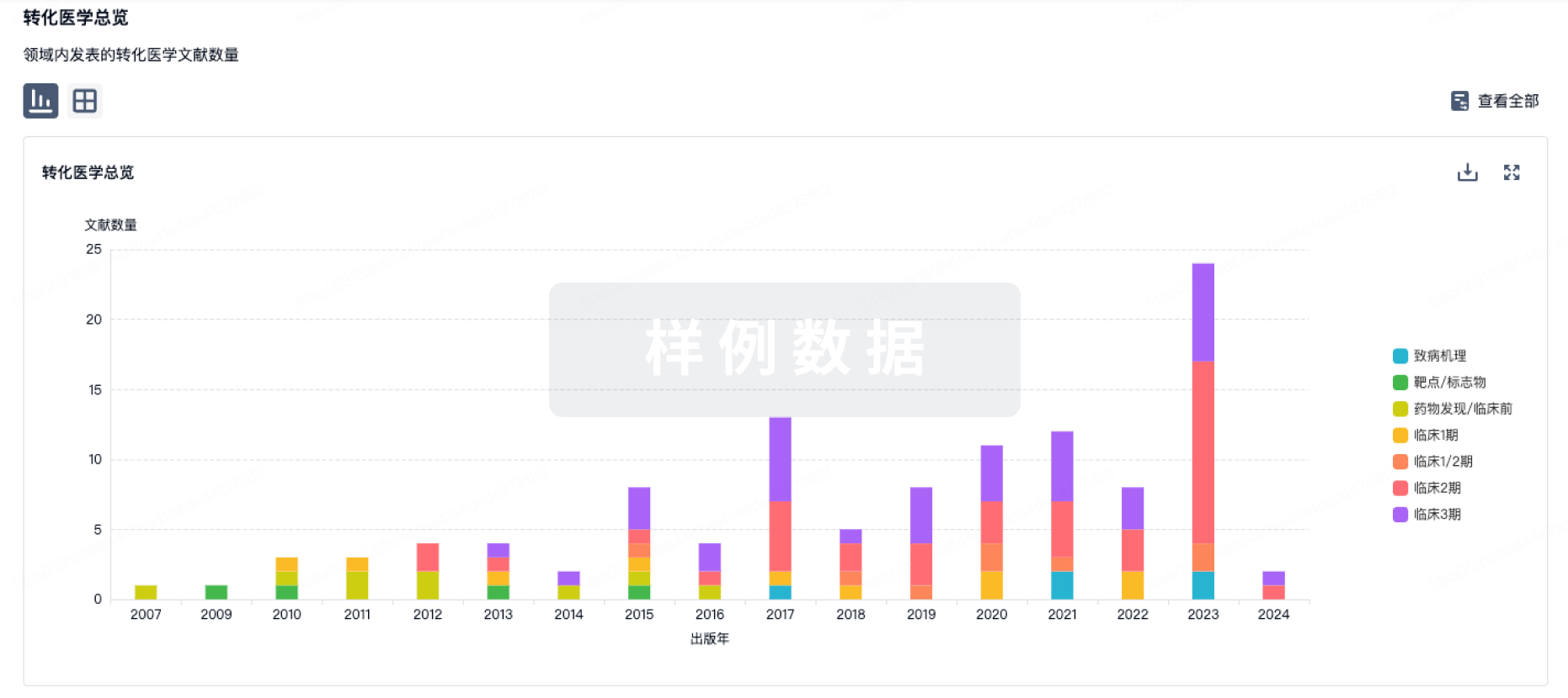
药物交易
使用我们的药物交易数据加速您的研究。
登录
或
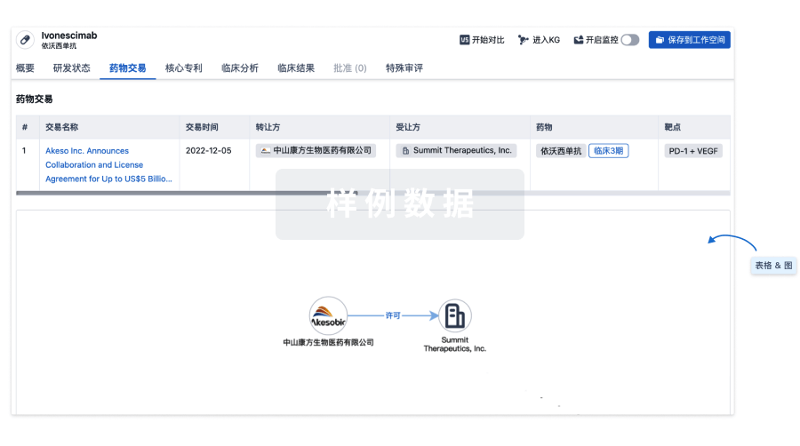
核心专利
使用我们的核心专利数据促进您的研究。
登录
或
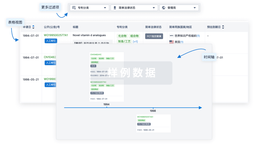
临床分析
紧跟全球注册中心的最新临床试验。
登录
或
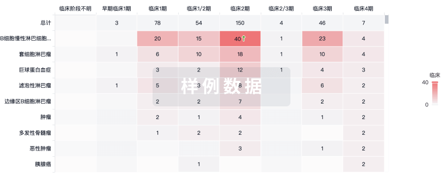
批准
利用最新的监管批准信息加速您的研究。
登录
或
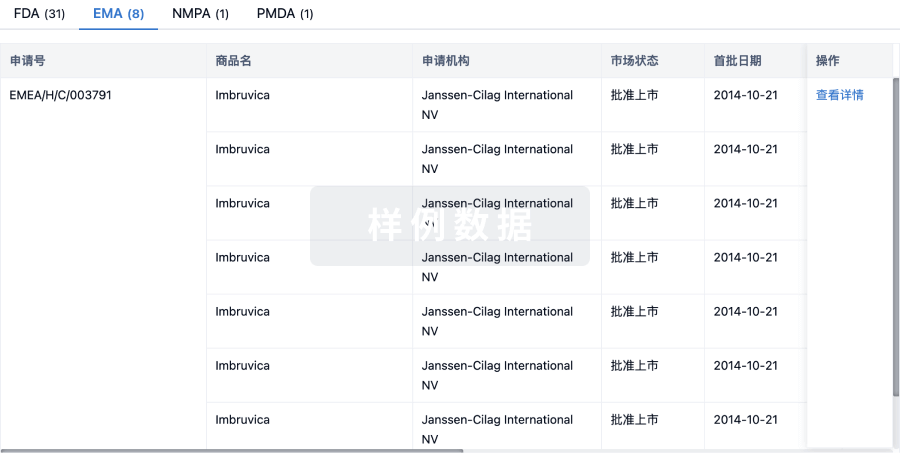
特殊审评
只需点击几下即可了解关键药物信息。
登录
或
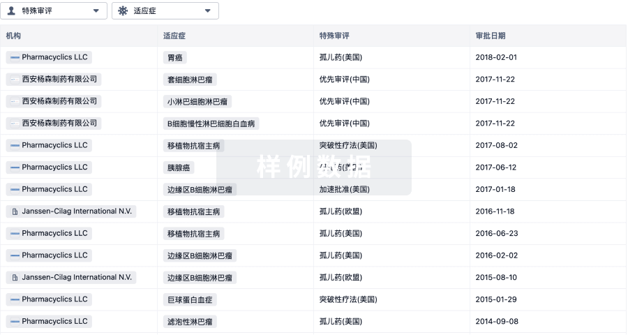
生物医药百科问答
全新生物医药AI Agent 覆盖科研全链路,让突破性发现快人一步
立即开始免费试用!
智慧芽新药情报库是智慧芽专为生命科学人士构建的基于AI的创新药情报平台,助您全方位提升您的研发与决策效率。
立即开始数据试用!
智慧芽新药库数据也通过智慧芽数据服务平台,以API或者数据包形式对外开放,助您更加充分利用智慧芽新药情报信息。
生物序列数据库
生物药研发创新
免费使用
化学结构数据库
小分子化药研发创新
免费使用