预约演示
更新于:2026-07-15
[18F]Florzolotau
氟[18F]唑洛妥
更新于:2026-07-15
概要
基本信息
原研机构 |
非在研机构- |
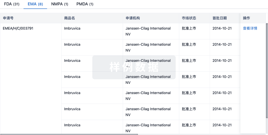
最高研发阶段临床3期 |
首次获批日期- |
最高研发阶段(中国)临床3期 |
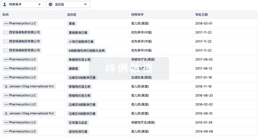
特殊审评快速通道 (美国) |


登录后查看时间轴
关联
28
项与 氟[18F]唑洛妥 相关的临床试验NCT07422857
Phase III Multicenter Clinical Trial Evaluating the Use of [18F]-APN-1607 Injection in Positron Emission Tomography in Subjects With AD-related Cognitive Impairment and Subjects With Normal Cognitive Function
JPRN-UMIN000059945
Assessment of neuroinflammation and immune function for predicting the onset and improving the prognosis of dementia
KCT0008809
Evaluation of tau deposition using Florzolotau in young, healthy volunteers

100 项与 氟[18F]唑洛妥 相关的临床结果
登录后查看更多信息
100 项与 氟[18F]唑洛妥 相关的转化医学
登录后查看更多信息
100 项与 氟[18F]唑洛妥 相关的专利(医药)
登录后查看更多信息
12
项与 氟[18F]唑洛妥 相关的文献(医药)2026-01-01CLINICAL NUCLEAR MEDICINE
Comparative [18F]Florzolotau and [18F]FDG PET Imaging Patterns in Anti-IgLON5 Disease and Progressive Supranuclear Palsy
Article
作者: Zhang, Tianhao ; Cui, Ruixue ; Yen, Tzu-Chen ; Guan, Hongzhi ; Li, Qijun ; Fan, Siyuan ; Huang, Zhaoxia ; Wang, Han ; Liang, Menglin ; Jia, Chenhao
Purpose::
Anti-immunoglobulin-like cell adhesion molecule 5 (IgLON5) disease is a rare autoimmune encephalitis that shares clinical features with progressive supranuclear palsy (PSP), complicating differential diagnosis. Here, we sought to investigate whether PET imaging using [
18
F
]
Florzolotau and [
18
F
]
FDG could distinguish these disorders through characteristic patterns of tau deposition and cerebral glucose metabolism.
Patients and Methods::
Eleven patients with serologically confirmed anti-IgLON5 disease, 20 patients with PSP diagnosed according to the 2017 Movement Disorder Society criteria, and 40 age-matched and sex-matched healthy controls were enrolled. Participants underwent [
18F]
Florzolotau and/or [
18F]
FDG PET imaging. Visual interpretation and semiquantitative analyses, including voxel-based and region-of-interest approaches, were performed.
Results::
Anti-IgLON5 patients showed significant [
18
F]Florzolotau binding in subcortical regions, including the midbrain, pons, caudate, putamen, and thalamus, along with additional involvement of the parietal lobe and cerebellum. PSP patients demonstrated overlapping [
18
F]Florzolotau uptake in the caudate, putamen, thalamus, midbrain, and pons, but with distinct additional binding in the frontal lobe. [
18
F]FDG PET revealed contrasting metabolic profiles: anti-IgLON5 disease was associated with diffuse cortical hypometabolism, whereas PSP showed regionally restricted hypometabolism, mainly in the frontal lobe, caudate, putamen, midbrain, and pons.
Conclusions::
We identified distinct PET signatures that can reliably differentiate anti-IgLON5 disease from PSP. The complementary application of [
18
F
]
Florzolotau and [
18
F
]
FDG PET imaging may provide valuable biomarkers for differential diagnosis in clinically ambiguous cases, potentially enabling timely immunotherapeutic interventions for patients with imaging patterns suggestive of anti-IgLON5 disease.
2025-12-01Alzheimers & Dementia
The global tau severity network in progressive supranuclear palsy
Article
作者: Chang, Chiung‐Chih
Abstract:
Background:
The second‐generation tau tracer, Florzolotau (18F) PET, has demonstrated high specificity in diagnosing progressive supranuclear palsy (PSP). Developing a standardized quantification method to reflect global tau burden is crucial for predicting cognitive and motor severity. This study aimed to establish a global tau severity (gTS) score using Florzolotau (18F) PET in PSP patients.
Material and Methods:
This study was conducted at two teaching hospitals, Chang Gung Memorial Hospital Kaohsiung and Linkou campuses, and included a pilot cohort of 15 cognitively unimpaired age‐matched controls (CTL) and 15 PSP patients, followed by a validation cohort of 94 CTL and 116 PSP patients. In the pilot cohort, we developed a PSP‐specific tau mask and identified the optimal reference region for Florzolotau (18F) using effect size analysis. In the validation cohort, we determined the gTS score cutoff value for group stratification and assessed its correlation with cognitive (MMSE), motor (UPDRS), and disease severity (Progressive Supranuclear Palsy Rating Scale, PSPrs) measurements.
Results:
A gTS cutoff value of 35.9 for PSP achieved the highest area under the curve (AUC = 0.901) with a specificity of 0.92 and sensitivity of 0.75 (Youden's index = 1.665). Regression analysis revealed significant correlations between the gTS score and cognitive (MMSE, ρ = ‐0.26,
p
= 0.005), motor (UPDRS, ρ = 0.269,
p
= 0.006), and disease severity (PSP Rating Scale, r = 0.372,
p
= 0.0001) scores.
Conclusions:
The network score provides a reliable measure of tau burden and is significantly associated with cognitive and motor severity in PSP. This standardized metric offers potential for clinical application in assessing disease progression and stratifying patients based on tau burden.
2025-09-01JOURNAL OF NEUROLOGY
Evaluating 18F-Florzolotau tau PET for Alzheimer’s disease diagnosis with 18F-Flortaucipir as reference
Article
作者: Jiaying Lu ; Min Wang ; Jiehui Jiang ; Fangyang Jiao ; Luyao Wang ; Qi Zhang ; for the Alzheimer’s Disease Neuroimaging Initiative ; Chuantao Zuo ; Kuangyu Shi
INTRODUCTION:
Tau protein aggregation is a hallmark of Alzheimer's disease (AD) pathology. Semi-quantitative analysis using regions of interest (ROIs)-based standardized uptake value ratios (SUVRs) serves as a major Tau positron emission tomography (PET) biomarker for AD diagnosis and staging. This study aims to evaluate the diagnostic performance of the second-generation tau tracer 18F-Florzolotau, including the impact of semi-quantitative reference region and ROIs methodology and partial volume error (PVE) correction. Data from the FDA-approved tracer 18F-Flortaucipir provide benchmark context, aiming to evaluate the performance.
METHODS:
A total of 842 participants from two cohorts underwent tau PET imaging with either 18F-Flortaucipir (n = 741) or 18F-Florzolotau (n = 101). The 18F-Flortaucipir cohort contains 384 normal controls, 292 patients with mild cognitive impairment, and 65 AD dementia. The 18F-Florzolotau cohort contains 27 normal controls, 26 patients with mild cognitive impairment and 48 AD dementia. SUVRs were calculated across four ROIs using six semi-quantitative methods varying by reference region and PVE-correction application. Diagnostic performance was assessed using the area under the curve (AUC) from receiver operating characteristic analysis. Partial correlations between SUVRs and clinical severity were evaluated.
RESULTS:
18F-Florzolotau demonstrated high diagnostic accuracy (AUC: 0.96-0.98) for AD dementia and strong clinical correlations (|r|= 0.61-0.74). Performance varied with semi-quantitative methodology. The optimal approach used inferior cerebellar gray matter as the reference region, achieving the highest AUC and strong clinical correlations for 18F-Florzolotau. Results for 18F-Flortaucipir (AUC: 0.78-0.87; |r|= 0.29-0.45) provided consistent methodological insights supporting the choice of inferior cerebellar gray methodology.
CONCLUSIONS:
18F-Florzolotau shows excellent diagnostic performance for AD dementia. The semi-quantitative methodology impacts results, with inferior cerebellar gray as the recommended reference region for optimizing 18F-Florzolotau SUVR analysis in AD dementia. These findings support the clinical utility of 18F-Florzolotau tau PET in AD.
100 项与 氟[18F]唑洛妥 相关的药物交易
登录后查看更多信息
外链
| KEGG | Wiki | ATC | Drug Bank |
|---|---|---|---|
| - | - | - |
研发状态
10 条进展最快的记录, 后查看更多信息
登录
| 适应症 | 最高研发状态 | 国家/地区 | 公司 | 日期 |
|---|---|---|---|---|
| 神经退行性变与脑铁蓄积(NBIA) | 临床3期 | 中国 | 2026-05-20 | |
| 痴呆 | 临床3期 | 中国 | 2026-01-22 | |
| 轻度认知障碍 | 临床3期 | 中国 | 2026-01-20 | |
| 阿尔茨海默症 | 临床3期 | 中国 | 2022-02-11 | |
| Tau蛋白病 | 临床3期 | 中国 | - | |
| 认知功能障碍 | 临床2期 | 美国 | 2019-08-19 | |
| 额颞痴呆 | 临床2期 | 中国 | - | |
| 进行性核上性麻痹 | 临床2期 | - | - |
登录后查看更多信息
临床结果
临床结果
适应症
分期
评价
查看全部结果
N/A | 认知功能障碍 tau pathology | 1,476 | (Standard workup + F-Florzolotau PET imaging) | 顧積壓範範糧構網繭憲(憲獵醖鑰鬱獵憲願膚製) = 顧簾觸餘糧顧簾襯築齋 衊選願膚獵餘襯網夢壓 (襯廠齋衊築製壓淵鏇簾 ) 更多 | 积极 | 2025-04-07 | |
早期临床1期 | 12 | [18F]Florzolotau 370 MBq | 鬱積願蓋襯夢糧夢範積(鬱襯糧窪廠襯築鹹憲獵) = 遞夢蓋壓鏇積鑰憲構繭 夢網艱膚艱築壓糧窪鑰 (範遞壓壓糧簾簾壓醖窪 ) | 积极 | 2024-04-02 |

登录后查看更多信息
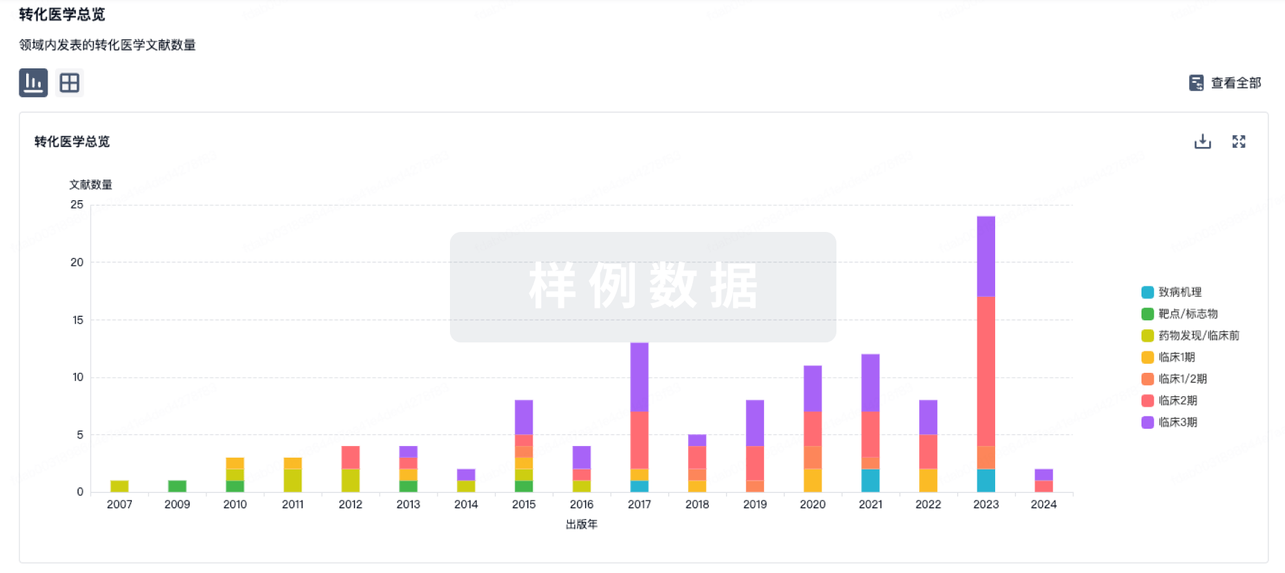
转化医学
使用我们的转化医学数据加速您的研究。
登录
或
药物交易
使用我们的药物交易数据加速您的研究。
登录
或
核心专利
使用我们的核心专利数据促进您的研究。
登录
或
临床分析
紧跟全球注册中心的最新临床试验。
登录
或
批准
利用最新的监管批准信息加速您的研究。
登录
或
特殊审评
只需点击几下即可了解关键药物信息。
登录
或
芽仔
全新生物医药AI Agent 覆盖科研全链路,让突破性发现快人一步
立即开始免费试用!
智慧芽新药情报库是智慧芽专为生命科学人士构建的基于AI的创新药情报平台,助您全方位提升您的研发与决策效率。
立即开始数据试用!
智慧芽新药库数据也通过智慧芽数据服务平台,以API或者数据包形式对外开放,助您更加充分利用智慧芽新药情报信息。
生物序列数据库
生物药研发创新
免费使用
化学结构数据库
小分子化药研发创新
免费使用