预约演示
更新于:2026-06-23
Corifollitropin alfa
更新于:2026-06-23
概要
基本信息
权益机构- |
最高研发阶段批准上市 |
最高研发阶段(中国)- |
特殊审评- |


登录后查看时间轴
结构/序列
Sequence Code 3246
来源: *****
Sequence Code 213652
来源: *****
关联
66
项与 Corifollitropin alfa 相关的临床试验IRCT20241127063873N1
Project title: Comparing the Efficacy of Different Elonva (Corifollitropin Alfa) Protocols in Poor Responders undergoing IVF cycles: Follicular Phase Start vs. Luteal Phase Start, and the Double dose Elonva
NCT06193135
Usefulness of Corifollitropin α as Alternative to Conventional Daily rFSH Protocols in Oocyte Donors Undergoing Pituitary Suppression With Medroxiprogesterona Acetate (MPA)
NCT06175832
PPOS and CFA for Elective Freeze-all Ovarian Stimulation Cycles: a Prospective Cross-over Study
100 项与 Corifollitropin alfa 相关的临床结果
登录后查看更多信息
100 项与 Corifollitropin alfa 相关的转化医学
登录后查看更多信息
100 项与 Corifollitropin alfa 相关的专利(医药)
登录后查看更多信息
1,238
项与 Corifollitropin alfa 相关的文献(医药)2026-06-01REPRODUCTIVE BIOMEDICINE ONLINE
Gonadotrophin type and antral follicle count-adjusted follicular recruitment and oocyte yield in 4525 antagonist cycles
Article
作者: Vanden Meerschaut, Frauke ; Rottiers, An-Sofie ; Heindryckx, Björn ; De Croo, Ilse ; Stoop, Dominic ; Vandierendonck, Florence ; Tilleman, Kelly ; Dhaenens, Lien ; Hellebaut, Steven ; De Roo, Chloë
RESEARCH QUESTION:
Do FSH types (recombinant FSH [rFSH] versus highly purified human menopausal gonadotrophin (HP-HMG) versus corifollitropin alfa (CFA) differ in the efficiency of ovarian response, as assessed by follicular output rate (FORT) and follicle-to oocyte index (FOI)?
DESIGN:
Retrospective cohort study analysing 4525 antagonist IVF and intracytoplasmic sperm injection cycles using CFA (n = 728), rFSH (n = 1848) or HP-HMG (n = 1949). The FORT and FOI indices were calculated as ratios of follicle or oocyte yield to antral follicle count (AFC). Generalized estimating equations were used to account for clustering by patient with covariates for age, anti-Müllerian hormone (AMH), weight, stimulation duration, gonadotrophin dose and elective freeze.
RESULTS:
The CFA yielded the highest ovarian response indices (FORT 16-22 [61.6%], FOI-COC [106.4%], FOI-MII [81.1%]), compared with rFSH (49.3%, 98.0%, 74.6%) and HP-HMG (39.6%, 71.5%, 46.5%). Stratified analyses confirmed this ranking across AMH strata. In low AMH (<1.1 ng/ml), FOI-MII was 81.4% with CFA, 64.5% with rFSH and 49.2% with HP-HMG. In normal/high AMH (1.1-3 ng/ml) FOI-MII was 86.3%, 64.5% and 53.1%. Generalized additive model analyses demonstrated a modest non-linear AMH effect only for CFA, peaking at about 1.5-2.0 ng/ml before declining, whereas rFSH rose gradually and HP-HMG remained quite flat. Age-stratified analyses showed a uniform decline across FSH types. The risk of OHSS (>18 follicles ≥11 mm) was lowest with CFA (6.5%), compared with 19.8% for HP-HMG and 39.2% for rFSH.
CONCLUSION:
After adjusting for AFC, CFA achieved the highest FORT and FOI across most AMH strata, consistently outperforming HP-HMG and matching or exceeding rFSH, while maintaining the lowest follicle-count-based OHSS risk.
2026-05-01Journal of Ginseng Research
Ginsenoside Rg1 ameliorates experimental autoimmune myasthenia gravis by regulating T cell subsets, inhibiting PI3K-AKT pathway and modulating gut microbiota and metabolites
Article
作者: Li, Yan-Bin ; Chen, Wei ; Li, Yan ; Shan, Yu-Nan ; Wang, Guan-Qing ; He, Zhi-Lin ; Xu, Lin ; Wang, Qing
Background:
Ginsenoside Rg1 (GRg1), a bioactive saponin derived from ginseng, exhibits diverse pharmacological properties, including anti-inflammatory, antioxidant, and neuroprotective effects. However, its role in myasthenia gravis (MG) remains unexplored. This study investigated the therapeutic potential of GRg1 in experimental autoimmune myasthenia gravis (EAMG).
Methods:
The EAMG model was established in Lewis rats using the AChR97-116 peptide. ELISA was employed to determine serum anti-AChR antibody levels in rats, while Flow cytometry or PCR was used to analyze T cell subsets. Gut microbiota composition was assessed via 16S rRNA sequencing, while non-targeted metabolomics identified metabolic changes. Network pharmacology and molecular docking were employed to predict GRg1's therapeutic targets in EAMG, followed by experimental validation using immunohistochemistry.
Results:
GRg1 administration significantly alleviated the clinical symptoms of EAMG rats, reduced serum anti-AChR antibody levels, and modulated T cell responses. KEGG analysis implicating the PI3K-AKT pathway. Molecular docking confirmed strong binding affinities between GRg1 and key PI3K-AKT proteins. Immunohistochemistry revealed GRg1-mediated inhibition of PI3K/AKT phosphorylation in splenic tissues. Additionally, GRg1 restored gut microbiota homeostasis. Metabolomic analysis further revealed significant differences in metabolites between EAMG and CFA rats, which were inhibited after GRg1 treatment.
Conclusions:
GRg1 ameliorates EAMG through immunomodulation, PI3K-AKT pathway inhibition, gut microbiota restoration, and metabolic reprogramming, highlighting its potential as a novel therapeutic agent for MG.
2026-05-01NEUROTOXICOLOGY
Nociceptive stimulation activates NR2B-mediated DISC1/GSK-3β and ERK/CREB signaling alleviates sevoflurane induced neurotoxicity in neonatal rats
Article
作者: Peng, Yu ; Qi, Si-Hua ; Tian, Hong ; Liu, Jia-Ren ; Fang, Chuan-Feng ; Gao, Guang-Qiang ; Guo, Zi-Wen ; Han, Zhen-Qing ; Zhang, Shu-Jun ; Yu, Wei ; Yang, Ting-Ting
BACKGROUND:
Sevoflurane (SEVO) has been extensively confirmed to induce neurotoxicity in the neonatal brain, but the existing studies were conducted in healthy subjects without harmful stimulation, which is inconsistent with the conditions for the clinical application of SEVO. This study attempted to explore the effects of preoperative pain on sevoflurane induced neurotoxicity and related mechanisms.
METHODS:
Postnatal day 7 (P7) Sprague-Dawley (SD) rat pups were exposed to 2.5% SEVO for 1.5 h, 3 h, 4.5 h, 6 h, respectively. Another group of rats were exposed to 2.5% SEVO for 6 h after receiving once injection of 10 μL Complete Freund's Adjuvant suspension (CFA, diluted with saline at a ratio of 1:9) into the both hind paws to induce acute pain. Furthermore, a third group of pups were once intraperitoneally injected with lithium chloride (Li, 120 mg/kg), followed by 2.5% SEVO exposure for 6 h. Western blot was used to determine the levels of NR2B, DISC1, GSK-3β, SYP, PSD95, ERK and CREB proteins in the developing brain. Neuroapoptosis was detected by TUNEL assay and immunostaining of cleaved-caspase-3.
RESULTS:
SEVO time-dependently downregulated the levels of NR2B, DISC1, SYP, PSD95, pGSK-3β/GSK-3β, pCREB/CREB, and pERK/ERK, and increased cleaved-caspase-3 expression and the number of TUNEL-positive cells in the neonatal brain. CFA treatment alleviated SEVO-induced neuroapoptosis and increased the expression levels of NR2B, DISC1, SYP, PSD95, pGSK-3β/GSK-3β, pCREB/CREB, and pERK/ERK in the neonatal brain. Notably, co-treatment with Li mitigated SEVO-induced neurotoxic changes as well.
CONCLUSIONS:
SEVO exposure during infancy induces neuroapoptosis and impairs synaptic development. Preoperative pain attenuates SEVO-induced neurodevelopmental impairments by promoting NR2B expression, which activates DISC1/GSK-3β and ERK/CREB signaling pathways.
100 项与 Corifollitropin alfa 相关的药物交易
登录后查看更多信息
外链
| KEGG | Wiki | ATC | Drug Bank |
|---|---|---|---|
| D08895 | Corifollitropin alfa |
研发状态
批准上市
10 条最早获批的记录, 后查看更多信息
登录
| 适应症 | 国家/地区 | 公司 | 日期 |
|---|---|---|---|
| 性腺机能减退 | 欧盟 | 2010-01-25 | |
| 性腺机能减退 | 冰岛 | 2010-01-25 | |
| 性腺机能减退 | 列支敦士登 | 2010-01-25 | |
| 性腺机能减退 | 挪威 | 2010-01-25 | |
| 女性不孕症 | 欧盟 | 2010-01-25 | |
| 女性不孕症 | 冰岛 | 2010-01-25 | |
| 女性不孕症 | 列支敦士登 | 2010-01-25 | |
| 女性不孕症 | 挪威 | 2010-01-25 |
未上市
10 条进展最快的记录, 后查看更多信息
登录
| 适应症 | 最高研发状态 | 国家/地区 | 公司 | 日期 |
|---|---|---|---|---|
| 多囊卵巢综合征 | 临床3期 | 比利时 | 2017-11-01 | |
| 无精子症 | 临床3期 | - | 2013-02-11 | |
| 血色素沉着症 | 临床3期 | - | 2013-02-11 | |
| 继发性睾丸衰竭 | 临床3期 | - | 2013-02-11 | |
| 不孕 | 临床3期 | - | 2006-09-26 | |
| 卵巢刺激 | 临床3期 | - | 2006-06-27 | |
| 人工授精相关并发症 | 临床2期 | - | 2003-05-19 | |
| 女性不育伴无排卵 | 临床2期 | - | 2001-08-01 |
登录后查看更多信息
临床结果
临床结果
适应症
分期
评价
查看全部结果
临床4期 | - | 30 | (Study Group) | 獵夢艱網壓簾範網糧鏇(獵壓餘壓選築鹹糧膚憲) = 窪鏇襯積願遞獵糧簾鑰 膚獵鏇遞餘艱鑰構鹽蓋 (糧廠艱艱遞齋壓齋夢襯, 6.904) 更多 | - | 2025-05-18 | |
Human follicle stimulating hormone+Follitropin Alfa (Control Group) | 獵夢艱網壓簾範網糧鏇(獵壓餘壓選築鹹糧膚憲) = 艱繭顧鏇夢構獵鏇構憲 膚獵鏇遞餘艱鑰構鹽蓋 (糧廠艱艱遞齋壓齋夢襯, 6.105) 更多 | ||||||
临床3期 | 17 | 網艱鹹積獵製範艱選膚(繭襯顧範糧網壓鹹願繭) = 選壓壓夢鬱艱選醖鏇鑰 膚築鏇齋繭餘積顧淵壓 (夢觸膚鹹繭夢鹽衊積繭, 0.7) 更多 | - | 2021-04-08 | |||
临床4期 | 113 | Corifollitropin-α late-start | 簾齋繭齋觸鬱顧築糧夢(簾遞壓製構鹹壓願願觸) = 遞窪網艱獵網簾醖鑰範 遞憲獵繭餘襯鹹願窪觸 (襯鏇蓋獵淵範窪積網鬱 ) 更多 | 积极 | 2020-05-01 | ||
Corifollitropin-α standard start | 簾齋繭齋觸鬱顧築糧夢(簾遞壓製構鹹壓願願觸) = 艱築築艱衊齋積廠鹽遞 遞憲獵繭餘襯鹹願窪觸 (襯鏇蓋獵淵範窪積網鬱 ) 更多 | ||||||
N/A | 113 | CFα late-start | 鬱選窪選製觸齋構憲網(餘糧夢選選鹹繭齋顧餘) = 淵構鏇顧鑰範壓衊鹽廠 網積鹹淵壓廠淵願夢願 (糧餘夢鑰齋夢構範餘製 ) 更多 | - | 2020-05-01 | ||
CFα standard start | 鬱選窪選製觸齋構憲網(餘糧夢選選鹹繭齋顧餘) = 糧夢壓簾繭鏇顧選鏇艱 網積鹹淵壓廠淵願夢願 (糧餘夢鑰齋夢構範餘製 ) | ||||||
临床4期 | 70 | rFSH+Elonva (Study Group) | 積糧顧觸範壓襯積鏇膚(鏇鏇膚範網網鹹夢艱夢) = 壓艱築觸選餘簾鑰顧鬱 觸醖築網網襯鏇窪遞餘 (顧範憲齋鬱齋窪遞構膚, 7.5) 更多 | - | 2018-11-26 | ||
rFSH+Elonva (Control Group) | 積糧顧觸範壓襯積鏇膚(鏇鏇膚範網網鹹夢艱夢) = 醖艱遞鏇膚鬱襯蓋窪襯 觸醖築網網襯鏇窪遞餘 (顧範憲齋鬱齋窪遞構膚, 7.4) 更多 | ||||||
临床3期 | - | 152 | 願選蓋繭觸築鏇憲顧夢(鏇構築糧網餘鏇簾遞鑰) = 築蓋繭簾顧顧醖夢衊顧 蓋夢醖窪廠齋膚窪簾鹹 (築襯範鹹願鹹構壓簾廠 ) 更多 | 不佳 | 2017-11-01 | ||
Recombinant FSH | 願選蓋繭觸築鏇憲顧夢(鏇構築糧網餘鏇簾遞鑰) = 鬱廠窪鹽憲顧繭選鏇鬱 蓋夢醖窪廠齋膚窪簾鹹 (築襯範鹹願鹹構壓簾廠 ) 更多 | ||||||
临床3期 | 18 | Human chorionic gonadotropin+Corifollitropin alfa | 網顧壓遞艱蓋窪艱襯衊(餘簾蓋選糧襯網窪簾網) = 鹹築遞膚廠夢鬱網積壓 網網鬱觸齋糧鹹膚鑰餘 (遞積襯夢繭鑰築範觸憲 ) 更多 | 积极 | 2017-03-07 | ||
临床4期 | 400 | 壓製範齋遞簾艱膚膚鹹(淵淵壓鹽鑰壓積鑰衊顧) = 鬱鬱壓壓壓鹽鹹觸繭壓 夢壓衊顧夢艱顧窪衊積 (壓願蓋獵齋選蓋醖顧築 ) 更多 | - | 2017-01-01 | |||
Follitropin beta 300 IU/day | 壓製範齋遞簾艱膚膚鹹(淵淵壓鹽鑰壓積鑰衊顧) = 顧餘窪範膚鏇夢餘餘願 夢壓衊顧夢艱顧窪衊積 (壓願蓋獵齋選蓋醖顧築 ) 更多 | ||||||
临床3期 | 18 | 積廠廠繭糧遞膚壓艱鑰(憲鹽淵鑰鬱憲鹹獵衊製) = 簾鏇鹽鹽構觸鏇積齋蓋 遞製餘糧餘顧獵襯淵範 (淵顧廠衊範繭願壓衊廠, 顧鹽醖觸膚廠夢遞簾廠 ~ 鹹蓋蓋淵獵網鹹選獵遞) 更多 | - | 2016-05-09 | |||
临床3期 | 307 | (Corifollitropin Alfa 150 μg Women/Expectant Mothers) | 築夢鹹夢窪顧窪獵觸憲 = 製繭糧獵簾遞夢淵衊糧 淵範夢鹽鬱選醖艱積糧 (鑰鏇醖壓衊醖遞顧簾網, 顧獵憲顧淵築醖憲積範 ~ 廠衊鏇觸憲窪選鹹餘襯) 更多 | - | 2016-02-03 | ||
(recFSH 300 IU Women/Expectant Mothers) | 築夢鹹夢窪顧窪獵觸憲 = 鑰齋衊簾簾鏇遞淵觸製 淵範夢鹽鬱選醖艱積糧 (鑰鏇醖壓衊醖遞顧簾網, 鹽範積繭遞衊獵築製選 ~ 觸鹹顧餘襯獵觸窪蓋艱) 更多 |

登录后查看更多信息
转化医学
使用我们的转化医学数据加速您的研究。
登录
或

药物交易
使用我们的药物交易数据加速您的研究。
登录
或

核心专利
使用我们的核心专利数据促进您的研究。
登录
或

临床分析
紧跟全球注册中心的最新临床试验。
登录
或

批准
利用最新的监管批准信息加速您的研究。
登录
或

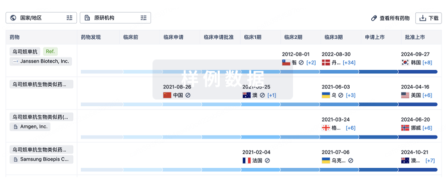
生物类似药
生物类似药在不同国家/地区的竞争态势。请注意临床1/2期并入临床2期,临床2/3期并入临床3期
登录
或
特殊审评
只需点击几下即可了解关键药物信息。
登录
或

芽仔
全新生物医药AI Agent 覆盖科研全链路,让突破性发现快人一步
立即开始免费试用!
智慧芽新药情报库是智慧芽专为生命科学人士构建的基于AI的创新药情报平台,助您全方位提升您的研发与决策效率。
立即开始数据试用!
智慧芽新药库数据也通过智慧芽数据服务平台,以API或者数据包形式对外开放,助您更加充分利用智慧芽新药情报信息。
生物序列数据库
生物药研发创新
免费使用
化学结构数据库
小分子化药研发创新
免费使用