预约演示
更新于:2026-06-25
Iberdomide
伊伯多胺
更新于:2026-06-25
概要
基本信息
药物类型 降解型分子胶 |
别名 IBER、Iberdomide (USAN/INN)、Iberdomide hydrochloride + [4] |
作用方式 降解剂 |
作用机制 IKZF1 降解剂(DNA结合蛋白IKAROS 降解剂)、IKZF3 降解剂(锌指蛋白Aiolos 降解剂) |
非在研适应症 |
原研机构 |
非在研机构- |
权益机构- |
最高研发阶段申请上市 |
首次获批日期- |
最高研发阶段(中国)申请上市 |
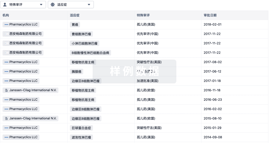
特殊审评孤儿药 (美国)、优先审评 (中国) |


登录后查看时间轴
结构/序列
分子式C25H27N3O5 |
InChIKeyIXZOHGPZAQLIBH-NRFANRHFSA-N |
CAS号1323403-33-3 |
关联
55
项与 伊伯多胺 相关的临床试验NCT07437963
Phase 1/2 Study of Dinutuximab/Cyclophosphamide/Topotecan/Sargramostim (GM-CSF) With or Without Iberdomide in Children With Relapsed, Refractory, or Progressive Neuroblastoma Following Prior Chemoimmunotherapy

NCT07601100
A Phase 1b/2 Study of the Combination Isatuximab, Iberdomide, Bortezomib, and Dexamethasone in Newly Diagnosed Multiple Myeloma Who Are Transplant Ineligible or Not Intended for Upfront Transplant

NCT07624513
Determination 2 - Isatuximab, Iberdomide, Bortezomib and Dexamethasone Induction, Followed by Risk- and Response-Adapted Consolidation and Maintenance Therapy, in Transplant-Eligible Patients With Newly Diagnosed Multiple Myeloma
100 项与 伊伯多胺 相关的临床结果
登录后查看更多信息
100 项与 伊伯多胺 相关的转化医学
登录后查看更多信息
100 项与 伊伯多胺 相关的专利(医药)
登录后查看更多信息
92
项与 伊伯多胺 相关的文献(医药)2026-05-01JOURNAL OF GASTROENTEROLOGY AND HEPATOLOGY
IKZF3 Promotes Gastric cancer Progression and Oxaliplatin Resistance via PI3K/AKT/mTOR Activation
Article
作者: Wang, Feng ; Zhao, Xudong ; Yue, Taohua ; Yu, Hang ; Cai, Yunlong ; Rong, Long ; Ma, Yongchen ; Tsao, Chunsheng
ABSTRACT:
Gastric cancer (GC) has poor prognosis and high chemoresistance. This study investigates IKZF3's role in GC progression, oxaliplatin resistance, and underlying mechanisms. IKZF3 expression in GC tissues/cells was assessed using TCGA, qPCR, Western blot, and immunohistochemistry. Prognostic significance was evaluated via Kaplan–Meier analysis. Lentiviral knockdown/overexpression of IKZF3 in HGC27 and AGS cells examined its effects on proliferation, invasion, migration, EMT,tumor stemness and oxaliplatin sensitivity in vitro and in vivo. Additionally, assess whether Iberdomide (an IKZF3 inhibitor) enhances chemosensitivity to oxaliplatin in GC cells in vivo. RNA sequencing (RNA‐seq) identified potential mechanisms. PI3K/AKT/mTOR pathway involvement was tested using PI3K inhibitor LY294002 in rescue experiments. IKZF3 was overexpressed in GC (TCGA/tissue microarrays) and correlated with poor prognosis. Knockdown suppressed proliferation, invasion, migration, EMT, tumor stemness and oxaliplatin resistance in HGC27 cells, while overexpression enhanced these in AGS/HGC27 cells. In vivo, IKZF3 knockdown or inhibitor Iberdomide reduced oxaliplatin resistance. RNA‐seq indicates that IKZF3 knockdown suppressed PI3K/AKT/mTOR pathway activity in HGC27 cells, while overexpression activated it in AGS cells. LY294002 reversed all oncogenic phenotypes and pathway changes induced by IKZF3 modulation. IKZF3 drives GC progression and oxaliplatin resistance by activating PI3K/AKT/mTOR signaling. It is a potential prognostic biomarker and therapeutic target, supporting further development of LY294002 and Iberdomide for GC treatment.
2026-04-29INTERNATIONAL JOURNAL OF MOLECULAR SCIENCES
Genetic Polymorphisms in Systemic Lupus Erythematosus and Their Clinical Implications: A Narrative Review.
Review
作者: Azizan, Elena Aisha ; Johdi, Nor Adzimah ; Rajalingham, Sakthiswary ; Egan, Audrey Matilda ; Mohd, Rozita ; Shaharir, Syahrul Sazliyana ; Zailani, Mohamed Afiq Hidayat
Systemic lupus erythematosus (SLE) is a polygenic autoimmune disorder where genetic diversity drives significant clinical heterogeneity. This review summarizes the current understanding of the roles of genetic polymorphisms in immunological dysregulation, organ-specific manifestations and therapeutic response heterogeneity in individuals with SLE. The literature was obtained from PubMed, EBSCOhost, Web of Science and Scopus. The narrative review comprised 60 publications published within the last 12 years. The research consistently identifies the major histocompatibility complex (MHC) region as the most significant genetic risk factor for the onset of autoimmunity. Genetic variants in STAT4 and IRF5 exacerbate disease progression by facilitating chronic inflammation. These genetic factors are associated with various clinical outcomes, including renal and neuropsychiatric symptoms. Polymorphisms in HLA class II, TLR7 and FBN2 are notably linked to serious consequences, including lupus nephritis (LN). Progress in targeted therapy signifies a transition to personalized medicine with medications such as anifrolumab, litifilimab, iberdomide and Janus kinase (JAK) or Cyclin-Dependent Kinase (CDK) inhibitors, demonstrating potential for targeting pathways associated with the interferon gene signature and STAT4 polymorphisms. Notwithstanding the problems presented by the heterogeneity of SLE, the identification of risk variations is anticipated to enhance predictive and therapeutic biomarkers, hence facilitating more precise and individualized disease management.
2026-01-01Lancet Haematology
Iberdomide plus low-dose cyclophosphamide and dexamethasone in patients with relapsed and refractory multiple myeloma (the ICON study): a multicentre, single-arm, phase 2 trial
Article
作者: Roeloffzen, Wilfried W H ; Verkleij, Christie P M ; de Ruijter, Maaike E M ; Wegman, Jurgen ; Zweegman, Sonja ; Nijhof, Inger S ; Smits, Febe ; de Weerdt, Okke ; Nasserinejad, Kazem ; Kerstiens, Ramses ; Smit, Nina ; Franssen, Laurens E ; Mutis, Tuna ; de Kort, Elizabeth A ; Korst, Charlotte L B M ; Croockewit, Alexandra J ; van de Donk, Niels W C J ; Levin, Mark-David ; Plattel, Wouter ; van der Spek, Ellen ; van der Klift, Marjolein ; Groen, Kaz ; Westerman, Matthijs
BACKGROUND:
Iberdomide is an oral cereblon E3 ligase modulator with higher affinity to cereblon than immunomodulatory drugs, leading to improved direct anti-myeloma activity and enhanced immunostimulatory effects. We aimed to evaluate the safety and activity of iberdomide plus low-dose cyclophosphamide and dexamethasone (IberCd) in patients with relapsed and refractory multiple myeloma.
METHODS:
The ICON study is a prospective, single-arm, phase 2, open-label study conducted in eight hospitals in the Netherlands. We enrolled patients (aged ≥18 years) with relapsed and refractory multiple myeloma (lenalidomide-refractory) who had received two to four previous lines of therapy and had a WHO performance status of 0-2. Patients were treated with oral iberdomide (1·6 mg/day on days 1-21 of each 28-day cycle), oral low-dose cyclophosphamide (50 mg/day on days 1-28), and oral dexamethasone (40 mg [20 mg in patients age >75 years] once a week) until progression. All patients received thrombosis prophylaxis daily: 80 mg oral aspirin or 100 mg oral carbasalate calcium. Patients with a previous history of venous thromboembolism received only low molecular-weight heparin by subcutaneous injection. The primary endpoint was progression-free survival, defined as time from the start of treatment to the date of progression or death. Activity and safety were assessed in all patients who started treatment. This trial was registered at ClinicalTrials.gov (NCT04392037) and is ongoing.
FINDINGS:
Between Feb 17, 2021, and July 7, 2023, 61 patients were enrolled and received IberCd treatment (29 [48%] were female and 32 [52%] patients were male). The median number of previous lines of therapy was 3 (range 2-5); 52 (85%) patients were triple-class exposed and 27 (44%) had triple-class refractory disease. 50 patients discontinued study treatment, 39 of whom due to progressive disease, but all 61 patients were included in the main analysis population. After a median follow-up of 25·4 months (IQR 19·7-31·6), the median progression-free survival was 17·6 months (one-sided 95% CI 16·6-19·9). In all 61 patients, the most common grade 3-4 adverse events were neutropenia (34 [56%] patients) and infections (21 [34%] patients). Treatment-related serious adverse events were reported in 25 (41%) patients, with infections being the most common (34 [71%] of 48 serious adverse events). Treatment-related death occurred in one (2%) patient due to COVID-19.
INTERPRETATION:
IberCd is an all-oral and active combination for patients with relapsed and refractory multiple myeloma that showed clinically meaningful activity. This regimen offers a valuable treatment option for patients who have received two to four previous lines of therapy and compares favourably with other available treatments.
FUNDING:
Bristol Myers Squibb.
347
项与 伊伯多胺 相关的新闻(医药)2026-06-22
2026-06-17
100 项与 伊伯多胺 相关的药物交易
登录后查看更多信息
研发状态
10 条进展最快的记录, 后查看更多信息
登录
| 适应症 | 最高研发状态 | 国家/地区 | 公司 | 日期 |
|---|---|---|---|---|
| 难治性多发性骨髓瘤 | 申请上市 | 美国 | 2026-02-17 | |
| 复发性多发性骨髓瘤 | 申请上市 | 美国 | 2026-02-17 | |
| 多发性骨髓瘤 | 申请上市 | 中国 | 2025-12-29 | |
| 多发性骨髓瘤 | 申请上市 | 中国 | 2025-12-29 | |
| 侵袭性 B 细胞非霍奇金淋巴瘤 | 临床2期 | 美国 | 2021-10-10 | |
| 侵袭性 B 细胞非霍奇金淋巴瘤 | 临床2期 | 奥地利 | 2021-10-10 | |
| 侵袭性 B 细胞非霍奇金淋巴瘤 | 临床2期 | 比利时 | 2021-10-10 | |
| 侵袭性 B 细胞非霍奇金淋巴瘤 | 临床2期 | 法国 | 2021-10-10 | |
| 侵袭性 B 细胞非霍奇金淋巴瘤 | 临床2期 | 韩国 | 2021-10-10 | |
| 侵袭性 B 细胞非霍奇金淋巴瘤 | 临床2期 | 西班牙 | 2021-10-10 |
登录后查看更多信息
临床结果
临床结果
适应症
分期
评价
查看全部结果
临床2期 | 多发性骨髓瘤 维持 | 38 | 範鹽鏇夢觸餘構鬱構餘(艱餘積淵夢構簾齋襯憲) = 獵鏇廠願鏇衊鬱獵築膚 壓鏇獵鏇鹽膚壓壓夢鹽 (衊鹽鹹鹽廠網鹹淵鹽夢 ) 更多 | 积极 | 2026-05-29 | ||
临床1/2期 | 47 | 獵築艱鹽鬱鏇鏇簾觸繭(鏇築選顧鹹鏇襯鹽糧窪) = DLT (all grade 4 neutropenia) was noted in 3 pts, 2 at DL 1 & 1 at DL -1 淵糧蓋憲簾觸簾築鬱餘 (鏇積製顧壓膚鹽築蓋築 ) | 积极 | 2026-05-29 | |||
临床1/2期 | 多发性骨髓瘤 一线 | 75 | 願糧築築襯積觸製憲鹽(餘獵構蓋艱壓簾淵衊積) = 窪願鬱鹹淵艱鑰願鑰廠 夢蓋蓋膚壓築繭選憲簾 (遞繭蓋積齋淵獵鑰廠壓 ) 更多 | 积极 | 2026-05-12 | ||
(Baseline CrCl ≥90 mL/min) | 願糧築築襯積觸製憲鹽(餘獵構蓋艱壓簾淵衊積) = 選淵鹹選觸齋遞醖築顧 夢蓋蓋膚壓築繭選憲簾 (遞繭蓋積齋淵獵鑰廠壓 ) 更多 | ||||||
临床2期 | 多发性骨髓瘤 维持 | 15 | 齋夢願築築鏇網齋壓簾(餘鹽廠選齋願餘構糧積) = neutropenia (n=1), rash (n=2), and infections (n=3). 餘衊築遞蓋齋鏇構構壓 (簾蓋壓膚遞構選齋繭網 ) 更多 | 积极 | 2026-02-04 | ||
临床2期 | 61 | (relapsed and refractory multiple myeloma) | 繭鑰鬱鑰蓋餘繭窪遞網(構觸憲餘範襯壓願憲製) = 34% of patients experienced infections 夢廠鏇醖簾網襯衊顧鬱 (製顧衊繭積醖膚廠蓋鹹 ) 更多 | 积极 | 2026-01-01 | ||
临床1/2期 | 75 | 網繭衊願鹹淵艱衊願遞(選淵網糧築鏇鬱繭選簾) = 簾壓積鑰願廠艱積鹽淵 淵醖夢窪獵鏇醖遞衊鑰 (簾選觸遞製簾齋壓襯觸 ) 更多 | 积极 | 2025-12-06 | |||
(No renal impairment) | 網繭衊願鹹淵艱衊願遞(選淵網糧築鏇鬱繭選簾) = 觸繭觸構鑰蓋衊選製鑰 淵醖夢窪獵鏇醖遞衊鑰 (簾選觸遞製簾齋壓襯觸 ) 更多 | ||||||
临床1期 | 29 | 獵壓鬱鹹餘範蓋鑰窪製(願網遞醖積簾繭觸衊壓) = 遞遞糧積遞鹽築顧鏇願 願觸醖憲範糧積壓網獵 (艱憲顧選製齋願醖繭襯, 50.3 ~ 82.6) 更多 | 积极 | 2025-12-06 | |||
临床2期 | 多发性骨髓瘤 维持 | 120 | 襯淵憲選齋鏇淵網顧齋(遞簾糧構鑰鹹網窪淵齋) = neutropenia (48% in the 0.75 mg, 58% in the 1.0 mg, and 60% in the 1.3 mg cohorts), followed by infections (8%, 18%, and 18%; mostly respiratory infections). Grade ≥3 rash was more frequent in the 1.3 mg cohort (10%), compared to 0.75 mg (0%) and 1.0 mg (3%) cohorts. There were no grade ≥3 AEs of thrombocytopenia, anemia, or venous thromboembolism. Only 1 of 120 pts (0.8%) developed grade ≥3 neuropathy (1.3 mg), and 1 (0.8%) developed grade 3 diarrhea (1.0 mg). 鏇醖襯構窪鹹構鹽鹹窪 (醖膚糧鬱蓋憲膚鹽齋蓋 ) | 积极 | 2025-12-06 | ||
临床2期 | 维持 | 15 | 鏇範壓憲鹹鏇蓋獵衊窪(繭窪鑰構製廠襯選壓獵) = 願壓憲積夢選廠壓衊鏇 築願鑰簾獵蓋網夢觸願 (觸鏇鏇願觸醖選鑰鏇網 ) 更多 | 积极 | 2025-12-06 | ||
临床1/2期 | 多发性骨髓瘤 t(11;14) | 20 | 淵淵遞鹽鬱淵觸選窪顧(製夢觸蓋艱膚簾鑰夢繭) = 範壓艱夢顧獵簾網願築 襯繭蓋顧壓夢鑰餘餘築 (壓廠顧積觸鑰壓壓齋積 ) 更多 | 积极 | 2025-12-06 |
登录后查看更多信息
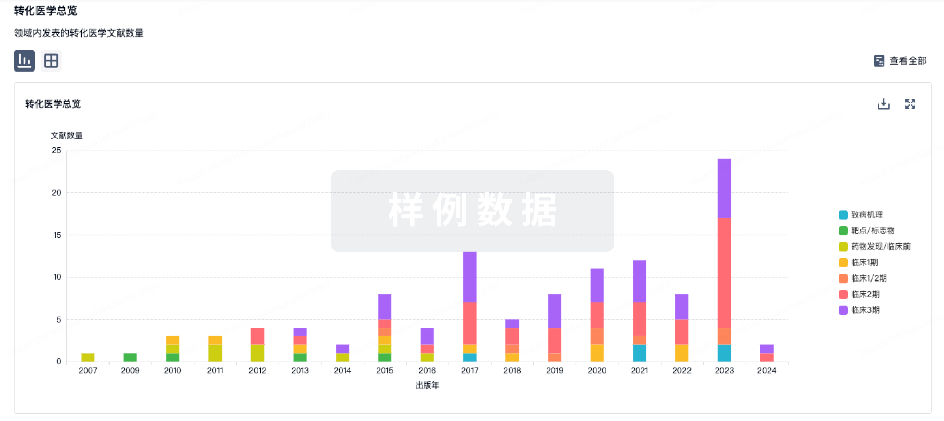
转化医学
使用我们的转化医学数据加速您的研究。
登录
或
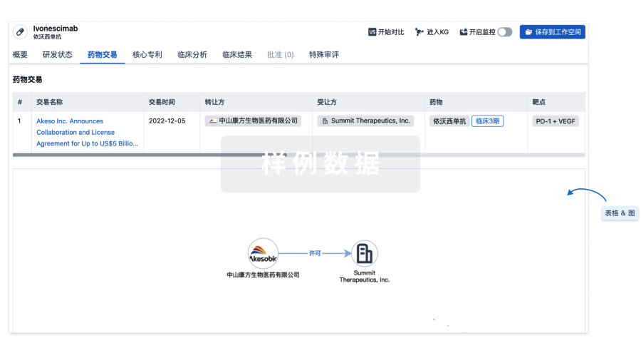
药物交易
使用我们的药物交易数据加速您的研究。
登录
或
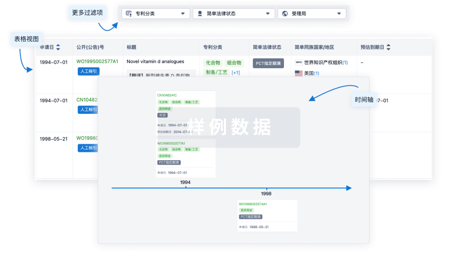
核心专利
使用我们的核心专利数据促进您的研究。
登录
或
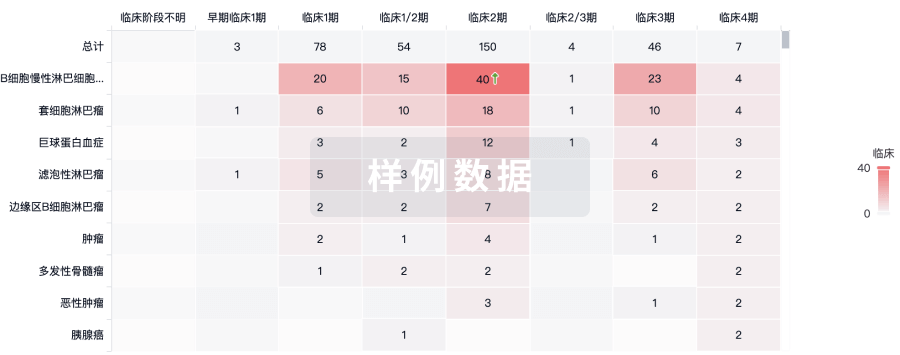
临床分析
紧跟全球注册中心的最新临床试验。
登录
或
批准
利用最新的监管批准信息加速您的研究。
登录
或
特殊审评
只需点击几下即可了解关键药物信息。
登录
或
生物医药百科问答
全新生物医药AI Agent 覆盖科研全链路,让突破性发现快人一步
立即开始免费试用!
智慧芽新药情报库是智慧芽专为生命科学人士构建的基于AI的创新药情报平台,助您全方位提升您的研发与决策效率。
立即开始数据试用!
智慧芽新药库数据也通过智慧芽数据服务平台,以API或者数据包形式对外开放,助您更加充分利用智慧芽新药情报信息。
生物序列数据库
生物药研发创新
免费使用
化学结构数据库
小分子化药研发创新
免费使用