预约演示
更新于:2026-06-15
Sichuan University
更新于:2026-06-15
概览
标签
肿瘤
消化系统疾病
其他疾病
小分子化药
蛋白水解靶向嵌合体(PROTAC)
放射与诊断药物
疾病领域得分
一眼洞穿机构专注的疾病领域

技术平台
公司药物应用最多的技术
靶点
公司最常开发的靶点
关联
靶点 |
作用机制 |
在研机构 |
原研机构 |
在研适应症 |
非在研适应症 |
最高研发阶段 |
首次获批国家/地区 |
首次获批日期 |
靶点 |
作用机制 |
在研机构 |
原研机构 |
在研适应症 |
非在研适应症 |
最高研发阶段 |
首次获批国家/地区 |
首次获批日期 |
靶点 |
作用机制 |
在研机构 |
原研机构 |
在研适应症 |
非在研适应症 |
最高研发阶段 |
首次获批国家/地区 |
首次获批日期 |
NCT07258147
Clinical Study on the Efficacy and Safety of Radiotherapy Combined With Immunotherapy and Chemotherapy for Pre-treated Patients With Small Cell Lung Cancer and Liver Metastases
NCT07637786
An Exploratory Clinical Study of Peptide Nanovaccine (ONVAX-01) and Anti-PD-1 Antibody Combined With Chemotherapy for the Treatment of Advanced Pancreatic Cancer
NCT07519486
A Prospective Randomized Controlled Clinical Trial of Intensive Nutritional Support in Conversion Therapy for Locally Advanced Unresectable Esophageal Squamous Cell Carcinoma
100 项与 四川大学 相关的临床结果
登录后查看更多信息
登录后查看更多信息
2026-12-31Human Vaccines & Immunotherapeutics
HPV vaccine awareness and uptake among women attending cervical screening in health-resource-limited areas of China: A multicenter cross-sectional study
Article
作者: Hu, Shangyin ; Shi, Jingyi ; Wang, Junling ; Feng, Ruimei ; Duan, Rufei ; Jia, Xinhua ; Zhang, Tai ; Yang, Chunxia ; Zhang, Hongyun ; Qiao, Youlin ; Li, Zhifang ; Zhao, Yuqian ; Da, Xi’ao
Evidence shows HPV vaccination reduces infection, precancer and cervical cancer, yet coverage in health-resource-limited of China remains uncertain. We assessed awareness, uptake and correlates among women attending cervical screening, and examined associations with screening outcomes. We conducted a cross-sectional study in eight county sites in 2023-2024 among women aged 35-64 y. A standardized questionnaire captured sociodemographic factors, awareness and vaccination. Cervical samples were tested for hrHPV. Outcomes were awareness, vaccination, hrHPV, HPV16/18 and CIN2+. Associations were estimated using modified Poisson models with site fixed effects and HC3 robust errors. Adjusted prevalence ratios (aPRs) and covariate-standardized marginal estimates were reported. We included 93,027 unique participants. Awareness was 45.15% and vaccination 6.73%. hrHPV prevalence was 11.38% and CIN2+ detection was 0.70%. In 2024 versus 2023, awareness was lower (40.66% vs 51.34%) while vaccination was higher (7.61% vs 5.53%; aPR 1.25, 95% CI 1.18-1.32). Awareness and uptake declined with age; coverage was 23.23% at ages 35-39 and 0.29% at ages 60-64. Urban residence and higher education were associated with uptake (urban aPR 1.19, 95% CI 1.11-1.27; bachelor's or higher aPR 1.73, 95% CI 1.58-1.90). The age-by-year interaction was significant, with standardized gains concentrated at ages 35-49. Vaccination was associated with lower HPV16/18 infection (aPR 0.66, 95% CI 0.52-0.85) but not with overall hrHPV or CIN2+. HPV vaccine awareness and uptake were low among women aged 35-64 y in health-resource-limited areas, with strong age, educational and urban-rural gradients and marked site heterogeneity. Uptake increased in 2024, mainly at ages 35-49, and vaccination was associated with a lower prevalence of HPV16/18 infection.
2026-12-31ANNALS OF MEDICINE
Research progress of mesenchymal stem cells/stromal cells and their derivatives against cell senescence in the treatment of osteoarthritis
Review
作者: Tan, Chunyu ; Wu, Tong ; Huang, Deying ; Liu, Yi ; Li, Yanhong ; Deng, Daihua ; Liang, Xiuping ; Wu, Yinlan
BACKGROUND:
Cellular senescence plays a critical role in the pathogenesis and progression of osteoarthritis (OA), contributing to articular cartilage degradation, chronic inflammation, and joint function impairment. Mesenchymal stem cells/stromal cells (MSCs) and their derivatives have emerged as potential targets for novel therapeutic strategies against cell senescence in OA, as they exert anti-aging effects in repairing damaged cartilage through multiple mechanisms-including regulating age-related signaling pathways, reducing the secretion of pro-inflammatory cytokines, and improving mitochondrial function.
DISCUSSION:
In recent years, a growing body of in-depth studies has focused on various types of MSCsand their derivatives, revealing subtle differences in their anti-cell senescence pathways and therapeutic effects. This review summarizes the latest cutting-edge research, systematically exploring the multi-dimensional roles of these MSCs and their derivatives in ameliorating senescent cell-related pathology in OA, and highlighting their potential clinical application value.
CONCLUSIONS:
Despite the promising anti-aging effects of MSCs and their derivatives in OA treatment, further molecular-level mechanistic studies and large-scale clinical trials are required to verify the efficacy and safety of therapies based on their anti-cell senescence properties. These efforts are expected to provide new breakthroughs in the clinical management of OA, offering more effective treatment options for patients.
2026-12-31ANNALS OF MEDICINE
The dynamics of glycosylation in trophoblast cells: development, function, and pregnancy-related disorders
Review
作者: Wu, Yilun ; Wang, Tiantong ; Ma, Fang ; Zhang, Yi ; Wang, Yan
BACKGROUND:
Trophoblast cells are essential for embryo implantation and placental development, with their functional versatility critically regulated by dynamic glycosylation modifications. These post-translational modifications create a spatiotemporally-defined molecular code that directs trophoblast behavior throughout pregnancy, though its comprehensive role in both physiological and pathological contexts remains to be fully elucidated.
DISCUSSION:
Glycosylation orchestrates key trophoblast functions in a stage-specific manner. During early pregnancy, specific glycan patterns mediate blastocyst adhesion and trophoblast invasion via modifications to integrins, cadherins, and selectins. As placental development progresses, spatially restricted glycosyltransferases guide cytotrophoblast differentiation into syncytiotrophoblasts and extravillous trophoblasts, modulating critical signaling pathways including Wnt, Notch, and FGF. Glycosylation further regulates maternal-fetal immune tolerance through cytokine receptor modifications and sialylated glycan interactions with inhibitory immune receptors. Disruptions to this spatiotemporal glycan landscape-manifested as aberrant sialylation, fucosylation, and N-glycan branching-are mechanistically linked to pregnancy disorders including preeclampsia, gestational diabetes, recurrent pregnancy loss, and choriocarcinoma. Emerging technologies such as single-cell glycomics and trophoblast organoids are now decoding this intricate regulatory system with unprecedented resolution, bridging molecular mechanisms with clinical phenotypes.
CONCLUSION:
Glycosylation constitutes a master regulatory code that coordinates trophoblast development and function across gestation. The spatiotemporal specificity of glycosylation patterns ensures proper placentation and maternal-fetal interface maintenance, while its dysregulation contributes significantly to pregnancy pathologies. Understanding this dynamic system opens new avenues for biomarker discovery and therapeutic interventions, with emerging glycomic technologies poised to translate these findings into clinical applications for managing complicated pregnancies.
100 项与 四川大学 相关的药物交易
登录后查看更多信息
100 项与 四川大学 相关的转化医学
登录后查看更多信息
组织架构
使用我们的机构树数据加速您的研究。
登录
或
管线布局
2026年07月28日管线快照
管线布局中药物为当前组织机构及其子机构作为药物机构进行统计,早期临床1期并入临床1期,临床1/2期并入临床2期,临床2/3期并入临床3期
药物发现
44
213
临床前
临床1期
10
4
临床2期
临床3期
2
1
批准上市
其他
48
登录后查看更多信息
当前项目
登录后查看更多信息
药物交易
使用我们的药物交易数据加速您的研究。
登录
或
转化医学
使用我们的转化医学数据加速您的研究。
登录
或
营收
使用 Synapse 探索超过 36 万个组织的财务状况。
登录
或
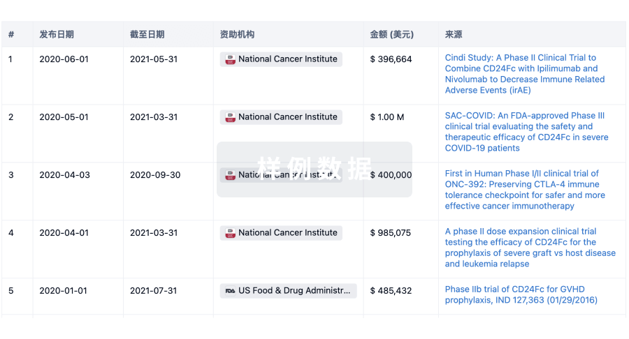
科研基金(NIH)
访问超过 200 万项资助和基金信息,以提升您的研究之旅。
登录
或
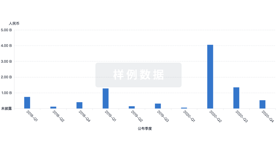
投资
深入了解从初创企业到成熟企业的最新公司投资动态。
登录
或
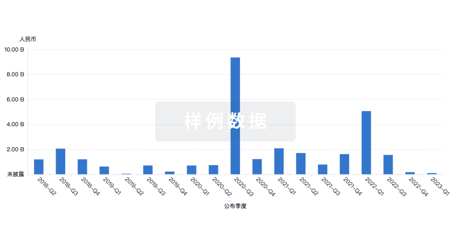
融资
发掘融资趋势以验证和推进您的投资机会。
登录
或
芽仔
全新生物医药AI Agent 覆盖科研全链路,让突破性发现快人一步
立即开始免费试用!
智慧芽新药情报库是智慧芽专为生命科学人士构建的基于AI的创新药情报平台,助您全方位提升您的研发与决策效率。
立即开始数据试用!
智慧芽新药库数据也通过智慧芽数据服务平台,以API或者数据包形式对外开放,助您更加充分利用智慧芽新药情报信息。
生物序列数据库
生物药研发创新
免费使用
化学结构数据库
小分子化药研发创新
免费使用