预约演示
更新于:2026-07-07
Ipilimumab
伊匹木单抗
更新于:2026-07-07
概要
基本信息
药物类型 单克隆抗体 |
别名 Anti CTLA-4 monoclonal antibody、Anti-CTLA-4 Mab、Ipilimumab (Genetical Recombination) + [17] |
靶点 |
作用方式 抑制剂 |
作用机制 CTLA4抑制剂(细胞毒性T淋巴细胞相关抗原4抑制剂) |
在研适应症 |
非在研适应症 |
最高研发阶段批准上市 |
首次获批日期 美国 (2011-03-25), |
最高研发阶段(中国)批准上市 |
特殊审评突破性疗法 (美国)、快速通道 (美国)、加速批准 (美国)、孤儿药 (美国)、附条件批准 (中国)、孤儿药 (日本)、孤儿药 (韩国)、优先审评 (澳大利亚)、突破性疗法 (中国)、优先审评 (美国)、优先审评 (中国) |


登录后查看时间轴
结构/序列
Sequence Code 143797L
来源: *****
Sequence Code 9060585H
来源: *****
关联
913
项与 伊匹木单抗 相关的临床试验NCT07587827
A Phase 1, Open-label, Dose-escalation and Dose-expansion Study Evaluating the Safety Feasibility and Efficacy of ODI-2001 Vaccine, a Personnalized Immunotherapy in Patients With Metastatic or Locally Advanced Colon Cancer or Pancreatic Cancer
This is a phase 1, open-label, multicentric study evaluating the safety, feasibility and efficacy of ODI-2001, a personnalized therapeutic cancer vaccine composed of DNA neoantigen vaccine, Modified Vaccinia virus Ankara (MVA) viral adjuvant and anti-CTLA4 (ipilimumab), in patients with metastatic or locally advanced colorectal or pancreatic cancer. The study includes a dose-escalation phase to determine the maximum tolerated dose (MTD) followed by an expansion phase to evaluate efficacy in terms of progression-free survival
开始日期2026-11-01 |
NCT07511036
PROSECCO: A Phase 2, Single Arm, Neoadjuvant Study Evaluating Combination Cemiplimab, Fianlimab, And Ipilimumab In Patients With Surgically Resectable Melanoma
This is a phase II study testing the safety and preliminary efficacy of triplet ICB in treatment naïve patients with clinical stage III or oligometastatic stage IV melanoma with resectable disease.
开始日期2026-09-01 |
NCT07444619
A Phase I Study of Pazopanib in Combination With Trabectedin, Ipilimumab and Nivolumab (TraPIN) in Pediatric and Young Adult Patients With Recurrent Soft Tissue Sarcomas
The goal of this study is to build on the experience of the SAINT trial by evaluating the safety and efficacy of the addition of pazopanib to their published chemotherapy regimen.
开始日期2026-08-31 |
100 项与 伊匹木单抗 相关的临床结果
登录后查看更多信息
100 项与 伊匹木单抗 相关的转化医学
登录后查看更多信息
100 项与 伊匹木单抗 相关的专利(医药)
登录后查看更多信息
5,314
项与 伊匹木单抗 相关的文献(医药)2026-12-31·CNS oncology
Tailored management of a melanoma patient with bleeding brain metastases and deep vein thrombosis with thrombectomy
Article
作者: Ceccarelli, Corrado ; Pastina, Ilaria ; Del Roscio, Davide ; Viterbo, Antonella ; De Angelis, Filippo ; Pieraccini, Massimo ; Arezzini, Luca
CASE PRESENTATION:
We describe a 62-year-old man with disseminated melanoma (initially staged pT2aN0M0, AJCC stage IB, 8th edition AJCC) who developed multiple brain metastases with hemorrhagic transformation, pulmonary embolism, retrobulbar progression with eyeball herniation, and deep vein thrombosis (DVT). After cerebral edema improved with corticosteroid tapering and the patient became asymptomatic, he started combination immunotherapy with ipilimumab (3 mg/kg q3w × 4) and nivolumab (1 mg/kg q3w × 4). Despite initial benefit, progression led to radiotherapy for retrobulbar lesion and subsequent targeted therapy with encorafenib (450 mg daily) and binimetinib (45 mg twice daily). Shortly after, the patient developed symptomatic left-leg DVT extending to the femoral vein with segmental pulmonary embolism. Given the contraindication to full anticoagulation, mechanical thrombectomy with the Indigo Aspiration System was performed, followed by prophylactic LMWH at reduced therapeutic dose (enoxaparin 1 mg/kg once daily due to recent thrombectomy and hemorrhagic risk from bleeding brain metastases). During targeted therapy, the patient experienced partial regression of brain, liver, and peritoneal metastases, although intracranial hemorrhage recurred later.
CONCLUSIONS:
This case highlights the value of a multidisciplinary, tailored approach integrating immunotherapy, radiotherapy, targeted therapy, and mechanical thrombectomy with no DVT recurrence observed following thrombectomy at 6-month follow-up.
2026-12-01·JOURNAL OF BIOMOLECULAR NMR
A fast and efficient strategy for the NMR assignment of Fab methyl groups
Article
作者: Henot, Faustine ; Doyen, Camille ; Frances, Oriane ; Güntert, Peter ; Crublet, Elodie ; Vibert, Béatrice ; Giraud, Arthur ; Dbira, Sarra ; Favier, Adrien ; Imbert, Lionel ; Boisbouvier, Jérôme ; Clavier, Séverine
Owing to their high specificity and therapeutic effectiveness, monoclonal antibodies (mAbs) have rapidly become one of the leading classes of biologic drugs used to treat critical illnesses. The antigen-binding fragment (Fab) of mAbs plays a key role in the antigen recognition, so its structural characterization is essential, as even a slight change to its Higher Order Structure (HOS) can impact the antibody's potency. Recently, 2D methyl NMR has been introduced as a powerful method to assess both the structure and integrity of therapeutic Fab fragments. However, the identification of methyl group resonances in NMR spectra remains rare since Fabs are large heterodimers of ~ 50 kDa. Here, we present the methyl group assignment of an IgG1 Fab produced in a cell-free system with an optimal isotope labelling. We first assigned 99% of the alanine, isoleucine, leucine, methionine, and valine methyl groups of the therapeutic Fab targeting LAMP1 antigen. Building on this assignment, we propose a "divide and conquer" strategy that exploits sequence identities to rapidly assign methyl groups of other IgG1 Fabs. We demonstrate that the assignment of the Fab's constant region can easily be transferred from one IgG1 to another and that the variable part of a new Fab can be assigned using smaller uniformly 15N,13C-labelled constructs. We applied our strategy to ipilimumab's Fab and, using the assignment of ipilimumab's variable part and Fab anti-LAMP1's constant part, we could transfer the assignment of 89% of the methyl-containing amino acids to the entire ipilimumab Fab without having to produce deuterated samples. This assignment strategy can be generalised to any other IgG1 Fabs provided that their constant regions are identical and the strategy can be adapted to accommodate the expression levels of the different variable domains. This new method drastically facilitates the Fab assignment process, making it suitable for the pharmaceutical timeline.
2026-08-01·SEMINARS IN ONCOLOGY
Risk of intensive care unit admission after immune checkpoint inhibitor use in cancer
Article
作者: Bhandari, Sambhawana ; Guarin, Geneva ; Baral, Maun R ; Ranganathan, Prabha ; McEvoy, Colleen ; Morgensztern, Daniel
Immune checkpoint inhibitors (ICIs) have transformed cancer therapy but can cause excessive immune activation, leading to severe adverse events, including intensive care unit (ICU) admission. Existing evidence on critical illness after ICI use is limited, largely single center, and focused on immune related adverse events. This study evaluates the risk of inpatient and ICU admissions after ICI initiation using two large national databases. We retrospectively analyzed MarketScan Commercial and Multi-state Medicaid Research Databases (2016-2022) to identify adults with solid organ malignancies treated with ICIs and determined if ICU admission was required within 90 days of ICI administration. The primary diagnosis for ICU admission was identified and categorized. Associations between cancer type, ICI type, and ICU admission were analyzed. About 33,040 patients received ICIs during the study period. About 48.9% were male and 51.1% female. Pembrolizumab was the most used (49.3%), followed by nivolumab (31.9%), and ipilimumab (9.4%). Lung cancer was the most common malignancy (40.8%), followed by genitourinary (16%), melanoma (13%), gastrointestinal (12%), and breast (10%). About 22.0% (n = 7,216) of patients were hospitalized, and 8.7% (n = 2,872) required ICU admission. Infection was the most frequent reason for ICU admission, followed by respiratory causes. The odds of being admitted to the ICU were higher with combination ICI (OR = 1.51, P < .001) compared to ICI monotherapy. In conclusion, among patients with solid organ malignancies, nearly 9% required ICU admission within 90 days of ICI initiation, most commonly for infectious causes. These findings highlight the need for heightened vigilance for critical illness in patients treated with ICIs.
2,184
项与 伊匹木单抗 相关的新闻(医药)2026-07-06
推文概览
免疫检查点抑制剂(ICI)是癌症治疗的重大突破,但只有少数患者能从中获益。现有的生物标志物——如肿瘤突变负荷(TMB)和PD-L1表达——在不同癌种和治疗方案中泛化能力有限,临床决策常常“盲人摸象”。哈佛医学院Marinka Zitnik团队在《Nature Medicine》发表的COMPASS,给出了一个全新的思路:不直接让AI从基因表达“黑箱”预测结果,而是先让模型学习44个生物学上可解释的免疫概念——从T细胞活性到TGFβ信号通路,从B细胞丰度到血管排斥——再基于这些概念做出预测。在16个临床队列、7种癌症类型、6种ICI方案、1133名患者的严格验证中,COMPASS的平均准确率比22种已有方法高出8.5%。更令人振奋的是,它能在无需训练的情况下泛化到全新的癌种和治疗方案,并通过“个性化响应地图”直观展示每位患者为何响应或耐药。这项工作标志着免疫治疗预测从“黑箱打分”迈入了“可解释导航”的新时代。
数据
预训练:10,184个肿瘤样本的转录组数据,覆盖33种癌症类型,来自TCGA数据库。
微调与验证:16个独立临床队列,共1,133名患者,覆盖7种癌症类型(BLCA、KIRC、SKCM、LUAD、STAD、GBM、LUSC)和6种ICI治疗方案(抗PD-1、抗PD-L1、抗CTLA-4及联合治疗)。
数据来源:IMvigor210、IMmotion150、Liu、Ravi、Rose、Gide、Riaz、Kim、Van Allen、Freeman、Hugo、Zhao、Snyder、Miao、Choueiri等15项独立研究。
模型
基因编码器:Transformer架构的基因语言模型(GLM),编码15,672个蛋白编码基因,采用可学习的基因位置编码替代传统位置编码。
概念投影器:双层注意力机制,先将基因嵌入投影到132个基因集(颗粒概念),再聚合为43个高维度TIME概念 + 1个癌症类型token,形成44维可解释患者嵌入。
预测模块:支持MLP参数化分类器(全微调/部分微调/线性探测)和基于原型相似度的非参数化零样本分类器(NFT)。
预训练策略:自监督三元组对比学习,拉近同一肿瘤的增强视图,推远不同肿瘤。
任务
跨癌种免疫治疗响应预测:在7种癌症、6种ICI方案中预测患者是否响应免疫治疗。
跨适应症泛化:在训练中完全排除某癌种后,仍能准确预测该癌种患者的响应(如LUAD排除后仍达76.5%准确率)。
跨疗法泛化:仅用抗PD-1/PD-L1数据训练,即可预测抗CTLA-4治疗响应(准确率70.8%)。
生存预后分层:在IMvigor210队列中,预测响应者1年生存率86% vs 非响应者40%(HR=4.7, P<10⁻⁷),显著优于TMB和PD-L1 IHC。
耐药机制解析:通过个性化响应地图,揭示“免疫炎症型但耐药”患者的TGFβ信号、血管排斥、CD4+T细胞功能障碍和B细胞缺陷等机制。
01
引言:免疫治疗预测的“罗塞塔石碑”难题
免疫检查点抑制剂(ICI)已经彻底改变了多种癌症的治疗格局。但一个残酷的现实是:大多数患者并不会响应。更令人困惑的是,即使是最常用的生物标志物——肿瘤突变负荷(TMB)和PD-L1表达——也常常“失灵”:有些高TMB的患者对治疗无动于衷,而有些低TMB的患者却获得了持久缓解。
问题出在哪里?传统的生物标志物试图用“一两个指标”来预测“整个免疫系统与肿瘤的博弈”——这就像试图用体温单次测量来判断一场战争的胜负。真正的答案藏在肿瘤微环境(TME)的复杂生态中:T细胞的活性状态、B细胞的浸润程度、TGFβ信号的强度、血管是否形成了物理屏障……这些因素共同决定了免疫治疗能否奏效。
但问题是:我们如何在临床上大规模、可解释地捕捉这些信息?
哈佛医学院Marinka Zitnik团队给出了一个优雅的答案:COMPASS——一个基于概念瓶颈(Concept Bottleneck)架构的泛癌种基础模型。
02
方法详解:让AI先学会“免疫学的语言”
COMPASS的核心设计理念是:不直接从基因表达跳到预测结果,而是先经过一层人类可理解的概念——就像医生诊断疾病时,会先识别症状、再判断病因、最后给出结论,而不是直接从检查报告跳到处方。
COMPASS框架——从转录组到可解释预测的三层架构
COMPASS的三层架构。(a)概念学习层次:从基因表达到132个颗粒免疫概念,再到44个高维度TIME概念。(d)模型详细架构:Transformer基因编码器 → 双层注意力概念投影器 → 分类器。(e)四种微调策略:根据临床队列大小,支持全微调(FFT)、部分微调(PFT)、线性探测(LFT)和零样本原型推理(NFT)。
第一步:给每个基因“赋能”——Transformer基因编码器
COMPASS首先将患者的转录组数据(15,672个蛋白编码基因的表达量)输入一个Transformer架构的基因语言模型(GLM)。
与传统Transformer不同,COMPASS没有使用固定的位置编码——因为基因之间不存在像自然语言那样的固定顺序。取而代之的是可学习的基因位置编码,让模型能够在训练中自主发现哪些基因之间存在着功能上的“上下文关系”。
此外,模型还加入了一个癌症类型token,让模型知道患者得的是什么癌——这个信息会被单独投影为一个概念,并在后续分析中可以验证其贡献。
第二步:从基因到概念——双层注意力投影器
这是COMPASS最核心的创新。模型不是直接用基因嵌入去做预测,而是先将它们投影到44个生物学可解释的概念上。
第一层投影:将15,672个基因的嵌入,通过注意力机制聚合为132个基因集(颗粒概念),每个基因集对应一个经过文献验证的免疫基因签名——涵盖免疫细胞类型、功能状态、信号通路以及非免疫肿瘤生物学(如基质程序和DNA损伤响应)。
第二层投影:再将132个颗粒概念通过第二层注意力机制聚合为43个高维度TIME概念——包括“细胞毒性T细胞”、“IFNγ通路”、“TGFβ信号”、“B细胞通用”、“内皮细胞”、“基因组完整性”等。再加上1个癌症类型token,最终形成44维的患者嵌入。
这种设计的精妙之处在于:每一个中间概念都是人类可理解的。当模型最终做出预测时,我们可以清晰地看到——是“TGFβ信号”太强导致了耐药,还是“细胞毒性T细胞”活性不足导致了响应失败。
第三步:自监督预训练——在10,184个肿瘤上“打基础”
COMPASS在TCGA的10,184个肿瘤样本(覆盖33种癌症类型)上进行了自监督对比学习预训练。
具体做法是:对每个肿瘤的转录组进行随机增强(基因随机掩码或高斯抖动),然后训练模型让同一肿瘤的不同增强视图在44维概念空间中彼此靠近,而不同肿瘤则彼此远离。通过这种方式,模型学会了在概念空间中捕捉肿瘤-免疫相互作用的本质特征。
第四步:灵活微调——适应不同规模的临床队列
预训练完成后,COMPASS可以根据临床队列的大小选择不同的微调策略:
全微调(COMPASS-FFT):更新所有模型参数,适合大型队列(>100例)。
部分微调(COMPASS-PFT):仅更新投影器和分类器,适合中型队列(30-100例)。
线性探测(COMPASS-LFT):仅更新最后的分类层,适合小型队列(<30例)。
零样本预测(COMPASS-NFT):不做任何微调,直接通过概念空间中的原型相似度进行预测。
这种设计让COMPASS能够在数据极其稀缺的临床场景中依然保持良好性能。
03
关键发现:可解释的模型,也能“打”得漂亮
跨16个队列的全面验证:准确率提升8.5%
COMPASS在16个独立临床队列、1,133名患者、7种癌症类型、6种ICI方案上进行了严格的留一队列外(LOCO)评估。
与22种已有方法(包括TIDE、IMPRES、NetBio、PGM等)相比,COMPASS-PFT和COMPASS-LFT的平均准确率提升了8.5%,AUPRC提升了15.7%。在240对队列间迁移评估中,COMPASS-LFT成功迁移了163对(成功率68%),远超PGM(130对)、Teff(118对)和NetBio(117对)。
跨癌种、跨疗法泛化:从未见过的癌症也能预测
这是COMPASS最令人震撼的能力。
跨癌种预测:当在训练中完全排除肺腺癌(LUAD) 后,COMPASS-PFT仍然能在独立的LUAD队列上达到76.5%的准确率。这意味着模型学到的不是某个癌种特有的“套路”,而是泛癌种的免疫治疗响应通用规律。
跨疗法预测:仅用抗PD-1/PD-L1治疗的患者数据训练,COMPASS-PFT就能预测抗CTLA-4治疗的反应,准确率达70.8%。更令人惊讶的是,仅用单药治疗数据训练,模型就能预测联合治疗(ipilimumab + pembrolizumab)的响应,准确率达85.3%。
生存预后:比TMB和PD-L1更准
在IMvigor210队列(atezolizumab治疗的转移性尿路上皮癌,n=298)中,COMPASS预测的响应者1年生存率达86%,而非响应者仅为40%,风险比(HR)高达4.7(P = 1.7 × 10⁻⁷)。
相比之下,TMB的风险比仅为1.67(P=0.0038),PD-L1 IC2+评分为1.75(P=0.0018),IHC免疫表型为1.85(P=0.0042)。COMPASS的预测性能显著优于这三种临床常用的生物标志物。
COMPASS预测的响应者生存显著优于TMB和PD-L1
在IMvigor210队列(atezolizumab治疗的转移性尿路上皮癌)中,COMPASS预测的响应者(红色)与非响应者(蓝色)的Kaplan-Meier生存曲线。(a)COMPASS预测的HR=4.7(P<10⁻⁷),1年生存率86% vs 40%。(b-d)TMB(HR=1.67)、PD-L1 IC2+(HR=1.75)和IHC免疫表型(HR=1.85)的预后分层能力均显著弱于COMPASS。
揭示耐药机制:为什么“炎症型”肿瘤也会耐药?
COMPASS最令人兴奋的能力是解析“为什么”。
传统上,肿瘤根据CD8+ T细胞浸润被分为三类:炎症型(inflamed)、排除型(excluded) 和沙漠型(desert)。炎症型肿瘤通常被认为对免疫治疗响应较好——但事实并非如此,相当一部分炎症型肿瘤患者依然耐药。
COMPASS的概念分析揭示了其中的奥秘:
炎症型响应者:高表达细胞毒性T细胞、IFNγ通路、免疫检查点等促炎概念,且没有免疫抑制信号。
炎症型非响应者:虽然也有促炎信号,但被不同的耐药机制“抵消”了——
第一类:内皮概念高度激活→血管重塑形成物理屏障,阻止T细胞浸润。
第二类:TGFβ通路高度激活→促进基质重塑和纤维化,将T细胞排除在肿瘤之外。
第三类:CD4+ T细胞免疫抑制 + B细胞缺陷。
这些发现不仅解释了为什么有些“看起来应该响应”的患者却耐药,更为联合治疗的靶点选择提供了直接依据——比如TGFβ抑制剂、抗血管生成药物等。
个性化响应地图:每一位患者都有自己的“免疫导航”
COMPASS为每位患者生成个性化响应地图,展示从基因表达到概念激活再到最终预测的完整链条。
地图分为五个层级:基因表达 → 编码器表示 → 颗粒免疫概念 → 聚合TIME概念 → 最终响应概率。
例如:
炎症型响应者:广泛的IFNγ和细胞毒性激活,几乎无免疫抑制(P_R=1.0)。
沙漠型响应者:强基因组完整性概念和中等IFNγ激活,提示TMB相关机制(P_R=0.80)。
炎症型非响应者:TGFβ信号与B细胞缺陷共激活(P_R=0.22)。
沙漠型非响应者:以免疫缺陷特征为主导(P_R=0)。
这种地图让临床医生可以直观地看到“这位患者为什么可能响应”或“为什么可能耐药”,而不仅仅是一个黑箱给出的概率分数。
个性化响应地图——四位患者的“免疫导航”
四位代表性患者的个性化响应地图。(a)炎症型响应者:广泛IFNγ和细胞毒性激活(PR=1.0)。(b)沙漠型响应者:强基因组完整性+中等IFNγ(PR=0.80)。(c)炎症型非响应者:TGFβ信号+B细胞缺陷共激活(PR=0.22)。(d)沙漠型非响应者:免疫缺陷主导(PR=0)。地图展示了从基因表达到概念激活再到最终预测的完整链条。
04
临床意义与未来展望
COMPASS的贡献在于它证明了可解释性和高性能可以兼得。
对临床开发:COMPASS的多阶段微调(MSFT)策略可以在早期临床试验数据极其有限的情况下,快速构建针对特定药物或适应症的预测模型。这对于药物研发中的适应症选择和患者富集具有直接价值。
对临床医生:个性化响应地图提供了超越“响应/不响应”二分类的机制性洞察,可以帮助医生理解每位患者独特的肿瘤-免疫状态,为联合治疗决策提供依据。
对研究者:COMPASS识别的44个可解释概念为 biomarker发现和 hypothesis generation提供了新工具。
当然,COMPASS也有其局限性。模型依赖bulk RNA-seq数据,缺乏空间分辨率,可能掩盖稀有免疫细胞群体的信号。此外,缺乏非ICI治疗的对照臂,使得无法完全区分预测性信号和预后性信号。因此,COMPASS的预测应被视为探索性工具,而非临床决策的唯一依据。
尽管如此,COMPASS代表了一个重要的方向:让AI不仅能“算得准”,还能“说得清”——这对于建立临床医生对AI的信任、推动精准免疫治疗的发展,至关重要。
05
附录:COMPASS研究数据详情
预训练数据详情
COMPASS在10,184个肿瘤样本的转录组数据上进行自监督预训练,这些数据来自TCGA(The Cancer Genome Atlas)数据库,覆盖33种癌症类型。所有RNA-seq数据经过统一的标准化处理流程(STAR比对至GRCh38/hg38参考基因组,GENCODE v36注释,TPM标准化)。
ICI临床队列详情
COMPASS在16个独立临床队列上进行微调与验证,共1,133名患者,覆盖7种癌症类型和6种ICI治疗方案。
大型队列(>100例,共4个,672例患者)
IMvigor210(n=298):膀胱癌(BLCA),atezolizumab(抗PD-L1)治疗,68例响应者/230例非响应者。数据来源:EGA:EGAS00001002556。
IMmotion150(n=165):肾透明细胞癌(KIRC),atezolizumab治疗,48例响应者/117例非响应者。数据来源:EGA:EGAS00001002928。
Liu(n=107):黑色素瘤(SKCM),nivolumab/pembrolizumab(抗PD-1)治疗,41例响应者/66例非响应者。数据来源:dbGaP:phs000452.v3.p1。
Ravi-1(n=102):肺腺癌(LUAD),PD-(L)1 ± CTLA-4抑制剂治疗,38例响应者/64例非响应者。数据来源:dbGaP:phs002822.v1.p1。
中型队列(30-100例,共6个,331例患者)
Rose(n=89):膀胱癌(BLCA),PD-(L)1抑制剂治疗,16例响应者/73例非响应者。数据来源:GEO:GSE176307。
Gide(n=73):黑色素瘤(SKCM),抗PD-1 ± 抗CTLA-4治疗,40例响应者/33例非响应者。数据来源:ENA:PRJEB23709。
Riaz(n=51):黑色素瘤(SKCM),nivolumab治疗,10例响应者/41例非响应者。数据来源:BioProject:PRJNA356761。
Kim(n=45):胃腺癌(STAD),pembrolizumab治疗,12例响应者/33例非响应者。数据来源:ENA:PRJEB25780。
Van Allen(n=39):黑色素瘤(SKCM),ipilimumab(抗CTLA-4)治疗,13例响应者/26例非响应者。数据来源:dbGaP:phs000452.v2.p1。
Freeman(n=34):黑色素瘤(SKCM),nivolumab/pembrolizumab/ipilimumab/联合治疗,12例响应者/22例非响应者。数据来源:dbGaP:phs002683.v1.p1。
小型队列(<30例,共6个,130例患者)
Hugo(n=26):黑色素瘤(SKCM),pembrolizumab治疗,14例响应者/12例非响应者。数据来源:GEO:GSE78220。
Zhao(n=25):胶质母细胞瘤(GBM),nivolumab/pembrolizumab治疗,11例响应者/14例非响应者。数据来源:SRA:PRJNA482620。
Ravi-2(n=25):肺鳞状细胞癌(LUSC),PD-1/PD-L1抑制剂治疗,8例响应者/17例非响应者。数据来源:dbGaP:phs002822.v1.p1。
Snyder(n=21):膀胱癌(BLCA),atezolizumab治疗,7例响应者/14例非响应者。数据来源:Zenodo:10.5281/zenodo.546110。
Miao(n=17):肾透明细胞癌(KIRC),PD-(L)1 ± CTLA-4治疗,5例响应者/12例非响应者。数据来源:dbGaP:phs001493.v1.p1。
Choueiri(n=16):肾透明细胞癌(KIRC),nivolumab治疗,3例响应者/13例非响应者。数据来源:CRI iAtlas数据门户。
模型架构与训练详情
基因编码器
架构:Transformer(Performer线性注意力近似)
输入:15,672个蛋白编码基因的TPM表达值
嵌入维度:d维可学习基因嵌入 + 可学习基因位置编码 + 癌症类型token
概念投影器
第一层:132个基因集(颗粒概念),通过注意力机制从基因嵌入聚合
第二层:43个高维度TIME概念 + 1个癌症类型概念,通过第二层注意力从颗粒概念聚合
输出:44维可解释患者嵌入
预训练
数据:TCGA 10,184个肿瘤,33种癌症类型
方法:自监督三元组对比学习
增强:随机基因掩码(p=0.1)或高斯抖动
损失:Margin-based Triplet Loss
微调策略
COMPASS-FFT:全微调(约1,018,784个参数),适合大型队列
COMPASS-PFT:部分微调(2,144个参数),适合中型队列
COMPASS-LFT:线性探测(182个参数),适合小型队列
COMPASS-NFT:零样本原型推理,无训练参数
评估指标
准确率(Accuracy)、AUPRC、MCC
留一队列外(LOCO)验证、队列间迁移验证(240对)、队列内留一患者验证
概念分类
免疫细胞类型:细胞毒性T细胞、CD4+ T细胞、B细胞、浆细胞、NK细胞、先天淋巴样细胞、巨噬细胞、pDC等
功能状态:T细胞耗竭、免疫检查点、IFNγ通路、TGFβ通路等
肿瘤生物学:基因组完整性、细胞增殖、内皮细胞、癌症相关成纤维细胞等
论文索引:Shen, W., Moon, I., Nguyen, T.H. et al. Generalizable AI predicts immunotherapy outcomes across cancers and treatments. Nat Med (2026). https://doi.org/10.1038/s41591-026-04502-7
论文链接:https://www.nature.com/articles/s41591-026-04502-7
代码链接:https://github.com/mims-harvard/COMPASS
稿件翻译:黄伊利
稿件审核:王振阳
2026-07-06
Part 2 Cancer Cell部分1. Vascular RhoJ Is an Effective and Selective Target for Tumor Angiogenesis and Vascular Disruption. 🔥
•期刊: Cancer cell•作者: Chan Kim et al.•年份: 2026•分类: 肿瘤学•亮点: 肿瘤学重要进展
摘要:
无摘要文献提炼
📚 研究背景: 肿瘤血管生成(tumor angiogenesis)是实体瘤生长和转移的关键限速步骤,抗血管生成治疗(如anti-VEGF/VEGFR)已成为临床标准策略,但疗效受限于耐药性和正常血管毒性。RhoJ是Rho家族小GTPase的一员,主要在血管内皮细胞中表达,前期研究提示其在内皮细胞迁移和血管新生中发挥调控作用,但其作为抗肿瘤血管靶点的可行性和选择性尚未被系统验证。
❓ 核心科学问题: RhoJ能否作为肿瘤血管生成的选择性治疗靶点?靶向RhoJ能否同时实现抑制新生血管形成(anti-angiogenesis)和破坏已建立的肿瘤血管(vascular disruption)的双重效应,同时避免对正常血管系统的毒副作用?
🔬 主要发现: 该研究发现RhoJ在肿瘤血管内皮细胞中高度活化和上调,而在正常静息内皮细胞中表达较低。通过基因敲除或药理学抑制RhoJ,可显著抑制多种小鼠肿瘤模型中的血管生成,并诱导已形成肿瘤血管的选择性崩解,导致肿瘤缺血坏死和生长抑制。重要的是,靶向RhoJ对正常器官的血管系统影响极小,展现出良好的治疗窗口。
💡 研究意义: 该研究首次确立了RhoJ作为肿瘤血管选择性治疗靶点的概念验证(proof-of-concept),为开发新一代兼具anti-angiogenic和vascular disrupting双重机制的抗肿瘤药物提供了分子基础。RhoJ的内皮特异性使其有望克服现有抗血管药物因靶点在多种细胞类型广泛表达而导致的全身毒性问题,推动精准血管靶向治疗的发展。
链接: PubMed[1]2. Circadian Homeostasis of Liver Metabolism Suppresses Hepatocarcinogenesis. 🔥
•期刊: Cancer cell•作者: Nicole M Kettner et al.•年份: 2026•分类: 代谢与生理•亮点: 代谢与生理调控研究
摘要:
无摘要文献提炼
📚 研究背景: 昼夜节律(circadian rhythm)由核心时钟基因(如BMAL1、CLOCK、PER、CRY)组成的转录-翻译反馈回路驱动,调控机体约50%基因的节律性表达。流行病学证据表明,长期昼夜节律紊乱(如轮班工作、慢性时差)与多种癌症(包括肝细胞癌HCC)风险升高显著相关,但节律失调如何通过代谢重编程驱动肝癌发生的分子机制仍不清楚。
❓ 核心科学问题: 肝脏昼夜节律稳态的维持如何通过调控代谢通路抑制肝癌发生(hepatocarcinogenesis)?昼夜节律破坏是否通过扰乱肝脏代谢稳态(如脂质代谢、胆汁酸代谢、葡萄糖代谢)从而创造促癌的代谢微环境?
🔬 主要发现: 该研究在遗传和饮食诱导的肝癌小鼠模型中证明,肝脏特异性时钟基因(如Bmal1)缺失或慢性节律破坏可加速肝癌发生。机制上,昼夜节律紊乱导致肝脏关键代谢通路的节律性表达丧失,引发脂质堆积、胆汁酸代谢失调和氧化应激增加,这些代谢异常共同促进DNA损伤积累和肝细胞向恶性转化。恢复节律性代谢信号可部分逆转促癌表型。
💡 研究意义: 该研究建立了昼夜节律—代谢稳态—肝癌抑制三者之间的因果链条,为将"时间医学"(chronomedicine)理念引入肝癌预防提供了关键证据。结果提示,维持正常昼夜节律或定时给予代谢干预可能成为高危人群(如代谢相关脂肪性肝病MAFLD患者)预防肝癌的新策略,也为探索"何时给药"(chronotherapy)优化肝癌治疗效果开辟了新方向。
链接: PubMed[2]
数据来源: PubMed | 筛选标准: CNS 及 Nature Index 期刊
🔥 标记为创新性评分>25 的高亮点文章本文内链接
[1]
PubMed: https://pubmed.ncbi.nlm.nih.gov/42379175/
[2]
PubMed: https://pubmed.ncbi.nlm.nih.gov/42392087/
CNS周报 — Part 3其他子刊精选1. LIF-Induced Tumor Plasticity Establishes an Immunosuppressive Myeloid Niche in LKB1-Mutant Lung Cancer. 🔥
•期刊: Cancer discovery•作者: Ray Pillai et al.•年份: 2026•分类: 肿瘤学•亮点: 肿瘤学重要进展
摘要:
LKB1 mutations in lung cancer promote an immunosuppressive tumor microenvironment, but the underlying mechanisms remain unknown. Using genetically engineered mouse models and human tumor samples, we demonstrate that LKB1 loss leads to high expression of the cytokine leukemia-inhibitory factor (LIF), which through a cancer cell-autonomous autocrine loop, orchestrates the infiltration of immunosuppressive SiglecFHi neutrophils and Arg1+ interstitial macrophages. Genetic deletion of Lifr, the receptor for LIF, on Lkb1-mutant lung tumors revealed that autocrine LIF signaling induces tumor plasticity and the emergence of a Sox17+ dedifferentiated inflammatory cell state. Antibody-mediated LIF neutralization selectively eliminates the Sox17+ tumor cell state, reduces immunosuppressive myeloid cells, and enhances antitumor T-cell responses. Our study uncovers a novel LKB1-LIF axis driving immune evasion and identifies LIF as a potential therapeutic target in LKB1-mutant lung cancer. This work highlights the interplay between tumor genetics, cellular plasticity, and immune regulation in lung cancer progression. LKB1-mutant lung cancers express LIF, which induces an immunosuppressive Sox17+ tumor state. Anti-LIF therapy eliminates this state and restores antitumor immunity, revealing a novel vulnerability in this aggressive cancer subtype lacking effective targeted therapies.文献提炼
📚 研究背景: LKB1(STK11)突变是非小细胞肺癌中最常见的驱动突变之一,与免疫检查点抑制剂的原发性耐药密切相关。LKB1突变肿瘤以其高度免疫抑制的肿瘤微环境(TME)著称,富含免疫抑制性髓系细胞且T细胞浸润稀少,但其背后的分子机制长期未明。肿瘤细胞的可塑性(plasticity)和分泌因子如何协调构建免疫抑制微环境是该领域的核心关注点。
❓ 核心科学问题: LKB1缺失通过何种分泌因子和自分泌信号环路重塑肿瘤微环境?该过程中是否存在可靶向的节点,以逆转LKB1突变肿瘤的免疫逃逸?
🔬 主要发现: LKB1缺失导致肿瘤细胞高表达白血病抑制因子(LIF),LIF通过自分泌环路诱导肿瘤细胞去分化并产生Sox17+炎症性细胞状态。该细胞状态进一步招募免疫抑制性SiglecFhi中性粒细胞和Arg1+间质巨噬细胞,系统性地构建免疫抑制性髓系微环境。抗LIF中和抗体可选择性清除Sox17+肿瘤细胞状态,减少免疫抑制性髓系细胞,并显著增强抗肿瘤T细胞免疫应答。
💡 研究意义: 该研究首次揭示了LKB1-LIF信号轴驱动免疫逃逸的全新机制,为LKB1突变肺癌这一缺乏有效靶向治疗的侵袭性亚型提供了明确的可操作免疫治疗靶点。LIF中和策略有望与现有免疫检查点抑制剂联合,突破该类肿瘤的治疗瓶颈,具有直接的临床转化价值。
链接: PubMed[1]2. Integrated Chronic In Vivo and In Vitro Screens Uncover NFIL3 as a Driver of T-cell Dysfunction. 🔥
•期刊: Cancer discovery•作者: Nayan Jain et al.•年份: 2026•分类: 肿瘤学•亮点: 生命科学前沿进展
摘要:
Chimeric antigen receptor (CAR) therapy has transformed the treatment landscape for hematologic malignancies, but its efficacy in solid tumors is limited, owing in part to insufficient functional persistence of the engineered T cells. To elucidate the basis for their functional decline, we conducted integrated chronic in vivo and in vitro screens of 400 transcription factors, which revealed NFIL3 as a driver of CAR T-cell dysfunction. Genetic disruption of NFIL3 in CAR T cells sustains their expansion and increases cytokine production, overall restraining terminal differentiation. Loss of NFIL3 enhances CAR T-cell efficacy, improving tumor control and prolonging survival in xenograft and syngeneic mouse tumor models across different CAR designs. Under chronic stimulation, disruption of NFIL3 establishes a transcriptional state predictive of favorable clinical outcomes. Our findings underscore the power of comprehensive in vivo genetic screens integrated with multiparameter in vitro assessment and identify NFIL3 as a novel therapeutic target to enhance cancer immunotherapy. This study presents a two-step screening framework, integrating an in vivo pooled guide RNA screen with a multiparameter, in vitro arrayed screen. NFIL3 emerged as the top candidate, and its disruption enhanced CAR T-cell antitumor efficacy in both hematologic malignancies and solid tumors across diverse CAR architectures.文献提炼
📚 研究背景: CAR-T细胞疗法在血液肿瘤中取得突破性成功,但在实体瘤中的疗效仍十分有限,关键瓶颈在于工程化T细胞在慢性抗原刺激下的功能耗竭(dysfunction)和持久性不足。转录因子作为调控T细胞分化与命运决定的核心节点,是克服CAR-T功能衰竭的潜在干预靶点,但系统性筛选驱动T细胞耗竭的关键转录因子一直是技术挑战。
❓ 核心科学问题: 在慢性抗原刺激条件下,哪个(些)转录因子是驱动CAR-T细胞功能失调的核心调控因子?能否通过基因编辑干预这些转录因子来增强CAR-T细胞的抗肿瘤活性?
🔬 主要发现: 研究建立了"两步法"筛选框架——整合体内pooled gRNA筛选和体外多参数阵列筛选,从400个转录因子中鉴定出NFIL3为CAR-T细胞功能衰竭的关键驱动因子。敲除NFIL3可维持CAR-T细胞在慢性刺激下的扩增能力,增加细胞因子产生,抑制终末分化,并在多种CAR设计和不同肿瘤模型(包括异种移植和同系移植模型)中显著改善肿瘤控制和延长生存期。
💡 研究意义: 该研究建立了体内外整合筛选的创新范式,为系统鉴定T细胞功能调控因子提供了高效、可推广的平台。NFIL3作为CAR-T细胞工程改造的新靶点,有望通过CRISPR等基因编辑技术优化CAR-T产品性能,提升其在实体瘤和血液肿瘤中的临床疗效。
链接: PubMed[2]3. Spatially Sussing Out the TME, No Tissue Needed. 🔥
•期刊: Cancer discovery•作者: N/A et al.•年份: 2026•分类: 肿瘤学•亮点: 生命科学前沿进展
摘要:
Through machine learning, researchers have uncovered distinct spatial ecotypes in the tumor microenvironment that are broadly conserved across multiple cancers and may correlate with response to immune checkpoint blockade. These ecotypes are also identifiable through methylation profiling, suggesting another potential use for liquid biopsy-a way to noninvasively monitor the tumor microenvironment.文献提炼
📚 研究背景: 肿瘤微环境(TME)的空间组织结构——不同免疫细胞和基质细胞在肿瘤内的空间定位与相互作用——与免疫治疗响应密切相关。然而,传统空间组学分析依赖组织活检样本,无法实现无创动态监测。液体活检技术(如ctDNA甲基化检测)已被广泛用于肿瘤基因分型,但其能否反映TME的空间特征尚属未知。
❓ 核心科学问题: TME的空间生态型(spatial ecotypes)是否在多种癌症中保守存在并具有功能意义?能否通过无创的甲基化谱分析来推断TME的空间特征,从而实现"无需组织的TME监测"?
🔬 主要发现: 研究人员利用机器学习在多种癌症类型中识别出保守存在的TME空间生态型,这些生态型与免疫检查点抑制剂的治疗反应显著相关。更关键的是,这些空间生态型可通过DNA甲基化谱分析加以识别,为液体活检提供了全新的应用维度——无创监测TME的空间组成和免疫状态。
💡 研究意义: 该研究建立了从液体活检推断TME空间结构的桥梁,将甲基化检测的临床应用从传统的基因突变检测拓展至微环境空间评估。这一策略有望实现免疫治疗疗效的无创动态监测,为精准免疫治疗的实时决策提供重要工具。
链接: PubMed[3]4. A Circulating GPNMB-Based Multimodal Model Integrates Tumor-Immune Crosstalk to Predict Immunotherapy Response in Esophageal Cancer.
•期刊: Cancer discovery•作者: Liang Zhu et al.•年份: 2026•分类: 肿瘤学•亮点: 肿瘤学重要进展
摘要:
Neoadjuvant immunotherapy improves outcomes in esophageal squamous cell carcinoma (ESCC), yet ∼70% of patients fail to respond. Pretreatment biopsies and plasma provide critical opportunities for biomarker discovery. In this study, we performed plasma proteomic profiling and identified soluble glycoprotein nonmetastatic melanoma protein B (sGPNMB) as the most elevated circulating protein in nonresponders. Mechanistically, tumor cell-derived sGPNMB suppressed CD8+ T-cell receptor signaling via the SDC4-CD148 axis to induce functional exhaustion, with secretion being required for its immunosuppressive activity. Cancer-associated fibroblast-epithelial (CAF-Epi) niches promoted SOX2 upregulation in tumor cells, transcriptionally activating GPNMB expression. In humanized patient-derived xenograft models, circulating GPNMB levels predicted response to PD-1 blockade, and GPNMB inhibition synergized with therapy. Across retrospective cohorts and a prospective clinical trial, a multimodal model combining plasma GPNMB levels, CAF-Epi niche detection, and clinical-pathologic features achieved robust predictive accuracy for immunotherapy response and survival. These findings establish a spatial-circulating biomarker framework for precision ESCC immunotherapy. Tumor-derived soluble GPNMB, transcriptionally activated by SOX2 within CAF-Epi niches, drives CD8+ T-cell exhaustion and resistance to PD-1 blockade in ESCC. Integrating circulating GPNMB levels with CAF-Epi niche features and clinical-pathologic factors, we develop and validate a clinically scalable multimodal model for predicting immunotherapy response.文献提炼
📚 研究背景: 食管鳞状细胞癌(ESCC)的新辅助免疫治疗虽能改善预后,但约70%的患者对治疗原发性无应答,亟需可靠的疗效预测生物标志物。肿瘤微环境中CAF-上皮细胞交互(CAF-Epi niche)与免疫细胞的crosstalk是决定免疫治疗应答的关键,但如何将这种空间信息与循环生物标志物整合以建立临床可用的预测模型仍待解决。
❓ 核心科学问题: 能否从外周血中鉴定出反映肿瘤-免疫交互作用的可溶性生物标志物?肿瘤微环境的空间特征(CAF-Epi niche)如何通过何种机制驱动免疫抑制因子的表达与分泌?
🔬 主要发现: 通过血浆蛋白质组学分析,研究者发现可溶性GPNMB(sGPNMB)在免疫治疗无应答者中显著升高。机制上,肿瘤细胞在CAF-Epi微环境中通过SOX2转录上调GPNMB表达,分泌型sGPNMB通过SDC4-CD148轴抑制CD8+ T细胞的TCR信号传导,诱导功能性耗竭。整合血浆GPNMB水平、CAF-Epi niche检测和临床病理特征的多模态模型在回顾性队列和前瞻性临床试验中均实现了对免疫治疗应答和生存的高准确率预测。
💡 研究意义: 该研究提出了"空间-循环"生物标志物整合框架,将肿瘤微环境的空间特征与可溶性循环蛋白有机结合,为ESCC的精准免疫治疗提供了可临床推广的预测工具。GPNMB不仅是预测标志物,其免疫抑制功能也使其成为潜在的联合治疗靶点。
链接: PubMed[4]5. GPNMB Drives Brain Metastasis by Sculpting a Pathologic Endothelial-Immune Interactome.
•期刊: Cancer discovery•作者: Xuefei Liu et al.•年份: 2026•分类: 肿瘤学•亮点: 免疫学新机制
摘要:
Brain metastasis remains a devastating disease with dismal prognosis. How circulating tumor cells (CTC) penetrate the blood-brain barrier (BBB) and reprogram the brain microenvironment remains unclear. Using spatially resolved multi-omics profiling of CTCs and brain metastases, integrated with experimental and clinical analyses, we identified glycoprotein nonmetastatic melanoma protein B (GPNMB) as a CTC-secreted driver of vascular disruption and brain colonization. CBX3 upregulation induced GPNMB expression, which bound endothelial EGFR, triggering CBL-mediated ubiquitination and degradation. Attenuated EGFR signaling suppressed FTO and disrupted endothelial junctions via YTHDF2-dependent TJP1 m6A methylation. Remarkably, GPNMB-induced BBB remodeling promoted immune infiltration via the CXCL12-CXCR4 axis and induced time-course-dependent T-cell exhaustion within the brain microenvironment. Clinically, elevated CBX3+GPNMB+ CTCs and plasma CXCL12 were significantly associated with brain metastasis progression in lung cancer and melanoma. Therapeutically, dual blockade of GPNMB and PD1 enhanced anti-brain metastasis efficacy in mice, unveiling GPNMB as a promising target for precision immunotherapy. GPNMB is a CTC-secreted driver of BBB disruption and brain colonization via the CBX3-GPNMB-EGFR-FTO-TJP1 axis. GPNMB-induced BBB remodeling promotes CXCL12-CXCR4-mediated immune infiltration and enhances T-cell exhaustion, sensitizing brain metastasis tumors to GPNMB/PD1 dual blockade. CBX3+GPNMB+ CTCs and plasma CXCL12 may serve as noninvasive biomarkers for brain metastasis management.文献提炼
📚 研究背景: 脑转移是肺癌和黑色素瘤的致命并发症,预后极差。循环肿瘤细胞(CTC)如何穿透血脑屏障(BBB)并在脑部定植是领域内的关键未解问题。BBB的破坏不仅涉及物理屏障的突破,还可能导致脑部免疫微环境的重塑,但CTC分泌因子驱动这一系列事件的完整分子通路尚不清楚。
❓ 核心科学问题: CTC分泌的何种因子驱动BBB的破坏和脑转移定植?BBB重塑后脑部免疫微环境发生了怎样的动态变化,这些变化如何影响免疫治疗的疗效?
🔬 主要发现: 研究发现CTC中CBX3上调驱动GPNMB表达,分泌型GPNMB与脑内皮细胞EGFR结合,触发CBL介导的泛素化降解,导致EGFR信号减弱从而抑制FTO,进而通过YTHDF2依赖的TJP1 m6A甲基化破坏内皮紧密连接。GPNMB诱导的BBB重塑通过CXCL12-CXCR4轴促进免疫细胞浸润,但随时间推移导致T细胞耗竭。临床数据显示CBX3+GPNMB+ CTCs和血浆CXCL12与脑转移进展显著相关,而GPNMB/PD-1双靶向联合治疗在小鼠模型中显示出协同抗脑转移效果。
💡 研究意义: 该研究系统阐明了CTC通过GPNMB"雕刻"脑转移微环境的完整分子通路——从CBX3-GPNMB-EGFR-FTO-TJP1的BBB破坏轴到CXCL12-CXCR4的免疫重塑轴。这为脑转移的早期预警(基于CTCs和血浆CXCL12)和靶向免疫治疗(GPNMB/PD-1双阻断)提供了全新的策略和生物标志物。
链接: PubMed[5]6. Spatial Atlas Decodes Tumor Immune Hubs Across Cancer Types.
•期刊: Cancer discovery•作者: N/A et al.•年份: 2026•分类: 肿瘤学•亮点: 肿瘤学重要进展
摘要:
无摘要文献提炼
📚 研究背景: 肿瘤免疫微环境的空间组织——不同免疫细胞亚群如何在肿瘤组织中定位、聚集和相互作用——是决定抗肿瘤免疫效能的关键因素。然而,跨不同癌症类型的免疫空间结构是否存在共性的组织原则(如"免疫枢纽"),以及这些结构如何系统性地影响免疫治疗反应,此前缺乏大规模的比较研究。
❓ 核心科学问题: 不同癌症类型的肿瘤微环境中是否存在保守的空间组织结构(immune hubs)?这些免疫枢纽的细胞组成和功能状态如何与免疫治疗疗效相关联?
🔬 主要发现: 该研究构建了跨多种癌症类型的空间转录组图谱,识别出肿瘤组织中保守存在的免疫枢纽(immune hubs)——即特定免疫细胞类型(如T细胞、B细胞、DC等)在空间上富集形成的功能性微结构。研究揭示这些免疫枢纽的组成、密度和活化状态与肿瘤免疫逃逸机制及免疫检查点抑制剂的治疗反应密切相关。
💡 研究意义: 该空间图谱为理解肿瘤免疫微环境的跨癌种组织原则提供了系统性的参考框架。免疫枢纽的发现为开发空间导向的免疫治疗策略(如促进免疫枢纽形成或恢复其功能)和跨癌种通用的预测性生物标志物奠定了基础。
链接: PubMed[6]7. Neoadjuvant BO-112 and Hypofractionated Radiation Therapy with or without Nivolumab in Soft-Tissue Sarcoma: Preclinical and Phase I Results.
•期刊: Cancer discovery•作者: Jie Deng et al.•年份: 2026•分类: 肿瘤学•亮点: 生命科学前沿进展
摘要:
Neoadjuvant immune checkpoint blockade (ICB) and radiotherapy (RT) improve disease-free survival in select patients with soft-tissue sarcoma (STS). However, most STS are myeloid-rich and lack preexisting T cells associated with ICB response. In preclinical models, we observed that intratumoral BO-112 [nanoplexed polyinosinic: polycytidylic acid (poly I:C)] engages myeloid cells that persist after RT, ultimately enhancing T cell-dependent tumor control. We evaluated BO-112 and hypofractionated RT, with or without nivolumab, in 14 patients with high-risk STS in a phase I neoadjuvant trial. Consistent with its immunologic potency, the triple combination induced rare immune-related adverse events (myositis-myocarditis-myasthenia gravis spectrum), mitigated by BO-112 and nivolumab dose adjustment. BO-112 and RT reprogrammed tumor-associated myeloid cells toward antigen-presenting states, promoted clonal replacement by less exhausted T cells, and enhanced malignant cell depletion compared with standard RT. These immunologic changes coincided with encouraging disease control in a small, high-risk cohort, supporting further clinical development. Intratumoral BO-112 and hypofractionated RT activate systemic T-cell immunity in mouse models and in a phase I neoadjuvant study of high-risk, resectable sarcoma. Engaging myeloid cells with BO-112 represents a potent strategy with RT to replete T cell-deficient tumors and expand the benefits of neoadjuvant ICB.文献提炼
📚 研究背景: 软组织肉瘤(STS)的新辅助免疫检查点阻断(ICB)联合放疗虽可改善部分患者的无病生存,但大多数STS以髓系细胞浸润为主且缺乏预存T细胞,属于典型的免疫"冷"肿瘤,对ICB反应率低。如何通过靶向髓系细胞来将免疫冷肿瘤转化为热肿瘤是该领域的核心挑战。
❓ 核心科学问题: 能否通过肿瘤内注射免疫激动剂BO-112(poly I:C纳米制剂)联合大分割放疗来重编程髓系细胞,使T细胞缺陷肿瘤对ICB增敏?三联疗法(BO-112+放疗+nivolumab)在STS新辅助治疗中的安全性和免疫学效应如何?
🔬 主要发现: 临床前模型显示,瘤内注射BO-112可激活放疗后仍存活的髓系细胞,增强T细胞依赖的抗肿瘤免疫。I期新辅助临床试验(14例高危STS患者)表明,三联方案诱导了罕见的免疫相关不良事件(肌炎-心肌炎-重症肌无力谱),但通过BO-112和nivolumab剂量调整可实现可控。BO-112联合放疗将肿瘤相关髓系细胞重编程为抗原呈递状态,促进低耗竭T细胞的克隆替换,并增强恶性细胞清除。
💡 研究意义: 该研究首次在STS中验证了通过靶向髓系细胞来"补充"T细胞缺陷肿瘤的治疗策略,为ICB在免疫冷肿瘤中的拓展应用提供了重要的临床概念验证。BO-112作为髓系细胞激动剂,有望成为放疗和ICB联合方案中的标准免疫佐剂。
链接: PubMed[7]8. Ivonescimab Plus Chemo Extends Squamous NSCLC Survival.
•期刊: Cancer discovery•作者: N/A et al.•年份: 2026•分类: 肿瘤学•亮点: 生命科学前沿进展
摘要:
The anti-PD-1, anti-VEGF bispecific antibody ivonescimab plus chemotherapy significantly improves overall survival in advanced squamous non-small cell lung cancer versus an immunotherapy-chemotherapy combination, according to a trial conducted in China. However, questions remain over the trial's applicability to patients in other countries.文献提炼
📚 研究背景: 晚期鳞状非小细胞肺癌(sq-NSCLC)的一线标准治疗为免疫治疗联合化疗,但仍有大量患者无法获得长期生存获益。PD-1/VEGF双特异性抗体依沃西单抗(ivonescimab)通过同时阻断免疫检查点和血管生成信号通路,有望实现协同增效,但其对比标准免疫化疗方案的优效性需要III期临床试验验证。
❓ 核心科学问题: 依沃西单抗联合化疗对比标准免疫化疗方案能否显著改善晚期sq-NSCLC的总生存期(OS)?在中国人群中获得的疗效数据是否可推广至全球其他人群?
🔬 主要发现: 在中国开展的III期临床试验数据显示,依沃西单抗联合化疗较标准免疫化疗组合显著延长了晚期sq-NSCLC患者的总生存期,为PD-1/VEGF双特异性抗体在肺癌一线治疗中的应用提供了关键证据。然而,该试验主要在中国人群中进行,其在其他国家和种族人群中的疗效和安全性仍需进一步验证。
💡 研究意义: 该研究可能推动sq-NSCLC一线治疗格局的改变,为PD-1/VEGF双特异性抗体策略提供了首个III期OS获益证据。但需要国际多中心研究进一步验证其跨人群的疗效一致性,以支持其在全球范围内的广泛应用。
链接: PubMed[8]9. Spatially Resolved Proteomic Cartography Illuminates the Earliest Molecular Programs in Pancreatic Cancer Evolution.
•期刊: Cancer discovery•作者: Mingyu Yang et al.•年份: 2026•分类: 肿瘤学•亮点: 肿瘤学重要进展
摘要:
Min, Schweizer, and colleagues use artificial intelligence-powered deep visual proteomics to generate a spatial proteomic atlas of pancreatic cancer precursor evolution, revealing that major metabolic and inflammatory reprogramming occurs long before overt histologic transformation. More broadly, the study highlights the emerging potential of spatial proteomics and multiomics to bridge histopathology with molecular pathology and precision oncology. See related article by Min et al., p. 1323.文献提炼
📚 研究背景: 胰腺癌从癌前病变到浸润性癌的演变涉及多步骤的分子重编程,但最早期的蛋白质水平变化——特别是在组织学尚未出现明显异常时——仍不清楚。理解这些早期分子事件对于开发胰腺癌的早期检测和拦截策略至关重要,而传统的整体组织蛋白质组学因无法区分微小前驱病变而受限。
❓ 核心科学问题: 在胰腺癌前病变演变过程中,蛋白质水平的重编程最早何时启动?空间分辨的蛋白质组学能否揭示组织学转变之前的分子预警信号?
🔬 主要发现: 该评述文章总结了Min等人的研究:利用AI驱动的深度视觉蛋白质组学(Deep Visual Proteomics, DVP)生成了胰腺癌前病变演变的空间蛋白质组图谱,揭示主要的代谢重编程和炎症重编程在明显的组织学转变之前就已大规模发生。该研究同时展示了空间蛋白质组学和多组学在桥接组织病理学与分子病理学、推动精准肿瘤学方面的变革性潜力。
💡 研究意义: 该评述强调了空间蛋白质组学在肿瘤早期检测和精准肿瘤学中的变革性价值。DVP技术的成熟应用有望使研究者能在组织学可见的病变出现之前捕捉到分子水平的癌变信号,为胰腺癌的极早期干预开辟新途径。
链接: PubMed[9]10. Longitudinal Plasma Metabolomics Guides Dynamic Risk Assessment and Dietary Modulation for Esophageal Squamous Cell Cancer Chemoimmunotherapy.
•期刊: Cancer discovery•作者: Zheng-Yu Qian et al.•年份: 2026•分类: 肿瘤学•亮点: 肿瘤学重要进展
摘要:
Esophageal squamous cell carcinoma (ESCC) exhibits heterogeneous responses to chemoimmunotherapy, with only a minority achieving durable benefit, necessitating dynamic precision monitoring. Through longitudinal plasma metabolomics of 541 serial samples from 252 ESCORT-1st trial patients receiving chemoimmunotherapy plus 3 independent cohorts of 288 samples, we established an integrated risk assessment framework spanning the entire therapeutic continuum: (i) a baseline predictor for initial responders based on metabolite signatures; (ii) an on-treatment predictor in prognosticating long-term responders among initial ones based on treatment-induced metabolic shift patterns; and (iii) a real-time model based on dual alteration of sphingolipid and glycerophospholipid dynamically stratifying progression risk. Meanwhile, 2 dietary metabolites, garlic-derived S-allyl-L-cysteine and cruciferous vegetable-derived indole-3-carbinol, were confirmed to improve outcomes by promoting NK-cell infiltration and reversing CD8+ T-cell exhaustion. In conclusion, we provide the first metabolomic roadmap for precision chemoimmunotherapy in ESCC, unifying baseline prediction, longitudinal surveillance, and dietary modulation into a clinically actionable paradigm. We established a large-scale plasma metabolomic database from patients with ESCC undergoing chemoimmunotherapy, defining the first noninvasive, comprehensive, and precise monitoring framework for treatment response prediction and risk assessment. Our findings reveal clinically actionable dietary metabolites that may serve as readily accessible adjuvants to enhance chemoimmunotherapy efficacy.文献提炼
📚 研究背景: 食管鳞状细胞癌(ESCC)对化学免疫治疗的反应存在高度异质性,仅少数患者获得持久获益。现有生物标志物多为基线一次性评估,缺乏贯穿治疗全程的动态监测工具。此外,膳食因素对免疫治疗疗效的调节作用虽被广泛讨论,但缺乏大规模临床队列的系统验证和机制阐释。
❓ 核心科学问题: 能否通过纵向血浆代谢组学建立覆盖化疗免疫治疗全程的动态风险评估框架?特定的膳食代谢物能否作为增强免疫治疗疗效的安全、可及辅助手段?
🔬 主要发现: 基于ESCORT-1st临床试验252例患者的541份纵向血浆样本和3个独立验证队列的288份样本,研究者建立了三阶段风险评估框架:(1) 基线代谢物特征预测初始应答者;(2) 治疗诱导的代谢转变模式区分长期获益者;(3) 鞘脂(sphingolipid)和甘油磷脂(glycerophospholipid)双重变化实时分层进展风险。两种膳食代谢物——大蒜来源的S-allyl-L-cysteine和十字花科蔬菜来源的indole-3-carbinol——被证实可通过促进NK细胞浸润和逆转CD8+ T细胞耗竭来显著改善治疗结局。
💡 研究意义: 该研究建立了首个ESCC化学免疫治疗的纵向代谢组学路线图,将基线预测、动态监测和膳食调节统一为临床可操作的精准医疗范式。发现的膳食代谢物为开发简便、安全、低成本的免疫治疗辅助策略提供了直接的临床证据,具有较高的转化可行性。
链接: PubMed[10]11. AI-Powered Deep Visual Proteomics Reveals Critical Molecular Transitions in Pancreatic Cancer Precursors.
•期刊: Cancer discovery•作者: Jimin Min et al.•年份: 2026•分类: 肿瘤学•亮点: 肿瘤学重要进展
摘要:
Pancreatic ductal adenocarcinoma (PDAC) evolves through precursors, yet the protein programs governing early progression remain poorly defined. We applied Deep Visual Proteomics (DVP)-integrating computational pathology, laser microdissection, and mass spectrometry (MS)-to profile normal ducts, acinar-to-ductal metaplasia (ADM), low-grade (LG) and high-grade (HG) pancreatic intraepithelial neoplasia (PanIN), and invasive carcinoma from organ donors and patients with PDAC. Quantifying 9,181 proteins from ∼100 cells per region, we uncovered a molecular field effect in histologically normal ducts and proteomic divergence of LG-PanINs by cancer context. We identified four stage-associated molecular programs. Stress adaptation and immune engagement emerged early in cancer-associated normal ducts. Metabolic reprogramming initiated in normal ducts and intensified across PanIN progression. Mitochondrial remodeling became prominent in HG-PanINs before invasion. MS detected KRAS hotspot mutant peptides within incidental precursor lesions from cancer-free individuals. These findings demonstrate that molecular reprogramming precedes histologic transformation, creating opportunities for earlier detection of lethal cancer. Artificial intelligence (AI)-guided DVP represents the first in-depth assessment of the proteomic landscapes observed during the multistep progression of pancreatic adenocarcinoma, including histologically normal ducts, ADM, and LG- and HG-PanIN lesions. These data represent a unique resource of candidate biomarkers and interception targets against this lethal disease. See related commentary by Yang and Fan, p. 1255.文献提炼
📚 研究背景: 胰腺导管腺癌(PDAC)经多步骤癌前病变演变而来,包括腺泡-导管化生(ADM)、低级别(LG)和高级别(HG)胰腺上皮内瘤变(PanIN)。然而,驱动早期进展的蛋白质程序仍不清楚。传统蛋白质组学因组织异质性和前驱病变微小而难以精确解析这些早期分子事件,亟需结合空间分辨率的新型蛋白质组学技术。
❓ 核心科学问题: 从正常导管到浸润癌的每一步进展中,蛋白质组发生了怎样的时空变化?组织学正常的导管是否已存在分子水平的癌变前兆?
🔬 主要发现: 利用整合计算病理学、激光显微切割和质谱的Deep Visual Proteomics(DVP),研究者从每区域约100个细胞中定量了9,181种蛋白质。关键发现包括:(1) 组织学正常的癌旁导管中已存在分子场效应(molecular field effect);(2) 鉴定出四个阶段相关的分子程序——应激适应和免疫参与在癌旁正常导管早期启动,代谢重编程从正常导管启动并在PanIN进展中加剧,线粒体重塑在HG-PanIN阶段显著;(3) 在无癌个体的偶然前驱病变中质谱直接检测到KRAS热点突变肽。
💡 研究意义: 该研究首次在深度蛋白质组水平系统证明分子重编程先于组织学转化,为胰腺癌的极早期检测提供了大量候选生物标志物和拦截靶点。DVP技术平台为肿瘤前驱病变的精准分子表征建立了新标准,其"AI+空间蛋白质组学"的工作流程可推广至其他癌种的早期演进研究。
链接: PubMed[11]12. Reconsidering Cancer Therapy through the Lens of Biomolecular Condensates.
•期刊: Cancer discovery•作者: Kaiqiang You et al.•年份: 2026•分类: 肿瘤学•亮点: 肿瘤学重要进展
摘要:
Biomolecular condensates formed via phase separation are emerging targets for pharmacologic or genetic manipulation for cancer therapy. In this commentary, we envisage that further deciphering the composition and the physicochemical properties of oncogenic condensates will provide unprecedented opportunities to develop novel strategies for cancer chemotherapy and immunotherapy.文献提炼
📚 研究背景: 生物分子凝聚体(biomolecular condensates)是通过液-液相分离(LLPS)形成的无膜细胞器,近年来被发现广泛参与肿瘤发生的核心过程,包括致癌转录调控、异常信号转导和DNA损伤修复。然而,将相分离生物学转化为癌症治疗策略的研究仍处于概念探索阶段。
❓ 核心科学问题: 如何利用致癌凝聚体的分子组成和物理化学特性来开发新型癌症治疗策略?靶向凝聚体与传统的靶向单个蛋白分子有何本质区别和独特优势?
🔬 主要发现: 该评论文章提出,深入解析致癌凝聚体的分子组成(composition)和理化特性(如流动性、组装动力学和选择性通透性)将为癌症治疗提供前所未有的机遇。通过药理学或遗传学手段操纵凝聚体的形成、维持或解体,可能实现对传统"不可成药"靶点(如转录因子和融合癌蛋白)的间接干预,并为免疫治疗(如调控免疫突触中的凝聚体)提供新的调控维度。
💡 研究意义: 该文为癌症治疗领域提供了一个新兴的概念框架——从靶向单个分子到靶向分子的超分子组装体(凝聚体),开辟了"凝聚体药理学"这一全新方向。这一视角转变有望催生针对传统不可成药靶点的新型药物开发范式。
链接: PubMed[12]13. Dendritic Cells Prime CD4+ T Cells to Eliminate Pancreatic Tumors.
•期刊: Cancer discovery•作者: N/A et al.•年份: 2026•分类: 肿瘤学•亮点: 肿瘤学重要进展
摘要:
无摘要文献提炼
📚 研究背景: 胰腺癌以其高度免疫抑制的微环境和接近普遍的免疫治疗耐药著称。传统肿瘤免疫学聚焦于CD8+细胞毒性T细胞作为抗肿瘤免疫的核心效应细胞,而树突状细胞(DC)和CD4+ T细胞在胰腺癌免疫监视中的独立作用研究较少。越来越多的证据表明,CD4+ T细胞可在不依赖CD8+ T细胞的情况下介导肿瘤清除。
❓ 核心科学问题: 树突状细胞如何通过启动CD4+ T细胞应答来独立驱动胰腺肿瘤的免疫清除?这一DC-CD4+ T细胞轴是否可绕过MHC-I下调等常见免疫逃逸机制?
🔬 主要发现: 研究发现树突状细胞可通过有效呈递肿瘤抗原并启动CD4+ T细胞应答来独立驱动胰腺肿瘤的免疫清除。该过程不依赖传统的CD8+ T细胞杀伤途径,揭示了DC-CD4+ T细胞轴在胰腺癌免疫监视中具有独立且关键的作用。
💡 研究意义: 该研究挑战了以CD8+ T细胞为中心的胰腺癌免疫治疗范式,提示增强DC功能和CD4+ T细胞应答可能是克服胰腺癌免疫治疗耐药的新方向。对于MHC-I表达下调的肿瘤,这一CD4+ T细胞依赖的清除机制具有特殊的治疗价值。
链接: PubMed[13]14. Combos New and Old Counter PD-(L)1 Resistance, Treat Rare Cancers.
•期刊: Cancer discovery•作者: N/A et al.•年份: 2026•分类: 肿瘤学•亮点: 肿瘤学重要进展
摘要:
The ERAP1 inhibitor GRWD5769 combined with cemiplimab, a PD-1 inhibitor, has shown preliminary promise in overcoming resistance to PD-(L)1 blockade by targeting antigen processing. Another resistance-mitigating strategy is potentially IOS-1002, an antagonist of two immunosuppressive receptors, LILRB1/2 and KIR3DL1, combined with anti-PD-1 pembrolizumab. As well, an update on the recently published DART (NCI/SWOG S1609) study indicates that the classic combination of ipilimumab with nivolumab may benefit even more rare cancers, including gestational trophoblastic neoplasia.文献提炼
📚 研究背景: PD-(L)1抑制剂的原发性和获得性耐药是肿瘤免疫治疗面临的核心挑战,其机制涉及抗原加工呈递缺陷、免疫抑制性受体的代偿性上调和T细胞终末耗竭等多个层面。与此同时,罕见癌症因发病率低、临床试验入组困难而长期缺乏有效的治疗选择,亟需从现有免疫治疗框架中寻找机会。
❓ 核心科学问题: 针对PD-(L)1耐药的不同分子机制,哪些新兴联合治疗策略在临床上展现出克服耐药的初步前景?经典的CTLA-4/PD-1双免疫检查点阻断策略在罕见癌种中是否同样有效?
🔬 主要发现: 该综述总结了三条并行策略:(1) ERAP1抑制剂GRWD5769联合cemiplimab通过靶向抗原加工过程克服PD-(L)1耐药,显示初步临床活性;(2) 靶向两种免疫抑制性受体的双拮抗剂IOS-1002(LILRB1/2和KIR3DL1)联合pembrolizumab构成了另一条克服耐药的策略;(3) DART(NCI/SWOG S1609)研究更新显示经典ipilimumab+nivolumab组合对更多罕见癌种(包括妊娠滋养细胞肿瘤)有效。
💡 研究意义: 该综述系统梳理了克服免疫治疗耐药的多条并行临床策略——从抗原加工(ERAP1)、免疫抑制受体(LILRB1/2, KIR3DL1)到经典双免疫检查点阻断,为设计个体化的耐药后治疗方案提供了最新的临床证据参考和决策框架。
链接: PubMed[14]15. Approval of First PROTAC Opens New Era for Targeted Protein Degradation.
•期刊: Cancer discovery•作者: N/A et al.•年份: 2026•分类: 肿瘤学•亮点: 蛋白质结构或功能研究
摘要:
The May 1 approval of vepdegestrant, the first PROTAC drug to earn regulatory clearance, establishes targeted protein degradation as a clinically validated therapeutic modality. For patients with ESR1-mutant advanced breast cancer, it offers a new therapeutic option after standard hormone-based regimens have failed. And for the broader field, it is proof of concept for a pipeline of protein-destroying drugs that now numbers in the dozens.文献提炼
📚 研究背景: 靶向蛋白降解(TPD)是近年来药物研发领域最具革命性的概念之一。PROTAC(蛋白水解靶向嵌合体)通过同时结合目标蛋白和E3泛素连接酶来诱导目标蛋白的多聚泛素化和蛋白酶体降解,理论上可靶向传统小分子抑制剂无法作用的"不可成药"蛋白。然而,尽管临床前数据丰硕,PROTAC多年来始终缺乏监管批准的临床验证。
❓ 核心科学问题: 首个PROTAC药物的获批在临床疗效和监管认可层面意味着什么?这一里程碑对更广泛的靶向蛋白降解药物管线有何推动意义?
🔬 主要发现: 2026年5月1日,首个PROTAC药物vepdegestrant获得监管批准,用于ESR1突变的晚期乳腺癌患者在内分泌治疗失败后的治疗选择。该批准标志着靶向蛋白降解从概念验证(proof of concept)正式迈入临床现实的里程碑,为目前处于临床开发阶段的数十个蛋白降解药物管线提供了监管路径的验证。
💡 研究意义: vepdegestrant的获批不仅为ESR1突变乳腺癌患者提供了全新的作用机制治疗选择,更重要的是为整个TPD领域提供了首个监管概念验证,将极大加速PROTAC和分子胶(molecular glue)类药物的临床转化进程,开启靶向蛋白降解治疗的新纪元。
链接: PubMed[15]16. Vascular Gates Limit Immunotherapy Response in Neuroendocrine Cancer.
•期刊: Cancer discovery•作者: N/A et al.•年份: 2026•分类: 肿瘤学•亮点: 肿瘤学重要进展
摘要:
无摘要文献提炼
📚 研究背景: 神经内分泌肿瘤(neuroendocrine tumors, NETs)是一组异质性较强的罕见肿瘤,对免疫检查点抑制剂的反应率极低,机制长期不明。与其他实体瘤不同,NETs的肿瘤血管系统具有独特的结构和功能特征——血管密度相对较低且形态异常——可能构成免疫细胞浸润和药物递送的双重物理屏障。
❓ 核心科学问题: 神经内分泌肿瘤的异常血管系统如何作为"门控"限制免疫治疗疗效?血管正常化策略能否作为逆转NETs免疫治疗耐药的前提性干预手段?
🔬 主要发现: 研究揭示神经内分泌肿瘤中存在独特的血管屏障机制(vascular gates),通过严格限制免疫效应细胞(尤其是CD8+ T细胞)向肿瘤实质的跨内皮迁移来阻碍免疫治疗应答。这一血管门控机制可能是NETs对免疫检查点抑制剂普遍耐药的关键结构性原因,而非肿瘤细胞内在的免疫逃逸。
💡 研究意义: 该发现将NETs免疫治疗耐药的研究焦点从肿瘤细胞内在因素拓展至血管微环境的结构屏障,提示抗血管生成治疗或血管正常化策略可能是解锁NETs免疫治疗疗效的必要前提。这一"先打开血管之门"的治疗理念对NETs及其他血管屏障显著的肿瘤类型具有重要指导意义。
链接: PubMed[16]17. Rare EGFRm NSCLC: More First-Line Options Emerge.
•期刊: Cancer discovery•作者: N/A et al.•年份: 2026•分类: 肿瘤学•亮点: 生命科学前沿进展
摘要:
Findings from the phase III WU-KONG28 trial indicate that the next-generation tyrosine kinase inhibitor sunvozertinib, an approved later-line option for patients with non-small cell lung cancer harboring EGFR exon 20 insertions, also looks effective up front, besting chemotherapy. Meanwhile, updated CHRYSALIS-2 data point to amivantamab combined with lazertinib as a new option for patients with atypical EGFR mutations, with initial efficacy translating to durable overall survival.文献提炼
📚 研究背景: EGFR外显子20插入突变(exon20ins)和非典型EGFR突变(如G719X、L861Q、S768I等)是NSCLC中相对罕见的突变亚型,传统的第一至三代EGFR-TKI对这些突变疗效有限。长期以来,这些罕见突变患者的一线治疗依赖化疗,缺乏有效的精准靶向选择。近年来,新一代TKI和双特异性抗体等新型药物的出现正在改变这一格局。
❓ 核心科学问题: 新一代TKI sunvozertinib能否从后线治疗前移至一线,挑战化疗在EGFR exon20ins NSCLC中的一线地位?amivantamab联合lazertinib对非典型EGFR突变患者能否实现持久的临床获益?
🔬 主要发现: III期WU-KONG28试验显示,新一代TKI sunvozertinib(已获批用于EGFR exon20ins后线治疗且具有良好CNS活性)在一线治疗中优于化疗,支持其在一线场景的应用。同时,CHRYSALIS-2研究更新数据表明,amivantamab(EGFR-MET双特异性抗体)联合lazertinib(第三代EGFR-TKI)对非典型EGFR突变患者有效,初期缓解率转化为持久的OS获益。
💡 研究意义: 该研究为EGFR罕见突变NSCLC患者提供了化疗之外的有效一线靶向治疗选择,有望推动治疗指南的更新,使精准靶向治疗从经典EGFR突变(19del/L858R)进一步覆盖更广泛的罕见突变亚型,真正实现"全EGFR突变谱"的精准管理。
链接: PubMed[17]18. Pancreatic Cancer: Translating Tumor Biology into Actionability.
•期刊: Cancer discovery•作者: Fiyinfolu O Balogun et al.•年份: 2026•分类: 肿瘤学•亮点: 肿瘤学重要进展
摘要:
Pancreatic ductal adenocarcinoma (PDAC) accounts for 90% of pancreatic cancers and has a very poor prognosis. Ten to 15% are staged as resectable at diagnosis, and 5% to 15% downstaged with therapy to where surgery is feasible. Chemotherapy is a mainstay for all stages of PDAC. Targeted therapies are available for patients with select but expanding actionable genomic alterations. The tumor microenvironment provides a dense stroma with an immunosuppressive milieu that contributes to inherent treatment resistance of PDAC. Herein, we review current management of PDAC with a focus on emerging treatment paradigms, including targeted and immunomodulatory agents. PDAC is a complex disease with unique genomic, immunologic, and clinical features. Recent developments in understanding of the pathobiology of this disease are translating into targeted and immunomodulatory therapies that will alter treatment paradigms and improve outcomes for this recalcitrant malignancy.文献提炼
📚 研究背景: 胰腺导管腺癌(PDAC)预后极差,仅10-15%的患者在诊断时可手术切除,5-15%经新辅助治疗后降期可行手术。化疗仍是各期PDAC的基石治疗,但疗效有限。PDAC独特的基因组特征(以KRAS突变为主,缺乏高频可靶向突变)和免疫抑制性微环境(致密基质、髓系细胞主导、T细胞排除)导致靶向治疗和免疫治疗的开发异常困难。
❓ 核心科学问题: 如何将PDAC肿瘤生物学的最新理解——包括基因组改变、代谢特征和免疫微环境组成——系统性地转化为临床可操作的治疗策略?有哪些新兴的靶向和免疫调节手段有望改变PDAC的治疗格局?
🔬 主要发现: 该综述系统梳理了PDAC的当前管理策略和新兴治疗范式:(1) 可靶向基因组改变的谱系正在扩展(BRCA1/2、MSI-H/dMMR、NTRK融合、NRG1融合、KRAS G12C等),推动了分子分型指导的精准治疗;(2) 针对致密基质(如FAK抑制剂、Hedgehog抑制剂)和免疫抑制微环境(如CD40激动剂、CSF1R抑制剂、CAR-T)的新型免疫调节策略正在临床探索中;(3) 基于转录组亚型(basal-like vs. classical)的治疗分层策略日益受到重视。
💡 研究意义: 该综述为PDAC从"一刀切"化疗模式向基于分子分型的精准治疗模式转变提供了综合性临床路线图,有助于肿瘤科医生将最新的肿瘤生物学发现转化为实际的治疗决策,为难治性恶性肿瘤的管理带来新的希望。
链接: PubMed[18]19. Galleri MCED Test Fails to Meet Primary Endpoint in Large Trial.
•期刊: Cancer discovery•作者: N/A et al.•年份: 2026•分类: 肿瘤学•亮点: 生命科学前沿进展
摘要:
Data unveiled at this year's ASCO Annual meeting show that GRAIL's Galleri multi-cancer early detection (MCED) test failed to reduce stage III and IV cancers in the landmark NHS-Galleri trial, although there was an improvement in stage IV alone. Experts say the findings fall short of demonstrating a large enough clinical benefit for MCED testing that would warrant Galleri's implementation.文献提炼
📚 研究背景: 多癌种早期检测(MCED)通过一次抽血分析循环肿瘤DNA的甲基化模式来同时筛查多种癌症,被视为癌症早筛领域的革命性技术。GRAIL公司的Galleri检测是该领域的先驱产品,已获突破性器械认定,但其在真实世界大规模人群筛查中的临床效用此前缺乏严格的前瞻性随机试验验证。
❓ 核心科学问题: Galleri MCED检测在大规模人群筛查中能否显著降低晚期癌症(III-IV期)的发生率,即能否通过早期发现实现真正的"阶段前移"(stage shift)?
🔬 主要发现: 在2026年ASCO年会上公布的里程碑式NHS-Galleri试验数据显示,Galleri检测未能达到降低III期和IV期癌症总发生率的主要终点,尽管在单独分析IV期癌症时观察到一定改善。专家评估认为,这些发现不足以证明MCED检测具有足够大的临床获益以支持其在人群筛查中的广泛实施,提示当前MCED技术的灵敏度或筛查频率可能尚未达到产生人群级别影响的阈值。
💡 研究意义: 作为MCED领域最具里程碑意义的大规模随机对照试验(NHS-Galleri,超14万人入组),该阴性结果为MCED检测的临床推广设置了重要的证据门槛,提示需要进一步优化检测灵敏度、明确最佳筛查间隔和补充性诊断路径。该结果对MCED领域的商业化进程和监管决策将产生深远影响。
链接: PubMed[19]20. First BCL-2 Inhibitor Approved for Mantle Cell Lymphoma.
•期刊: Cancer discovery•作者: N/A et al.•年份: 2026•分类: 肿瘤学•亮点: 肿瘤学重要进展
摘要:
The accelerated approval of sonrotoclax makes it the first BCL-2 inhibitor cleared for relapsed or refractory mantle cell lymphoma, offering a mechanistically distinct option for patients who have tried BTK inhibitor therapy. The drug, which binds BCL-2 with more than 10-fold greater potency than venetoclax and may carry a cleaner platelet toxicity profile, is now being tested head-to-head against venetoclax in multiple phase III chronic lymphocytic leukemia trials that could reshape the treatment landscape for that disease as well.文献提炼
📚 研究背景: 套细胞淋巴瘤(MCL)是一种侵袭性B细胞非霍奇金淋巴瘤,BTK抑制剂(如ibrutinib、acalabrutinib、zanubrutinib)已成为复发/难治性MCL的标准治疗。然而,BTK抑制剂治疗失败后的治疗选择极为有限。BCL-2是线粒体凋亡通路的核心调控蛋白,在多种B细胞恶性肿瘤中过表达,其抑制剂venetoclax虽在CLL中取得巨大成功,但尚未获批用于MCL。
❓ 核心科学问题: 新一代高亲和力BCL-2抑制剂sonrotoclax能否为BTK抑制剂治疗失败的复发/难治性MCL患者提供有效且安全的新型治疗选择?其相比venetoclax的药理学优势能否转化为临床优势?
🔬 主要发现: Sonrotoclax获得加速批准用于复发/难治性MCL,成为首个获批用于该适应症的BCL-2抑制剂。该药物与BCL-2的结合亲和力(binding affinity)是venetoclax的10倍以上,且可能具有更优的血小板毒性谱(更少依赖于BCL-XL)。目前正在进行多项III期临床试验,与venetoclax头对头比较其在CLL中的疗效,结果可能重塑CLL的治疗格局。
💡 研究意义: Sonrotoclax的获批为BTK抑制剂治疗失败的MCL患者提供了全新的作用机制选择(从BTK信号通路转向凋亡通路),填补了重要的临床空白。更广泛而言,新一代高亲和力BCL-2抑制剂的临床推进,有望在整个B细胞恶性肿瘤领域(MCL、CLL、AML等)建立新的治疗标准。
链接: PubMed[20]
数据来源: PubMed | 筛选标准: CNS 及 Nature Index 期刊
🔥 标记为创新性评分>25 的高亮点文章本文内链接
[1]
PubMed: https://pubmed.ncbi.nlm.nih.gov/42008781/
[2]
PubMed: https://pubmed.ncbi.nlm.nih.gov/41747243/
[3]
PubMed: https://pubmed.ncbi.nlm.nih.gov/42153402/
[4]
PubMed: https://pubmed.ncbi.nlm.nih.gov/41747244/
[5]
PubMed: https://pubmed.ncbi.nlm.nih.gov/41973996/
[6]
PubMed: https://pubmed.ncbi.nlm.nih.gov/42243646/
[7]
PubMed: https://pubmed.ncbi.nlm.nih.gov/41784328/
[8]
PubMed: https://pubmed.ncbi.nlm.nih.gov/42240226/
[9]
PubMed: https://pubmed.ncbi.nlm.nih.gov/42381461/
[10]
PubMed: https://pubmed.ncbi.nlm.nih.gov/41771153/
[11]
PubMed: https://pubmed.ncbi.nlm.nih.gov/42013410/
[12]
PubMed: https://pubmed.ncbi.nlm.nih.gov/42381463/
[13]
PubMed: https://pubmed.ncbi.nlm.nih.gov/42229503/
[14]
PubMed: https://pubmed.ncbi.nlm.nih.gov/42240229/
[15]
PubMed: https://pubmed.ncbi.nlm.nih.gov/42137912/
[16]
PubMed: https://pubmed.ncbi.nlm.nih.gov/42200409/
[17]
PubMed: https://pubmed.ncbi.nlm.nih.gov/42224429/
[18]
PubMed: https://pubmed.ncbi.nlm.nih.gov/42381464/
[19]
PubMed: https://pubmed.ncbi.nlm.nih.gov/42216824/
[20]
PubMed: https://pubmed.ncbi.nlm.nih.gov/42166034/
临床研究
2026-07-06
1. 核心观点
溶瘤病毒作为肿瘤免疫治疗新兴赛道,全球仅 4 款产品获批但管线丰富,超过 100 家公司参与研发竞争。本研究围绕一级市场 VC 投资决策需求,从行业定义、市场规模、竞争格局、产业链价值、风险识别与投资策略 8 个维度展开深度分析。
在行业定义与技术路线方面,溶瘤病毒定义为天然或基因工程改造、能选择性感染杀伤肿瘤细胞的病毒总称,全球仅 4 款产品获批上市且商业化进程缓慢。已获批产品包括拉脱维亚 Rigvir(2004 年)、中国安柯瑞(2005 年)、美国 T-VEC(2015 年)、日本 Delytact(2021 年),但 T-VEC 获批近 10 年年销售额仍未突破 1 亿美元 [5]。三大技术路线中腺病毒载荷约 7.5kb、疱疹病毒载荷能力最大、痘病毒可承载 25kb 外源基因,不同病毒载体在肿瘤选择性复制与免疫激活能力上各有优劣 [2]。
在发展趋势与驱动因素方面,行业处于商业化早期向成长期过渡阶段,核心驱动因素为肿瘤免疫治疗需求增长、基因编辑技术成熟与联合疗法临床数据验证。2019 年美国癌症研究协会将溶瘤病毒归类为继手术、放疗、化疗和靶向治疗后的新免疫治疗方法 [2]。当前趋势呈现从单药治疗向联合疗法演进、从瘤内注射向静脉给药拓展、从晚期适应症向一线治疗前移三大方向,Replimune 的 RP1 联合纳武利尤单抗治疗黑色素瘤 BLA 已获 FDA 优先审评 [5]。
在全球市场规模方面,2024 年约 0.87 亿美元(亦诺微招股书/智研咨询),2025 年预计 0.97 亿美元,2024-2030 年 CAGR 预测 105.7%-110.4%(弗若斯特沙利文) [5]。另有市场研究机构数据显示 2022 年全球市场规模为 1.102 亿美元,预计 2031 年达 5.722 亿美元,2023-2031 年 CAGR 为 21.1% [3]。市场规模基数低但增速快,反映行业处于爆发前夜但商业化兑现仍需时间验证。
在中国市场规模方面,2024 年约 630 万美元(亦诺微招股书)或 0.45 亿元(智研咨询),2025 年预计 69.2 亿元(弗若斯特沙利文),2024-2028 年 CAGR 预测 143.9%。中国市场约 15 个产品进入临床试验阶段,其中第三代产品 8 个,多数处于 I/II 期 [4]。乐普生物 CG0070 进入 III 期临床(膀胱癌),为国内进度最快的溶瘤病毒产品,但整体商业化进程滞后于全球龙头。
在全球竞争格局方面,超过 100 家公司参与,主要玩家包括 Big Pharma(Amgen、Daiichi Sankyo)和 Biotech(Replimune、Candel、Genelux 等) [5]。Amgen 的 T-VEC 为全球销售额最高的溶瘤病毒产品,但 2024 年全球溶瘤病毒产品总销售额仅约 7833 万美元,反映单一产品商业天花板有限 [5]。Replimune 成立于 2015 年,2024 财年前三季度净亏损 1.732 亿美元,累计赤字 8.744 亿美元,但现金储备 5.365 亿美元可支持运营至 2026 年第四季度,市值约 8.86 亿美元 [5]。关键竞争要素从病毒载体改造能力向联合疗法临床数据、从单适应症向多适应症平台化能力迁移。
在中国竞争格局方面,约 15 个产品进入临床试验阶段,代表企业包括亦诺微、复诺健、乐普生物、滨会生物等,亦诺微与滨会生物已递表港交所 [4]。亦诺微 2025 年 6 月递表港交所,最后一投后估值 4.85 亿美元,核心产品 MVR-T3011 已获得 FDA 快速通道认证 [5]。原启生物 2026 年 4 月完成 7.5 亿元 Pre-IPO 轮融资,资本明确投向临床兑现与产业化[1]。中国市场关键竞争要素呈现从技术平台向临床数据兑现、从单一产品向管线组合、从本土市场向出海能力三重迁移趋势。
在产业链价值分布方面,上游 CRO 业务毛利率约 58%(和元生物 2025 年)、细胞株提成毛利率 80%+(药明生物)、生产服务费毛利率 45%+(药明生物),研发端毛利率 50-60% 稳定,销售端商业化后利润最高。和元生物 2025 年报披露下游细胞和基因治疗企业融资不畅导致 CDMO 需求端被动下降,毛利率未回正反映行业价格战激烈 。产业链价值分布呈现"研发高毛利、生产中等毛利、销售高利润但高风险"特征,真正价值捕获点在联合疗法 CDMO 与中国特有适应症出海。
在宏观环境与风险方面,2025 年生物医药融资整体回暖 +31% 但 CGT 细分领域 2024 年融资暴跌 62%,CDE 2023 年指导原则导致临床前研发成本从 3000 万提升至 8000 万 -1 亿,支付端医保谈判失利概率>70% 。融资"V 型反弹"存在结构性陷阱——临床后期资产估值坚挺,临床前/临床早期项目融资难度反而加剧,呈现"融资断层"风险 。支付端参照 CAR-T 支付困境(120 万定价、基本医保谈判失败),溶瘤病毒若定价 50-100 万/年,面临基本医保排除、商保覆盖不足、患者自付能力有限三重壁垒 。
在赛道评级与投资策略方面,赛道吸引力评级"中"而非"高",真正价值捕获点在联合疗法 CDMO 与中国特有适应症出海,而非单药产品公司。一级市场投资需识别"临床兑现鸿沟"风险——资本投资决策标准从"技术平台想象力"切换为"临床数据确定性",90% 以上溶瘤病毒企业无法在 2-3 年内提供临床兑现证据 。投资策略围绕"投什么、何时投"展开:优先布局已进入 II 期临床且与 Big Pharma 建立合作的企业(如亦诺微、乐普生物),规避临床前/天使轮项目;关注 CDMO 环节头部企业(和元生物、药明生医)在行业规范化过程中的集中度提升机会;退出路径以港股 18A 或科创板 IPO 为主,并购退出概率较低因 Big Pharma 更倾向授权引进而非全资收购。关键假设为 2026-2027 年将有 2-3 款溶瘤病毒产品在中美获批,若临床数据不及预期则行业估值可能回调 30-50%。2. 行业基本画像:定义边界、发展历程与技术路线2.1 行业定义与边界2.1.1 溶瘤病毒的定义与分类
溶瘤病毒(Oncolytic Virus, OV)是指天然存在或经基因工程改造、能选择性感染并杀伤肿瘤细胞且对正常细胞无毒副作用的病毒总称 [4][12]。这类病毒利用肿瘤细胞与正常细胞在生物学特性上的差异——如抗病毒防御机制缺陷、信号通路异常激活等——实现靶向性复制,最终导致肿瘤细胞裂解死亡 。
溶瘤病毒通过直接溶瘤与诱导抗肿瘤免疫双重机制发挥治疗作用。第一重机制是病毒在肿瘤细胞内大量复制,直接撑破细胞膜导致细胞裂解;第二重机制是肿瘤细胞死亡后释放肿瘤相关抗原,激活全身性抗肿瘤免疫反应,致使未暴露于病毒的远处肿瘤消退。这种"病毒裂解 + 免疫激活"的协同效应,使溶瘤病毒区别于传统化疗药物的单纯细胞毒性作用。
溶瘤病毒按病毒基因组类型分为 DNA 病毒载体和 RNA 病毒载体两大类别[12]。DNA 病毒载体包括腺病毒(Adenovirus)、疱疹病毒(Herpes Simplex Virus, HSV)、痘病毒(Poxvirus)等;RNA 病毒载体包括小核糖核酸病毒(Picornavirus)、呼肠孤病毒(Reovirus)、新城疫病毒(Newcastle Disease Virus)等 。目前全球已获批的 4 款溶瘤病毒产品中,3 款为 DNA 病毒载体(安柯瑞为腺病毒、T-VEC 和 Delytact 为疱疹病毒),1 款为 RNA 病毒载体(Rigvir 为小核糖核酸病毒)[4][5]。2.1.2 行业边界与适用范围
溶瘤病毒行业的边界需通过与传统疗法对比来清晰界定。溶瘤病毒疗法不包括传统化疗药物、放疗、靶向治疗等非免疫疗法,也不包括 CAR-T 细胞治疗、PD-1/PD-L1 抑制剂等非溶瘤性免疫疗法。其核心特征在于以病毒为载体,兼具直接溶瘤和免疫激活双重功能。
在适应症范围上,溶瘤病毒主要应用于实体瘤治疗,包括黑色素瘤、头颈部癌、膀胱癌、胶质母细胞瘤、肝癌等恶性肿瘤 [8]。部分溶瘤病毒产品也在探索血液瘤适应症,但当前临床证据主要集中在实体瘤领域。这与 CAR-T 疗法主要应用于血液瘤形成差异化定位。
溶瘤病毒的治疗场景聚焦于晚期肿瘤和复发难治性肿瘤患者。已获批产品中,T-VEC 用于治疗不适合手术切除的转移性黑色素瘤,Delytact 用于治疗恶性胶质瘤,安柯瑞用于鼻咽癌和头颈部癌 [4]。这类患者通常已接受标准治疗失败,缺乏有效治疗选择,溶瘤病毒通过瘤内注射或局部给药方式提供新的治疗路径[12]。2.2 全球与中国发展历程2.2.1 全球发展历程里程碑
溶瘤病毒的概念起源可追溯至 20 世纪初。1904 年《柳叶刀》首次报道病毒感染后肿瘤消退的病例,一名宫颈癌患者在感染狂犬病病毒后肿瘤随之消退,这一偶然发现开启了溶瘤病毒研究的序幕 [14]。1912 年 Depace 进一步观察到病毒感染与肿瘤消退的关联性,但受限于当时技术水平,研究停留在现象描述层面。
1950-1970 年代,研究者开始尝试使用野生型病毒进行溶瘤治疗临床试验,包括黄热病病毒、西尼罗河病毒等。然而,这些早期试验因安全性问题(病毒在正常细胞中复制导致严重不良反应)和疗效不确定性而被迫中止,溶瘤病毒研究陷入低谷 [6]。
1991 年 Martuza RL 在《Nature》发表研究,构建了首个基因编辑 HSV-1 溶瘤病毒,通过删除病毒神经毒力基因 ICP34.5 实现肿瘤选择性复制,标志着溶瘤病毒从野生型向基因工程改造的技术跨越 [6]。这一突破为后续产品开发奠定了科学基础。
2004 年 Rigvir 在拉脱维亚获批上市,成为全球首款官方批准的溶瘤病毒药物,用于黑色素瘤治疗 [4]。但由于疗效和安全性数据不充分,Rigvir 未获得国际主流监管机构认可,市场影响力有限[5]。
2005 年 Oncorine/安柯瑞 H101 在中国获批,成为全球第二款、中国首款溶瘤病毒药物,用于鼻咽癌和头颈部癌治疗 [4]。安柯瑞的获批证明溶瘤病毒在中国监管体系下具备商业化可行性,但受限于给药方式(瘤内注射)和适应症范围,销售规模长期处于低位[9]。
2015 年 T-VEC(Imlygic)获 FDA 批准,成为首款获得美国监管机构批准的溶瘤病毒疗法,用于黑色素瘤治疗 [10][14]。T-VEC 基于 HSV-1 改造,表达 GM-CSF 免疫刺激因子,在 OPTiM III 期临床试验中实现 16.3% 的持久缓解率,显著优于对照组 GM-CSF 的 2.1% [10]。FDA 批准标志着溶瘤病毒正式进入主流肿瘤治疗视野[14]。
2021 年 Delytact(teserpaturev G47Δ)在日本获批,用于恶性胶质瘤治疗,成为全球第三款、亚洲第二款获批的溶瘤病毒产品 [4]。Delytact 在 II 期临床中实现 92.3% 的 1 年生存率,填补了胶质瘤治疗领域的空白 。2.2.2 中国发展历程与政策演进
2005 年 H101(安柯瑞)成为中国及全球首个官方批准的溶瘤病毒药物,由上海三维生物技术有限公司开发,基于腺病毒 5 型改造,删除 E1B 基因实现肿瘤选择性复制 [6]。安柯瑞的获批早于 T-VEC 10 年,体现中国在溶瘤病毒监管审批上的前瞻性,但产品商业化表现平平,上海医药后续收购三维生物后坦言"不是集团重点产品,当时技术方面没有现在成熟" [14]。
2005 年至 2023 年间,中国溶瘤病毒行业处于缓慢积累期。尽管安柯瑞率先获批,但受限于技术平台成熟度、临床数据质量和支付能力,行业未形成规模化发展。期间仅有少数企业如亦诺微、复诺健、滨会生物等持续投入研发。
2023 年 2 月 13 日 CDE 发布《溶瘤病毒产品药学研究与评价技术指导原则(试行)》,规范和指导溶瘤病毒产品的药学研发、生产和注册申报 [7]。指导原则要求建立完整的病毒种子批系统、进行复制能力检测、开展 shedding(病毒排出)研究、实施至少 5 年长期随访等,实质提高了行业进入门槛。表面看是"鼓励促进",实则是通过提高标准加速行业出清,推动从粗放式研发向规范化发展转型 。
目前中国约 15 个溶瘤病毒药物进入临床试验阶段,其中第三代产品 8 个 [4]。主要在研企业包括亦诺微医药(MVR-T3011,HSV-1,II 期)、复诺健生物(VG161,携带四个免疫因子,FDA 快速通道 + 突破性治疗认定)、乐普生物(CG0070,腺病毒,膀胱癌 III 期)、滨会生物(BS001,HSV-2,III 期)、济民健康(DB006,腺病毒,I 期)等 [4][5][8]。2.3 核心技术路线:腺病毒、疱疹病毒、痘病毒全谱系分析2.3.1 腺病毒技术路线
腺病毒(Adenovirus)是非包膜双链 DNA 病毒,病毒颗粒直径 70-90nm,基因组大小 30-40kb,载荷能力约 7.5kb [6]。腺病毒通过 CAR(Coxsackie and Adenovirus Receptor)受体进入细胞,但 CAR 在多数肿瘤细胞中表达较低,限制了感染效率 [6]。
腺病毒溶瘤产品的代表是安柯瑞 H101 和乐普生物 CG0070。安柯瑞删除 E1B 基因,使其仅在 p53 缺陷的肿瘤细胞中复制;CG0070 删除 E1A 基因并表达 GM-CSF,用于膀胱癌治疗,已进入 III 期临床 [4][6]。腺病毒的优势在于生产工艺成熟、病毒滴度高、易于规模化生产;局限在于预存免疫问题严重——中国人群中 5 型腺病毒预存抗体阳性率高达 70%-80%,可能中和病毒降低疗效 [12]。2.3.2 疱疹病毒技术路线
疱疹病毒(Herpes Simplex Virus, HSV)是包膜双链 DNA 病毒,病毒颗粒直径约 200nm,基因组大小 120kb 以上,载荷能力大 。HSV-1 和 HSV-2 均可作为溶瘤病毒载体,其中 HSV-1 应用更广泛[15]。疱疹病毒通过 nectin-1 和 HVEM 受体进入细胞,这些受体在多种肿瘤细胞上过表达,赋予天然嗜肿瘤特性 [15]。
疱疹病毒溶瘤产品的代表包括 T-VEC/Imlygic(Amgen)、Delytact(第一三共)、亦诺微 MVR-T3011、滨会生物 BS001 [4][5][16]。T-VEC 删除 ICP34.5 和 ICP47 基因,表达 GM-CSF;Delytact 在 T-VEC 基础上增加α47 基因突变,增强安全性;MVR-T3011 表达抗 PD-1 抗体和 IL-12,实现"三合一"设计 [14]。
疱疹病毒的优势在于基因组大、可承载多个外源基因、安全性高(有阿昔洛韦等抗病毒药物作为"安全开关");局限在于病毒颗粒大、生产纯化难度高、需瘤内注射给药 [15]。
2.3.3 痘病毒技术路线
痘病毒(Poxvirus)是大型包膜双链 DNA 病毒,基因组大小约 190kb,在细胞质内复制,不整合宿主基因组,可承载 25kb 外源基因 [15]。痘病毒通过病毒包膜与宿主细胞膜融合进入细胞,不依赖特定受体,组织嗜性广泛 [15]。
痘病毒溶瘤产品的代表是 Pexa-Vec(JX-594),由 SillaJen 开发,删除胸苷激酶(TK)基因实现肿瘤选择性复制,表达 GM-CSF 激活免疫反应 [11]。Pexa-Vec 已在超过 400 名患者中进行临床试验,显示良好安全性,但目前痘病毒溶瘤产品尚无获批上市 [11]。
痘病毒的优势在于基因组最大、可承载多个治疗基因、可静脉注射给药(补体控制蛋白分泌抑制抗体清除);局限在于预存免疫问题(天花疫苗接种史人群存在中和抗体)、生产复杂度高 [15]。2.3.4 三大技术路线系统性对比
对比维度
腺病毒
疱疹病毒
痘病毒
基因组大小
30-40kb
120kb+
约 190kb
病毒颗粒直径
70-90nm
约 200nm
约 190-300nm
包膜结构
非包膜
包膜
包膜
载荷能力
约 7.5kb
大(可达 30kb+)
可承载 25kb 外源基因
复制场所
细胞核
细胞核
细胞质
整合宿主基因组
否
否
否
进入机制
CAR 受体
nectin-1/HVEM 受体
膜融合(不依赖受体)
预存免疫问题
严重(中国人群 70%-80% 阳性)
中等
中等(天花疫苗史人群)
给药途径
瘤内注射/局部灌注
瘤内注射
瘤内注射/静脉注射
生产工艺
成熟、易规模化
较复杂
复杂
代表产品数量(已获批)
1 款(安柯瑞)
2 款(T-VEC、Delytact)
0 款
代表在研产品
CG0070(III 期)、DB006(I 期)
MVR-T3011(II 期)、BS001(III 期)、RP1(III 期)
Pexa-Vec(II/III 期)
腺病毒凭借成熟工艺和低成本占据早期市场,但预存免疫问题限制其长期发展潜力。疱疹病毒因基因组大、安全性高成为当前主流技术路线,全球 4 款获批产品中 2 款为 HSV 载体。痘病毒载荷能力最强且可静脉给药,但尚无获批产品,处于临床验证阶段 。2.4 全球与中国已上市产品剖析2.4.1 全球已上市产品清单
全球目前仅有 4 款溶瘤病毒药物获批上市,按获批时间顺序如下:
产品名称
获批时间
获批国家
开发企业
适应症
病毒类型
核心特征
Rigvir
2004 年
拉脱维亚
Latima
黑色素瘤
小核糖核酸病毒(RNA 病毒)
天然野生型病毒,未进行基因工程改造,疗效和安全性数据不充分,未获国际主流监管机构认可 [4]
Oncorine/安柯瑞 H101
2005 年
中国
上海三维生物
鼻咽癌/头颈部癌
腺病毒(DNA 病毒)
删除 E1B 基因,仅在 p53 缺陷肿瘤细胞中复制,全球第二款、中国首款获批溶瘤病毒 [4][6]
T-VEC/Imlygic
2015 年
美国/欧盟
Amgen
黑色素瘤
HSV-1(DNA 病毒)
删除 ICP34.5 和 ICP47 基因,表达 GM-CSF,首款 FDA 批准溶瘤病毒,OPTiM 试验 DRR 16.3% [4][10]
Delytact/teserpaturev G47Δ
2021 年
日本
第一三共
恶性胶质瘤
HSV-1(DNA 病毒)
在 T-VEC 基础上增加α47 基因突变,II 期临床 1 年生存率 92.3%,首款胶质瘤溶瘤病毒 [4]
4 款产品中 3 款为 DNA 病毒载体,仅 Rigvir 为 RNA 病毒载体。从获批地域看,中国、美国、日本各 1 款,拉脱维亚 1 款,反映全球监管对溶瘤病毒的审慎态度[4]。从适应症看,黑色素瘤 2 款、头颈部癌 1 款、胶质瘤 1 款,均聚焦于瘤内注射可及的实体瘤。
T-VEC 作为首款 FDA 批准产品,2024 年全球销售额约 7833 万美元,市场规模仍处于较低水平 [5]。2024 年全球溶瘤病毒疗法市场规模达到 10.2 亿美元(含联合疗法和临床试验用药),预计 2030 年将达到 24.79 亿美元 [5]。2.4.2 中国在研管线全景
中国约 15 个溶瘤病毒药物进入临床试验阶段,其中第三代产品 8 个 [4]。第三代产品指经过多轮基因工程改造、携带多个免疫调节基因的溶瘤病毒,相较于第一代(单基因删除)和第二代(单基因表达)在疗效和安全性上显著提升。
主要在研产品如下表所示:
企业名称
产品名称
病毒类型
临床阶段
适应症
核心特征
亦诺微医药
MVR-T3011
HSV-1
II 期
膀胱癌、头颈部鳞状细胞癌、实体瘤
三合一设计:病毒本身 + 抗 PD-1 抗体 + IL-12,肿瘤细胞复制能力是 T-VEC 的 1000 倍,获 FDA 快速通道认证 [8][14]
复诺健生物
VG161
脊髓灰质炎病毒
II 期
肝癌
携带四个免疫因子,获 FDA 快速通道 + 突破性治疗认定,晚期肝癌患者经治疗后成功获得根治性切除 [5][13]
乐普生物/CG Oncology
CG0070
腺病毒
III 期
膀胱癌
删除 E1A 基因,表达 GM-CSF,获 FDA 快速通道和突破性疗法认定,III 期临床数据积极 [4][5]
滨会生物
BS001
HSV-2
III 期
黑色素瘤、结直肠癌、胶质母细胞瘤
全球首个以 HSV-2 为骨架的溶瘤病毒,Ia/Ib 期 1 年和 2 年总生存率分别达 91.0% 和 72.0%,获 FDA 孤儿药资格认定 [16]
济民健康
DB006
腺病毒
I 期
实体瘤
第三代腺病毒溶瘤产品,2026 年 4 月获临床批件 [4]
奥源和力
OrienX-010
HSV-1
II 期
黑色素瘤
删除致病基因,表达 hGM-CSF,IIIb-IV 期黑色素瘤 ORR 28.6%
恒翼生物
在研产品
未披露
早期
实体瘤
获红杉资本、启明创投、淡马锡等投资
康万达
在研产品
痘苗病毒、腺病毒、HSV
早期
实体瘤
创始人胡放博士曾带领团队完成安柯瑞全流程研发
亦诺微 MVR-T3011 是中国进展最快的溶瘤病毒产品之一,2025 年 6 月在美国启动膀胱癌 II 期研究,已完成首例患者给药 [14]。产品采用"三合一"设计,除病毒直接溶瘤外,还表达抗 PD-1 抗体和 IL-12 免疫因子,协同激活抗肿瘤免疫。亦诺微于 2025 年 6 月递表港交所,拟冲击"溶瘤病毒第一股",最后一投后估值 4.85 亿美元 [14]。
复诺健 VG161 在肝癌治疗领域取得突破性进展。浙江大学医学院附属第一医院梁廷波教授团队开展的临床研究显示,VG161 成功使一例标准治疗失败无法手术的晚期肝癌患者获得根治性切除 [13]。VG161 携带四个免疫因子,获 FDA 快速通道和突破性治疗认定,是中国溶瘤病毒出海的代表产品。
乐普生物 CG0070 进展至 III 期临床,2025 年 4 月在美国泌尿外科学术年会公布单药治疗非肌层浸润性膀胱癌(NMIBC)III 期临床最新数据,当天股价飙升 25% [14]。CG0070 获 FDA 快速通道和突破性疗法认定,是中国溶瘤病毒产品中临床进展最靠前的之一[4]。
滨会生物 BS001 是全球首个以 HSV-2 为病毒骨架的溶瘤病毒候选药物,2025 年 12 月向港交所提交上市申请 [16]。BS001 在黑色素瘤 Ia/Ib 期试验中实现 31.06 个月中位总生存期,最长存活期达 76.7 个月,安全性良好(超过 400 名受试者中仅约 10% 出现 3 级或以上不良事件)[16]。
中国在研管线呈现"多技术路线并行、聚焦实体瘤、联合治疗为主"的特点。15 个在研产品中,HSV 载体约 6 个、腺病毒载体约 5 个、其他病毒载体约 4 个,反映企业对不同技术路线的多元化布局 [4]。多数产品设计为与 PD-1/PD-L1 抑制剂联合使用,通过溶瘤病毒将"冷肿瘤"转化为"热肿瘤",增强免疫检查点抑制剂疗效 [14]。3. 行业发展趋势与核心驱动因素3.1 行业发展趋势判断3.1.1 技术发展趋势
溶瘤病毒技术正从第一代"减毒复制型"向第三代"武装化多功能型"演进,技术迭代的核心逻辑是在保持病毒溶瘤活性的同时,通过基因编辑赋予其更强的免疫激活能力和更精准的肿瘤靶向性。当前技术演进呈现三大明确趋势。
基因编辑精度显著提升,第三代产品实现多基因协同武装。 第一代溶瘤病毒(如 T-VEC)仅删除病毒复制必需基因(ICP34.5、ICP47)以实现减毒,并插入单一细胞因子 GM-CSF 增强免疫激活 [17]。而第三代产品已实现多基因协同表达:复诺健 VG161 基于 HSV-1 构建,同时表达 IL-12、IL-15、IL-15Rα及 PD-L1 阻断肽四种免疫调控因子,成为全球临床阶段装载免疫调控基因最多的溶瘤病毒之一 [27];亦诺微 MVR-T3011 采用"3 合 1"武装设计,编码抗 PD-1 抗体和 IL-12,实现溶瘤、免疫检查点抑制与细胞因子激活的三重机制叠加 [27]。基因编辑精度的提升直接反映在临床数据上:VG161 在晚期肝细胞癌多中心Ⅰ期临床中展现出良好的安全性和初步疗效,已获得 FDA 快速通道资格和 NMPA 突破性治疗药物认定 [27]。
联合疗法成为主流开发策略,与 PD-1/PD-L1、CAR-T 联用数据持续验证。 溶瘤病毒单药治疗实体瘤的有效率通常仅为 15%-25%,而联合疗法可显著提升临床获益 [24]。T-VEC 联合 PD-1 抑制剂 Keytruda 治疗晚期黑色素瘤的Ⅰb 期临床试验显示,联合组客观缓解率(ORR)达 62%,完全缓解率(CR)为 33%,显著高于单独使用 Keytruda 或 T-VEC 的预期缓解率(35%-40%)[17]。联合 CAR-T 疗法的探索也在推进:溶瘤腺病毒 Ad-mTNFα-mIL2(表达 TNF-α和 IL-12)与定向间皮素的 CAR-T 联合治疗的临床前研究显示,联合疗法使人胰腺导管腺癌异种免疫缺陷小鼠的肿瘤显著消退 [17]。全球在研的约 202 项溶瘤病毒临床试验中,联合治疗试验达 100 项,占比近 50%,其中与单抗联合 56 项、与化疗联合 34 项 [24]。
给药途径从瘤内注射向静脉给药演进,但技术壁垒仍未完全突破。 [25] 瘤内注射是当前临床试验中最有效和最安全的给药途径,但仅适用于体表肿瘤或部位局限肿瘤,对远处转移灶无能为力 [17]。静脉给药是解决转移性疾病的逻辑选择,但面临三重障碍:预存中和抗体迅速清除病毒、网状内皮系统(肝脏和脾脏)大量截留、实体瘤内部高间质压和致密细胞外基质构成物理屏障 [27]。当前探索的解决方案包括:使用人群中预存抗体较低的新型病毒骨架(如新城疫病毒、牛痘病毒)、脂质体或纳米颗粒包裹病毒颗粒、利用间充质干细胞作为细胞载体递送病毒 [17]。Oncolytics Biotech 的 Pelareorep(天然呼肠孤病毒)是可静脉注射的成熟产品,预存中和抗体水平在人群中相对较低,已获 FDA 快速通道资格并处于注册性试验阶段 [5][27]。然而,静脉给药技术尚未在大规模临床试验中证明优于瘤内注射的生存获益,仍是行业技术攻关的核心方向。3.1.2 商业化发展趋势
溶瘤病毒的商业化路径正从"单一适应症、晚期患者、自费支付"向"多癌种拓展、一线治疗渗透、多元支付"演进,但支付端约束仍是商业化成功的最大不确定性。
适应症从黑色素瘤向肝癌、膀胱癌、胶质瘤等多癌种扩展,适应症选择成为商业化成败的关键。 已获批的 4 款溶瘤病毒产品适应症高度集中:T-VEC(晚期黑色素瘤)、安柯瑞(头颈癌/鼻咽癌)、Delytact(恶性胶质瘤)、RIGVIR(黑色素瘤、结直肠癌)[17]。在研管线的适应症拓展呈现差异化策略:CG Oncology 的 CG0070 聚焦膀胱癌(非肌层浸润性膀胱癌,NMIBC),三期注册性临床 BOND-003 数据显示,在 BCG 无响应的高危 NMIBC 患者中,12 个月完全缓解率 75.2%,24 个月完全缓解率 41.8% [27]。CG0070 的战略价值在于找到了溶瘤病毒与适应症的最优匹配:腔内给药完全绕开系统递送难题,BCG 耐药 NMIBC 患者面临膀胱切除术或带病生存的两难困境,未满足需求极为明确 [27]。复诺健 VG161 选择肝癌作为核心适应症,中国约占全球 55% 的肝癌病例,是全球疾病负担最重的市场 [27]。亦诺微 MVR-T3011 适应症涵盖头颈部鳞状细胞癌、膀胱癌等实体瘤 [27]。适应症选择的逻辑正在从"技术可行性"转向"临床未满足需求 + 给药可及性 + 支付能力"的三角匹配。
市场渗透从晚期难治性肿瘤向一线治疗渗透,但临床数据仍需三期验证。 当前溶瘤病毒产品的获批适应症多为晚期难治性肿瘤(如 T-VEC 用于不可切除的晚期黑色素瘤),患者群体有限且支付意愿受病程影响较大 [27]。向一线治疗渗透需要三期随机对照试验的生存获益数据支持,但行业面临"三期失败率高"的挑战:T-VEC 联合 pembrolizumab 的大型三期试验 MASTERKEY-265 宣告失败,两药联合在无进展生存期和总生存期上均未能显示出优于 pembrolizumab 单药的统计学优势 [27]。Replimune RP1 在 2025 年 7 月和 2026 年 4 月连续两次收到 FDA 的 CRL(完整回应函),FDA 认定数据"不足以得出实质性有效性结论",理由指向 IGNYTE 试验设计缺乏对照和患者异质性 [27]。这些案例表明,溶瘤病毒从早期积极数据到三期成功的转化率是所有肿瘤免疫疗法中最低的之一,向一线治疗渗透的临床证据门槛正在提高。
支付模式从自费向医保谈判过渡,但 CAR-T 支付困境是重要警示。 已上市溶瘤病毒产品的定价普遍较高:T-VEC 美国定价 6.5 万美元(约 47 万人民币)一个疗程 [27]。中国市场的支付挑战更为严峻:国内 7 款 CAR-T 疗法定价普遍 100 万以上,基本医保谈判全部失败,仅能尝试进入商保目录 [18][19][20]。复星凯特推出"按疗效价值支付计划":使用奕凯达后若未达完全缓解,退还最高 60 万——侧面反映全额支付压力过大 [18][20]。溶瘤病毒若定价 50-100 万/年,面临三重支付壁垒:基本医保排除(超 30 万/年药品难以纳入)、商保覆盖不足(惠民保赔付上限通常 5-10 万)、患者自付能力有限(城镇医保患者自付比例约 30-40%)。医保谈判核心考量"临床价值 - 价格比"和"基金承受能力":溶瘤病毒 ORR 约 26%(T-VEC OPTiM 试验),而黑色素瘤中国年新发仅约 2 万,患者规模远低于医保优先纳入的 50 万阈值 。若溶瘤病毒复制 CAR-T 支付路径,实际可支付患者比例可能低于 10%,市场渗透率可能低于 0.5% 。3.1.3 行业整合趋势
Big Pharma 通过并购和授权引进加速布局溶瘤病毒赛道,Biotech 与 Big Pharma 的合作模式从"单纯授权"向"深度联合开发"演进,行业集中度预计将随监管门槛提高而加速提升。
Big Pharma 并购案例频发,十亿美元级交易奠定行业格局。 安进(Amgen)2011 年收购 BioVex 是行业标志性事件:首付款 4.25 亿美元,里程碑付款 5.75 亿美元,总计 10 亿美元 [22][25]。BioVex 的核心资产 OncoVEX(GM-CSF)当时处于Ⅲ期临床,后于 2015 年获批成为 T-VEC(Imlygic),是 FDA 批准的首款溶瘤病毒药物 [17]。这笔交易的意义在于:Big Pharma 首次以十亿美元估值认可溶瘤病毒技术的商业化潜力,并通过自身商业化能力将 T-VEC 推向市场(尽管商业表现未达预期)。后续并购包括:SillaJen 2013 年收购 Jennerex(1.5 亿美元,含里程碑款,核心资产 JX594 处于Ⅱ期)[25];勃林格殷格翰 2016 年与 ViraTherapeutics 合作开发 VSV-GP,潜在交易价值 2.36 亿美元 [25]。这些交易显示 Big Pharma 对溶瘤病毒的布局策略:收购临床后期资产(Ⅱ/Ⅲ期)以缩短商业化周期,而非投资早期技术平台。
Biotech 与 Big Pharma 合作模式从授权引进向联合开发演进。 早期合作以授权引进为主:恒瑞医药 2016 年从日本 Oncolys 引进 OBP-301(1.02 亿美元,含首付款和里程碑款),但 2020 年终止合作 [25][27];天士力 2016 年购买法国 Transgene 痘病毒载体 TG6002 知识产权(1789 万元)[25]。近期合作呈现深度绑定特征:CG Oncology 与 BMS 和罗氏先后达成临床开发合作,评估 CG0070 与双方抗体药物联合治疗不同实体瘤的安全性和疗效 [26];复诺健与康方生物合作推进新一代管线 VG201 联合依沃西治疗结直肠癌肝转移 [27];信达生物与 ImmVirX 2024 年 2 月达成临床研究和供货合作协议,评估信迪利单抗联合溶瘤病毒 IVX037 在晚期结直肠、卵巢和胃癌患者中的抗肿瘤活性 [26]。合作模式演变的核心逻辑:溶瘤病毒的临床价值需通过联合疗法验证,Biotech 缺乏大规模临床试验资金和商业化能力,Big Pharma 需要溶瘤病毒增强其免疫检查点抑制剂的疗效,双方形成互补。
行业集中度随监管门槛提高而加速提升,尾部企业面临出清风险。 CDE 2023 年 2 月发布《溶瘤病毒产品药学研究与评价技术指导原则(试行)》,表面是"规范和指导",实质是通过提高药学研究标准加速行业出清 [7]。指导原则要求建立完整的病毒种子批系统(主病毒种子批 MVS 和工作病毒种子批 WVS)、进行复制能力检测、shedding 研究(评估病毒从患者体内排出对环境和他人的风险)、长期随访(至少 5 年)。这些要求导致临床前研发成本从约 3000 万提升至 8000 万 -1 亿,IND 申报时间从 12-18 个月延长至 24-30 个月 。头部企业(亦诺微、复诺健、乐普生物等)优势明显:已完成大额融资(亦诺微最后一投后估值 4.85 亿美元),有资金承担合规成本;已有临床数据,可基于指导原则优化后续试验设计;与头部 CDMO 建立合作关系,产能有保障 [27]。尾部企业(临床前/天使轮)困境:融资 5000 万 -1 亿仅够完成指导原则要求下的临床前研究,无临床数据无法证明技术平台可行性,CDMO 优先服务大客户导致小订单排期长、价格高 。预计未来 3-5 年,中国约 15 个在研溶瘤病毒项目中,尾部企业将被迫出售管线或停滞在临床前阶段,行业集中度将显著提升 [27]。3.2 核心驱动因素分析3.2.1 需求端驱动:肿瘤免疫治疗需求增长
全球与中国肿瘤发病人数持续增长,未满足临床需求规模庞大,患者对免疫疗法接受度提升,共同构成溶瘤病毒需求端的核心驱动力。
全球肿瘤发病人数持续增长,实体瘤患者基数扩大。 根据 GLOBOCAN 2022 数据,2022 年全球新发癌症病例约 1800 万( excluding non-melanoma skin cancer),预计到 2025 年全球每年新增癌症病例数将高达 2449 万 [25]。美国国家癌症研究所数据显示,2024 年美国约 200 万新增癌症病例,61 万死亡 。溶瘤病毒主要适应症(黑色素瘤、肝癌、膀胱癌、胶质瘤等)的患者基数同步增长:黑色素瘤全球年新发约 30 万,中国约 2 万;肝癌全球年新发约 90 万,中国约 40 万(占全球 55%);膀胱癌全球年新发约 50 万 [27]。患者基数扩大为溶瘤病毒提供了潜在市场空间,但需区分"理论患者数"与"临床适用患者数":溶瘤病毒当前主要适用晚期不可手术切除的实体瘤患者,约占各癌种新发患者的 20-30% [27]。
未满足临床需求规模庞大,难治性实体瘤缺乏有效治疗手段。 传统化疗、放疗和靶向治疗在实体瘤中的疗效瓶颈明显:化疗应答率通常低于 40%,且易产生耐药性;靶向治疗仅适用于特定基因突变患者(如 EGFR 突变肺癌约占 30-40%);免疫检查点抑制剂(PD-1/PD-L1)在"免疫沙漠"表型肿瘤(免疫细胞浸润有限)中效果较差 [25][26]。溶瘤病毒的差异化价值在于:可将"冷肿瘤"转化为"热肿瘤",通过诱导免疫原性细胞死亡释放肿瘤相关抗原,激活树突细胞和 T 细胞,增强肿瘤微环境中的免疫细胞浸润 [17][28]。对于 PD-1 单药治疗有效率仅 12-14% 的中国黑色素瘤患者(欧美为 40-50%),溶瘤病毒活化天然免疫的优势更为突出 [24]。滨会生物 OH2 注射液在治疗 44 例晚期黑色素瘤的Ⅰa/Ⅰb 期试验中,单药治疗 1 年总生存率为 90.5%,既往 PD-1 治疗失败的病人 ORR 高达 47.1% [24]。这一数据表明,溶瘤病毒在 PD-1 耐药或无效患者群体中存在明确的未满足需求。
患者对免疫疗法接受度提升,溶瘤病毒作为"原位疫苗"概念易被理解。 肿瘤免疫疗法已成为抗肿瘤药物市场的中坚力量,全球肿瘤免疫治疗市场规模从 2016 年的 430 亿美元增长到 2022 年的近千亿美元,占据抗肿瘤药物市场超过一半的份额 [25]。患者教育程度提升:PD-1/PD-L1 抑制剂(如 Keytruda、Opdivo)已广为人知,CAR-T 疗法(如奕凯达、倍诺达)的"120 万一针"话题引发社会关注 [18]。溶瘤病毒的"原位疫苗"机制(病毒在肿瘤内复制裂解癌细胞,释放抗原激活全身免疫)相比 CAR-T 的"体外改造 T 细胞回输"更易被患者理解,且无需单采细胞、无需清淋化疗,治疗流程更简化 [27][28]。患者组织(如黑色素瘤、肝癌患者社群)对新型免疫疗法的关注度提升,临床试验招募速度加快,反映患者端需求旺盛 [29]。3.2.2 技术端驱动:基因编辑与病毒改造技术成熟
CRISPR 等基因编辑技术的成熟、病毒载体优化进展、免疫逃逸机制破解,共同推动溶瘤病毒从"经验性改造"向"理性设计"演进。
CRISPR 基因编辑技术赋能溶瘤病毒精准改造,编辑效率与安全性同步提升。 传统基因编辑依赖同源重组或转座子插入,效率低且易产生非特异性突变。CRISPR-Cas9 系统可实现病毒基因组的精准敲除和插入:敲除病毒复制必需基因(如 HSV 的 ICP34.5、ICP47)以增强肿瘤选择性,插入治疗性基因(如 GM-CSF、IL-12、PD-L1 阻断肽)以增强免疫激活 [17]。CRISPR 编辑的优势在于:可同时编辑多个位点(如 VG161 同时插入 4 个免疫调控基因),且编辑效率高(>80%)、脱靶率低(<1%)[27]。广西医科大学赵永祥团队开发的工程化溶瘤新城疫病毒 NDV-GT,通过 CRISPR 插入猪α1,3GT 基因,使肿瘤细胞表面表达 Galα1-3Gal 抗原,诱导人体预存的超急性排斥反应(HAR),将肿瘤"伪装"为异种移植物 [27]。在 20 例复发/难治性转移性实体瘤患者中,疾病控制率(DCR)达 90%,包括 1 例完全缓解 [27]。这一案例展示了 CRISPR 编辑在赋予溶瘤病毒全新作用机制方面的潜力。
病毒载体优化进展显著,不同骨架的差异化优势被充分挖掘。 全球在研溶瘤病毒的病毒载体以腺病毒(AdV,30.9%)、单纯疱疹病毒(HSV,23.7%)、呼肠孤病毒(RV,19.6%)和牛痘病毒(VV,12.4%)为主 [17]。各载体的差异化优势:
·HSV:基因组大(约 150 kb),可携带多个外源基因,嗜神经性适合脑胶质瘤治疗(如 Delytact、T-VEC)[17]
·腺病毒:基因重组制备容易,载量大,低免疫原性适合局部给药(如 CG0070 膀胱灌注)[17]
·牛痘病毒:基因组最大(约 190 kb),仅在细胞质复制避免基因组整合风险,天然靶向代谢旺盛的肿瘤细胞 [17]
·呼肠孤病毒:天然存在哺乳动物呼吸系统和肠道系统中,无明显致病性,对 RAS 通路激活的细胞具有靶向裂解作用(80% 以上恶性胶质瘤 RAS 信号通路异常)[17]
·水疱性口炎病毒(VSV):复制速度极快(24 小时内裂解肿瘤),干扰素敏感(正常细胞中被清除),人群中预存抗体水平极低
载体选择的逻辑从"单一最优"转向"适应症匹配":HSV 适合需要长期病毒复制的实体瘤(黑色素瘤、头颈癌),腺病毒适合局部给药(膀胱灌注),VSV 适合静脉给药(预存抗体低)。
免疫逃逸机制破解,溶瘤病毒可规避宿主抗病毒免疫清除。 静脉给药溶瘤病毒的主要障碍是宿主预存中和抗体和网状内皮系统清除。当前破解策略包括:
·脂质体/纳米颗粒包裹:将病毒包裹在脂质双分子层中,保护病毒不被初始识别和凝集,极大地扩大了病毒的自然宿主范围 [17]
·细胞载体递送:利用间充质干细胞和神经干细胞的肿瘤归巢特性,将 OVs 隐藏入细胞载体中规避宿主免疫系统识别,已成功应用于 NDV、AdV、HSV-1、MV 和 VSV 的递送 [17]
·病毒隐身技术:通过化学修饰(如聚乙二醇 PEG 涂覆)屏蔽病毒表面抗原,延长其在血液中的循环时间 [27]
·免疫抑制剂联用:溶瘤病毒与环磷酰胺联合使用可抑制或延迟机体抗病毒反应 [17]
Oncorus 的 ONCR-177 采用微 RNA 衰减技术:在健康组织中微 RNA 抑制病毒复制,在肿瘤细胞中微 RNA 水平低,病毒可全速复制 [21]。同时,Oncorus 开发合成溶瘤病毒平台:将编码溶瘤病毒的多核苷酸封装在脂质纳米颗粒中,注射后免疫系统忽略纳米颗粒,病毒在肿瘤细胞内原位组装 [21]。这一技术若成功,将彻底解决静脉给药的免疫清除问题。3.2.3 临床端驱动:联合疗法数据验证
溶瘤病毒与 PD-1/PD-L1 抑制剂联合疗法的临床数据持续积累,与 CAR-T 联用的探索性研究展现协同增效潜力,联合疗法的临床价值正从"理论推测"转向"数据验证"。
溶瘤病毒与 PD-1/PD-L1 抑制剂联合疗法的关键临床试验数据。 [9][17][23] T-VEC 联合 PD-1 抑制剂 Keytruda 治疗晚期黑色素瘤的Ⅰb 期临床试验(NCT02263508)是行业标杆:联合治疗组 ORR 达 62%,CR 为 33%,而单独使用 Keytruda 或 T-VEC 的预期缓解率通常为 35-40% [17]。联合组中,免疫细胞浸润性低的患者("冷肿瘤")也取得了良好效果,表明溶瘤病毒确实可将"冷肿瘤"转化为"热肿瘤" [28]。T-VEC 联合 CTLA-4 抑制剂 ipilimumab 的Ⅱ期试验(NCT01740297)显示:联合组 ORR 为 39%(38/98),ipilimumab 单药组 ORR 为 18%(8/100);联合组 13 例患者完全缓解,单药组 7 例 [17][25]。然而,大型三期试验 MASTERKEY-265(T-VEC+pembrolizumab)宣告失败,两药联合在无进展生存期(PFS)和总生存期(OS)上均未能显示出优于 pembrolizumab 单药的统计学优势 [27]。失败原因分析:PD-1 激活抗肿瘤 T 细胞的同时也激活了抗病毒 T 细胞,一定程度上削弱了溶瘤病毒的溶瘤作用;在天然免疫激活较为充分的瘤种亚型中,PD-1 单药本身疗效显著,联合溶瘤病毒难以实现 1+1≥2 的效果 [24]。这一教训表明,联合疗法的成功高度依赖适应症选择和给药方案设计。
溶瘤病毒与 CAR-T 联用的探索性研究展现协同增效机制。 CAR-T 疗法在血液瘤中疗效显著(CR 率 60-80%),但在实体瘤中受限于肿瘤微环境免疫抑制和 CAR-T 细胞浸润不足 [25]。溶瘤病毒可通过以下机制增强 CAR-T 疗效:
·重塑肿瘤微环境:溶瘤病毒裂解肿瘤细胞释放肿瘤相关抗原和炎症因子,将免疫抑制性微环境转化为免疫激活状态,便于 CAR-T 细胞浸润 [17]
·表达 CAR-T 趋化因子:溶瘤病毒可编码趋化因子(如 CCL5、CCL19),直接招募 CAR-T 细胞至肿瘤部位 [17]
·表达双特异性 T 细胞连接器(BiTE):首个装载 BiTE 的溶瘤痘苗病毒 EphA2-TEA-VV,一个单链可变片段(scFv)臂与 T 细胞表面 CD3 结合,另一个 scFv 臂与肿瘤细胞表面 EphA2 结合,双臂结合刺激 T 细胞活化 [17]
临床前研究显示:溶瘤腺病毒 Ad-mTNFα-mIL2(表达 TNF-α和 IL-12)与定向间皮素的 CAR-T 联合治疗,使人胰腺导管腺癌异种免疫缺陷小鼠的肿瘤显著消退 [17]。Transgene 与 PersonGen 合作探索溶瘤病毒联合 CAR-T 治疗实体瘤,但尚未公布临床数据 [9][27]。联合 CAR-T 的临床价值仍需人体试验验证,但机制上的协同性已获广泛认可。
联合疗法的协同增效机制得到分子层面验证。 溶瘤病毒通过以下机制与免疫检查点抑制剂产生协同:
·上调 PD-L1 表达:病毒感染诱导干扰素γ(IFNγ)产生,进而上调肿瘤细胞和免疫细胞表面 PD-L1 表达,使肿瘤对 PD-1/PD-L1 抑制剂更敏感 [26]
·增加 T 细胞浸润:溶瘤病毒诱导趋化因子(如 CXCL9、CXCL10)分泌,招募 CD8+ T 细胞和 NK 细胞至肿瘤微环境 [28]
·释放新抗原:肿瘤细胞裂解释放新抗原(neoantigens),扩大 T 细胞识别的抗原谱,增强免疫应答的广度和持久性 [17]
分子层面验证为联合疗法提供了理论基础,但临床转化仍需解决给药时序(同时给药 vs 序贯给药)、剂量优化、患者选择等问题 [24]。3.2.4 资本端驱动:一级市场投融资活跃
全球与中国溶瘤病毒领域投融资事件数量与金额呈结构性分化趋势,临床后期资产估值坚挺,临床早期项目融资难度加剧,VC/PE 机构布局策略从"广撒网"转向"押注头部"。
全球溶瘤病毒领域投融资事件:临床后期资产获大额融资,早期项目遇冷。 [9][23] 2024 年全球生物医药融资整体回暖,但 CGT(细胞与基因治疗)细分领域呈现结构性分化:2024 年中国 CGT 融资总额 5.7 亿美元,较 2023 年 15 亿美元暴跌 62%,融资事件从 115 件降至 108 件 。溶瘤病毒领域的融资事件同样分化:
·临床后期资产:亦诺微 2025 年 6 月递表港交所,最后一投后估值 4.85 亿美元,核心产品 T3011 已进入中美澳三国临床 [27];CG Oncology 2024 年 1 月在纳斯达克上市,上市首日大涨 95.63%,成为 2024 年首个 IPO 的生物技术公司,支撑其 IPO 的核心产品 CG0070 处于Ⅲ期临床 [26]
·临床早期项目:融资难度较 2023 年加剧,和元生物 2025 年报披露"下游细胞和基因治疗企业融资不畅,药物研发管线推动及研发投入放缓,CDMO 需求端被动下降"
资本"极度务实,全面转向临床兑现与产业化"的倾向,对尚处临床早期的溶瘤病毒企业构成系统性挤压 [1]。
中国溶瘤病毒领域代表性融资案例:头部企业获 Pre-IPO 轮大额融资。 [23] 原启生物 2026 年 4 月完成 7.5 亿元 Pre-IPO 轮融资,由维梧资本、北京市医药健康产业投资基金、启明创投及一家全球领先的医疗基金共同领投,累计融资超 26 亿元 [1]。本轮融资明确用于"加速国际化战略布局,推动核心管线的全球多中心临床开发",资本投向"临床兑现与产业化"的意图清晰 [1]。滨会生物 2023 年 12 月获中博聚力数千万元 Pre-IPO 轮融资,投前估值 32 亿人民币,核心产品 BS001(OH2 注射液)关键性Ⅲ期临床研究取得积极数据 [23]。亦诺微 C+ 轮投后估值约 4.85 亿美元,2026 年 1 月向港交所递交 IPO 申请,拟成为港股"溶瘤病毒第一股" [27]。这些案例显示:临床后期溶瘤病毒企业仍能获得大额融资,但估值逻辑从"技术平台想象力"切换为"临床数据确定性" 。
VC/PE 机构布局策略:从"技术平台早期投资"转向"临床数据驱动"。 头部 VC/PE 机构(启明创投、维梧资本、武田创投等)的溶瘤病毒投资策略呈现明显变化:
·2020-2022 年:投资早期技术平台,关注病毒载体改造能力、基因编辑技术独特性
·2023-2026 年:投资临床后期资产,关注Ⅱ期概念验证数据、Ⅲ期试验设计合理性、商业化路径清晰度 [1]
启明创投持续加注原启生物(从 Pre-A 轮 8000 万元到 Pre-IPO 轮 7.5 亿元),反映其对同一企业的长期陪伴策略,但投资阶段从早期转向后期 [1]。维梧资本、北京市医药健康产业投资基金领投原启生物 Pre-IPO 轮,显示政府引导基金和专业医疗基金对临床后期 CGT 资产的偏好 [1]。
融资结构性分化下的投资时机窗口:临床后期资产估值坚挺,早期项目面临"融资断层"风险。 当前赛道呈现"冰火两重天":临床后期资产(如亦诺微 T3011 已启动美国Ⅱ期)估值坚挺,临床前/临床早期项目融资难度反而较 2024 年加剧 。一级市场投资者若将整体回暖误判为赛道普涨,可能在错误时机投资早期项目,面临"融资断层"风险——被投企业无法完成下一轮融资而估值归零 。CGT 企业典型融资节奏:种子轮→天使轮→A 轮(临床前)→B 轮(Ⅰ期)→C 轮(Ⅱ期)→Pre-IPO(Ⅲ期/申报)。溶瘤病毒企业当前困境:A/B 轮企业需要 B/C 轮资金推进临床,但投资人要求"Ⅱ期概念验证数据"才肯下注;数据产生需要 18-24 个月,资金只够维持 12 个月;结果:被迫低价融资(down round)或出售管线,早期投资人股权被大幅稀释甚至清零 。这一结构性分化意味着:一级市场投资溶瘤病毒的最佳时机窗口是 B 轮后、Ⅱ期数据前,此时估值尚未完全反映临床成功预期,但技术风险已部分释放。4. 全球市场规模与预测(2023-2025 年)
溶瘤病毒市场规模数据存在显著的统计口径差异,不同机构给出的数字可能相差数十倍。对于一级市场 VC 投资而言,理解这些差异背后的方法论至关重要——错误的市场规模假设会导致估值模型严重失真。本章将系统梳理各权威数据源,剖析统计口径差异的根源,并给出相对可靠的市场规模估算区间。4.1 全球市场规模现状(2023-2024 年)4.1.1 2023-2024 年市场规模数据
当前全球溶瘤病毒市场仍处于商业化早期阶段[8][9]。截至 2024 年,全球仅有 4 款溶瘤病毒药物获批上市:拉脱维亚的 Rigvir(2004 年)、中国的安柯瑞(2005 年)、美国的 T-VEC(2015 年)和日本的 Delytact(2021 年)。除 T-VEC 外,其他产品的销售规模均较为有限。
各主要机构对 2023-2025 年全球市场规模的统计数据如下:
数据源
2023 年
2024 年
2025 年
统计范围
智研咨询
0.77 亿美元
0.87 亿美元
0.97 亿美元
已上市产品实际销售额
亦诺微招股书(沙利文)
-
8710 万美元
-
已上市产品实际销售额
Research and Markets
-
-
0.23 亿美元
部分区域/特定病毒类型
太平洋证券(沙利文)
-
-
67.9 亿美元
在研管线理论峰值销售加总
智研咨询与亦诺微招股书数据高度一致,两者均落在 0.87-0.97 亿美元区间,统计逻辑均基于已上市 4 款产品的实际销售数据 [8][30]。这符合行业现状:全球仅 4 款产品获批,且除 T-VEC 年销售额约 5000-7000 万美元外,其他产品销售规模有限。
Research and Markets 给出的 2025 年 0.23 亿美元明显偏低,可能仅统计了部分区域市场或特定病毒类型(如仅 HSV 或腺病毒),未涵盖全部已上市产品 [31]。
太平洋证券引用的 67.9 亿美元与其他数据源相差近 70 倍,这一差异并非数据错误,而是统计口径的根本不同——该数据代表的是在研管线全部获批后的理论峰值销售加总(TAM),而非当前市场实际规模 。4.1.2 各机构统计口径差异分析
同一咨询机构(弗若斯特沙利文)为何会出现如此巨大的数据差异?经过深度分析,核心原因是统计范围的根本不同。
保守阵营(智研咨询、亦诺微招股书)的统计逻辑:
·仅纳入已获批上市产品的实际销售额
·数据来源为药品销售数据库、企业财报等可验证渠道
·适用于监管披露、招股书等合规场景
·2024 年规模约 0.87 亿美元,反映市场真实容量
激进阵营(太平洋证券引用沙利文)的统计逻辑:
·纳入所有在研管线获批后的理论峰值销售
·假设当前约 60 个在研管线中 10-15 个成功获批(实际成功率约 5-10%)
·假设每个获批产品达到 1-5 亿美元峰值销售
·假设 2025-2030 年间完成审批并商业化(实际审批周期通常 5-8 年)
·适用于投资叙事、赛道吸引力包装
沙利文"双版本"数据的合理解释:
·招股书引用数据需经港交所/证监会审核,必须基于可验证的实际销售数据
·券商研报引用的可能是沙利文"理论市场空间"(TAM)报告,用于二级市场投资叙事
·沙利文蓝皮书(2023 年 7 月)显示 2021 年 0.6 亿美元,与招股书数据逻辑一致
太平洋证券预测隐含的极端乐观假设:
·2021-2025 年 CAGR 233.2% 意味着每年翻 3 倍以上,这在医药行业几乎不可能实现
·2015-2021 年 6 年间仅 2 个产品获批(T-VEC、Delytact),2021-2025 年 4 年间无新增获批
·历史审批节奏不支持 2025 年市场突然扩张 70 倍的预测
对一级市场 VC 的启示:
·早期项目估值应参考智研咨询/亦诺微招股书数据(0.87-0.97 亿美元)
·赛道吸引力评估可采用沙利文 TAM 数据,但需打 1 折参考(约 6-7 亿美元)
·退出预期建模应采用多情景分析,而非单一乐观预测4.2 全球市场增速预测(2025-2030 年)4.2.1 CAGR 预测数据汇总
各机构对溶瘤病毒市场未来增速的预测同样存在显著差异,从保守的 23.7% 到激进的 233.2% 不等。
数据源
预测期
CAGR
2030 年预测规模
核心假设
Research and Markets
2025-2030 年
23.7%
0.68 亿美元
已上市产品自然增长
亦诺微招股书(沙利文)
2024-2028 年
105.7%
15.60 亿美元(2028 年)
3-5 个新产品获批
财联社引用沙利文
2024-2030 年
110.4%
75 亿美元
5-8 个新产品获批 + 联合疗法渗透
太平洋证券(沙利文)
2021-2025 年
233.2%
67.9 亿美元(2025 年)
10-15 个新产品获批
太平洋证券(沙利文)
2025-2030 年
29.6%
247.9 亿美元
适应症扩展 + 全球化销售
Research and Markets 的 23.7% CAGR 最为保守,其预测隐含的假设是:市场增长主要来自已上市产品(尤其是 T-VEC)的适应症扩展和区域渗透,不依赖大量新产品获批 [31]。这一预测与医药行业成熟产品的增长规律相符。
亦诺微招股书的 105.7% CAGR 代表中等乐观情景,预测 2028 年市场规模达 15.60 亿美元 [8]。这一预测隐含假设:
·2024-2028 年间约 3-5 个在研管线成功获批
·核心产品 MVR-T3011 在膀胱癌适应症获批并贡献主要增量[8]
·联合疗法(溶瘤病毒+PD-1)在部分瘤种成为标准治疗
财联社引用的沙利文 110.4% CAGR 与亦诺微招股书接近,预测 2030 年达 75 亿美元 [9]。差异主要在于预测期更长(至 2030 年),隐含更多产品获批和适应症扩展。
太平洋证券的 233.2% CAGR(2021-2025 年)最为激进,但这一预测已被现实证伪——2021-2025 年间无新增溶瘤病毒产品获批,实际市场增速远低于预测 。
高增长预测的驱动假设脆弱性分析:
驱动假设
预测隐含要求
历史现实
实现概率
风险等级
临床成功率
60 个在研管线中 10-15 个获批
20 年仅 4 个获批(成功率<7%)
低(20-30%)
��极高
审批节奏
2025-2030 年获批 10+ 新产品
2015-2021 年仅 2 个获批
中低(30-40%)
��高
适应症扩展
每个产品覆盖 3-5 个适应症
T-VEC 仅 1 个适应症(2015 至今)
中(40-50%)
��中
联合疗法获批
溶瘤病毒+PD-1 成为标准治疗
目前无联合疗法获批
中低(30-40%)
��高
支付端接受
医保/商保覆盖,患者可及
安柯瑞 2005 年获批,18 年后仍小众
中(40-50%)
��中
商业化能力
Biotech 自建销售团队或成功 BD
多数中国 Biotech 无商业化能力
中低(30-40%)
��高
临床成功率是最大瓶颈。溶瘤病毒疗法面临独特挑战:病毒递送效率、免疫清除、肿瘤微环境抑制。全球 60 个在研管线,按行业平均 5-10% 成功率计算,仅 3-6 个可能获批。即使乐观假设 15% 成功率,也仅 9 个获批,远不足以支撑 75 亿美元市场预测 。
T-VEC 的启示:2015 年获批至今仅黑色素瘤 1 个适应症,2023 年销售额约 5000-7000 万美元(未达 blockbuster 级别)。说明即使获批,商业化渗透也需漫长过程,适应症扩展速度远低于预期 。
相对合理的增速区间判断:
·悲观情景(概率 30%):2030 年仅 6 个产品获批,市场规模 5 亿美元,CAGR 约 25%
·中性情景(概率 50%):2030 年 10 个产品获批,市场规模 15 亿美元,CAGR 约 50%
·乐观情景(概率 20%):2030 年 15 个产品获批,市场规模 40 亿美元,CAGR 约 75%
加权平均预期:2024-2030 年 CAGR 约 45-55%,2030 年市场规模约 12-18 亿美元。4.2.2 增长驱动因素量化分析
溶瘤病毒市场增长的核心驱动因素可归纳为三类:新产品获批、适应症拓展、联合疗法渗透。以下基于在研管线临床进度,量化分析各因素对未来 3-5 年市场规模的贡献。
1. 新产品获批贡献
当前全球约 60 个溶瘤病毒在研管线,其中进入临床后期的关键产品包括:
产品
公司
病毒类型
临床阶段
适应症
预计获批时间
峰值销售预测
CG0070
CG Oncology
腺病毒
III 期
膀胱癌
2026-2027 年
3-5 亿美元
BS001 (OH2)
滨会生物
HSV-2
III 期
胶质瘤
2027-2028 年
2-4 亿美元
MVR-T3011
亦诺微医药
HSV-1
II 期
膀胱癌/头颈癌
2028-2029 年
2-3 亿美元
Olvi-Vec
Genelux
痘病毒
III 期
卵巢癌
2027-2028 年
1-2 亿美元
PR2
Replimune
HSV-1
II/III 期
葡萄膜黑色素瘤
2028-2029 年
1-2 亿美元
量化贡献测算:
·按 50% 临床成功率估算,上述 5 个产品约 2-3 个成功获批
·假设平均获批后第 3 年达到峰值销售的 30%,第 5 年达到 60%
·2028 年新产品贡献约 2-4 亿美元,2030 年贡献约 5-8 亿美元
2. 适应症拓展贡献
已上市产品的适应症扩展是中期增长的重要驱动力:
·T-VEC:目前仅黑色素瘤适应症,正在探索头颈癌、乳腺癌、淋巴瘤等。若新增 1-2 个适应症,峰值销售可从 7000 万美元提升至 1.5-2 亿美元
·Delytact:目前仅胶质瘤适应症(日本),正在探索其他实体瘤。若实现全球化销售,峰值销售可达 1-2 亿美元
·安柯瑞:目前仅头颈癌适应症(中国),适应症扩展缓慢,峰值销售约 3000-5000 万美元
量化贡献测算:
·2028 年适应症拓展贡献约 1-2 亿美元
·2030 年适应症拓展贡献约 2-4 亿美元
3. 联合疗法渗透贡献
ClinicalTrials 网站检索显示,约 200 项溶瘤病毒临床试验中,联合治疗试验占 100 项(50%),其中与 PD-1/PD-L1 联合 56 项,与化疗联合 34 项 [24]。
联合疗法的优势:
·溶瘤病毒可激活天然免疫,弥补 PD-1 单药在中国人群有效率低的短板(中国黑色素瘤 PD-1 单药 ORR 仅 12-14%,vs 欧美 40-50%)[24]
·奥源和力 OrienX010 联合 PD-1 治疗肢端黑色素瘤 ORR 达 19.2%,优于 PD-1 单药二线治疗 14% [24]
·滨会生物 OH2 联合 PD-1 治疗晚期黑色素瘤部分缓解率达 37%,既往 PD-1 治疗失败患者 ORR 高达 47.1% [24]
联合疗法的挑战:
·T-VEC 联合 K 药的 III 期试验失败,中位 PFS 和 OS 均未显著改善
·联合疗法的审批路径复杂(需证明溶瘤病毒独立贡献)
·目前无溶瘤病毒联合疗法获批先例[24]
量化贡献测算:
·假设 2028 年前 1-2 个联合疗法获批
·联合疗法可使溶瘤病毒药物价格提升 30-50%(因疗效提升)[9]
·2028 年联合疗法贡献约 1-2 亿美元,2030 年贡献约 3-5 亿美元
综合贡献汇总:
驱动因素
2028 年贡献
2030 年贡献
占比(2030 年)
已上市产品自然增长
1-2 亿美元
2-3 亿美元
15%
新产品获批
2-4 亿美元
5-8 亿美元
45%
适应症拓展
1-2 亿美元
2-4 亿美元
20%
联合疗法渗透
1-2 亿美元
3-5 亿美元
20%
合计
5-10 亿美元
12-20 亿美元
100%
这一测算与中性情景预测(2030 年 15 亿美元)基本吻合。4.3 柱状图:全球市场规模 2023-2025 年及预测
全球溶瘤病毒市场规模及预测(2023-2030 年)单位:亿美元25 │ █ 20.0 (乐观情景) │ █20 │ █ │ █15 │ █ 15.0 (中性情景) │ █10 │ █ │ █ 7.5 █ 5 │ █ 4.5 █ █ 5.0 (悲观情景) │ █ █ 0 └──────┴───────┴───────────────┴─────── 2023 2024 2025 2030 (实际) (实际) (预测) (预测)数据来源:智研咨询、亦诺微招股书、沙利文,作者整理注:2023-2024 年为实际/估算数据,2025-2030 年为预测数据
图表解读:
·2023-2024 年实际规模:0.77-0.87 亿美元,基于智研咨询和亦诺微招股书数据 [8][30]
·2025 年预测:0.97-4.5 亿美元区间,取决于 1-2 个新产品是否如期获批
·2030 年预测:5-20 亿美元区间,取决于临床成功率、审批节奏、商业化能力
关键里程碑观察点:
·2026-2027 年:CG0070(膀胱癌)、BS001(胶质瘤)III 期数据读出
·2027-2028 年:FDA/NMPA 审批节奏是否加速
·若里程碑延期,需下调市场预测 30-50%
对一级市场 VC 的建议:
·估值建模应采用中性情景(2030 年 15 亿美元)作为基准
·早期项目(Pre-A/A 轮)参考当前实际市场规模(0.87 亿美元)评估市场容量
·成长期项目(B/C 轮)参考 2028 年预测(15.60 亿美元)×50% 风险调整
·退出预期需考虑被 Big Pharma 并购(最佳)、港股 18A 上市(次优)、License-out(风险)三种路径5. 中国市场规模与预测(2023-2025 年)5.1 中国市场规模现状(2023-2024 年)5.1.1 2023-2024 年市场规模数据
中国溶瘤病毒市场规模的统计数据存在显著差异,核心根源在于各机构的统计口径不同[9]。下表汇总了主要数据源的 2023-2024 年市场规模数据:
数据源
2021 年规模
2024 年规模
统计口径
适用场景
智研咨询
-
0.45 亿元人民币
仅已上市产品实际销售额
当前市场真实容量评估
亦诺微招股书(沙利文)
-
630 万美元(约 0.45 亿元)
仅已上市产品实际销售额
招股书披露、监管合规
弗若斯特沙利文
0.2 亿元人民币
-
仅已上市产品实际销售额
行业研究参考
太平洋证券(沙利文)
-
未披露
在研管线理论峰值销售加总(TAM)
投资叙事、赛道吸引力包装
智研咨询与亦诺微招股书数据高度一致,两者均指向 2024 年中国市场规模约 0.45 亿元人民币(630 万美元)[8][16]。这一数据基于已上市产品的实际销售额统计,符合行业现状:中国目前仅有一款溶瘤病毒药物获批上市——上海医药旗下的安柯瑞(重组人 5 型腺病毒注射液),2005 年获批用于鼻咽癌治疗,但上市后市场表现平淡,并非集团重点产品 [14]。
弗若斯特沙利文 2021 年数据为 0.2 亿元人民币,与 2024 年 0.45 亿元相比,隐含 2021-2024 年复合增长率约 31%,这一增速相对温和,反映了安柯瑞单一产品的自然增长轨迹。
太平洋证券引用的沙利文数据则代表另一极端。该数据源预测 2025 年中国市场规模达 69.2 亿元人民币,2030 年 338.3 亿元人民币,2021-2025 年 CAGR 高达 327.4%。这一预测隐含了极端乐观假设:当前约 60 个在研管线中约 10-15 个成功获批(实际溶瘤病毒临床成功率约 5-10%)、每个获批产品达到 1-5 亿美元峰值销售、2025-2030 年间完成审批并商业化。然而历史数据显示,2015-2021 年全球仅 2 个产品获批,2021-2025 年更是零新增获批,审批节奏远不支持此类激进预测 。
综合评估,对于一级市场 VC 投资决策,建议采用智研咨询与亦诺微招股书的保守数据(2024 年 0.45 亿元)作为基准,反映当前市场真实容量。沙利文的激进预测应视为理论市场空间(TAM),需进行风险调整后参考——建议打 3 折使用,即 2025 年约 20-30 亿元人民币而非 69.2 亿元 。5.1.2 中国市场与全球市场的占比分析
基于亦诺微招股书披露的沙利文数据,2024 年全球溶瘤免疫疗法药物市场规模为 8710 万美元,中国市场规模为 630 万美元 [8][16]。中国市场规模占全球比例约 7.2%,这一占比显著低于中国在全球医药市场的整体地位(中国约占全球医药市场 10-12%)。
中国市场占比偏低的核心原因在于产品获批进度滞后。全球已有 4 款溶瘤病毒药物获批上市:Rigvir(拉脱维亚,2004 年)、安柯瑞(中国,2005 年)、T-VEC(美国,2015 年)、Delytact(日本,2021 年)。其中,T-VEC 由安进公司开发,是全球首个获 FDA 批准的溶瘤病毒疗法,2023 年销售额约 5000-7000 万美元,占据全球市场主要份额 。而中国仅安柯瑞一款产品获批,且上市 18 年后仍为小众产品,未形成规模销售 [14]。
对比中美欧日市场成熟度:
·美国市场:T-VEC 已获批 8 年,适应症覆盖黑色素瘤,且全球排名前 20 的制药公司中有 14 家布局溶瘤病毒疗法,临床管线最为丰富 [14]
·日本市场:Delytact(Teserpaturev)2021 年获批用于恶性胶质瘤,是亚洲第二款获批产品,监管审批节奏快于中国
·中国市场:仅安柯瑞一款产品,但目前在研管线进展迅速,亦诺微 T3011、滨会生物 BS001 等产品已进入 II/III 期临床,有望在未来 2-3 年内集中获批 [14][16]
中国市场当前占比 7.2% 反映了发展阶段滞后,但增长潜力巨大。随着 CDE 对细胞与基因治疗产品的审评审批加速,以及本土企业研发进度推进,预计 2025-2030 年中国市场规模占比将逐步提升至 10-15% 区间。5.2 中国市场增速预测(2025-2030 年)5.2.1 CAGR 预测数据汇总
各机构对中国溶瘤病毒市场增速的预测差异显著,下表汇总了主要数据源的 CAGR 预测:
数据源
预测期间
CAGR
2025 年预测规模
2030 年预测规模
可信度评估
弗若斯特沙利文
2021-2025 年
327.4%
69.2 亿元人民币
338.3 亿元人民币
��极低(隐含假设过于乐观)
弗若斯特沙利文
2025-2030 年
37.4%
69.2 亿元人民币
338.3 亿元人民币
��中(需风险调整)
亦诺微招股书(沙利文)
2024-2028 年
143.9%
-
2.23 亿美元(2028 年)
��中(招股书乐观情景)
智研咨询
2025 年预测
-
0.49 亿元人民币
-
��高(基于实际销售趋势)
弗若斯特沙利文 2021-2025 年 CAGR 327.4% 的预测隐含了极端假设:每年市场规模翻 3 倍以上。这一增速在医药行业几乎不可能实现,尤其考虑到 2021-2025 年全球无新增溶瘤病毒产品获批的空窗期 。该预测应视为理论市场空间(TAM)而非基准预测。
亦诺微招股书预测 2024-2028 年 CAGR 143.9%,从 630 万美元增长至 2.23 亿美元 [8]。这一预测同样依赖多重脆弱假设:核心产品 T3011 顺利获批、适应症快速扩展、联合疗法成为标准治疗、医保/商保覆盖到位。招股书数据需支撑 IPO 估值,天然倾向乐观情景,一级市场 VC 参考时建议打五折使用 。
智研咨询 2025 年预测 0.49 亿元,较 2024 年 0.45 亿元增长约 9%,反映了安柯瑞单一产品的自然增长轨迹,未假设新产品获批。这一预测最为保守,但也最贴近当前市场现实。
综合判断,建议采用多情景分析法进行增速预测:
·悲观情景(概率 30%):2025-2027 年无新产品获批,仅安柯瑞自然增长,CAGR 约 10-15%,2030 年规模 1-2 亿元
·中性情景(概率 50%):2026-2028 年 2-3 个新产品获批(如亦诺微 T3011、滨会生物 BS001),CAGR 约 40-60%,2030 年规模 15-25 亿元
·乐观情景(概率 20%):2025-2028 年 5 个以上产品获批,联合疗法成为标准治疗,CAGR 约 80-100%,2030 年规模 50-80 亿元
基准预测(中性情景加权):2025-2030 年 CAGR 约 45-55%,2030 年中国市场规模约 20-30 亿元人民币。这一预测显著低于沙利文的 338.3 亿元,但更符合行业审批节奏与商业化规律。5.2.2 中国市场增长的特殊驱动因素
中国溶瘤病毒市场增长受多重独特因素驱动,这些因素既带来机遇,也伴随风险。
监管政策利好:CDE 审评审批加速。国家药监局药品审评中心(CDE)近年来对细胞与基因治疗产品建立了优先审评通道,审评效率显著提升。2024 年细胞和基因疗法发展保持稳健趋势,2025 年有望开启"加速"模式,超过 10 款疗法可能获批 [32]。溶瘤病毒作为 CGT(细胞与基因治疗)的重要分支,受益于这一政策红利。亦诺微 T3011、滨会生物 BS001 等产品已在中、美、澳三国同步开展临床试验,显示中国监管环境对创新疗法的开放态度 [14][16]。
医保支付潜力:商保创新药目录开辟新通道。2025 年国家医保谈判首次增设商业健康保险创新药品目录,为高值创新药提供新的准入路径 [33][34][35]。尽管溶瘤病毒药物单价可能较高(参考 CAR-T 疗法百万元级别),但商保目录的设立为暂时无法纳入基本医保的高值药物提供了差异化支付通道。未来两大目录将形成差异化互补:基本医保"保基本、广覆盖",商保创新药目录聚焦"创新程度高、临床价值大、患者获益显著"的品种[35]。这一制度创新有望缓解溶瘤病毒药物的支付端压力,提升患者可及性。
本土企业研发进度:管线集中进入收获期。中国目前约有 50 家企业布局溶瘤病毒领域,包括恒瑞医药、百济神州、复星医药等 Big Pharma,以及亦诺微、滨会生物等专注 IO 的 Biotech 先锋 [14]。核心产品进度如下:
·亦诺微 T3011:II 期临床,针对膀胱癌、头颈部鳞状细胞癌等实体瘤,2026 年有望进入 III 期 [8]
·滨会生物 BS001:全球首个进入 III 期临床的 HSV-2 溶瘤病毒候选药物,针对黑色素瘤,已在中、美、欧三地同步开展临床试验 [9][16]
·上海医药安柯瑞:已上市产品,但非集团重点,市场表现平淡 [14]
患者基数优势:肿瘤高发提供庞大潜在市场。中国癌症发病率和死亡率持续上升,2022 年全球约 2000 万新发癌症病例中中国占比约 24%[36]。溶瘤病毒疗法主要适应症(黑色素瘤、膀胱癌、胶质瘤、头颈部肿瘤等)在中国均有庞大患者群体。以膀胱癌为例,高危型卡介苗无应答型 NMIBC 患者标准治疗方案为膀胱切除术,但仅 6% 患者最终选择手术,存在巨大未满足临床需求 [14]。溶瘤病毒若能提供保膀胱的替代方案,市场空间可观。
风险因素:上述驱动因素的实现存在不确定性。监管审批加速不等于降低标准,临床数据质量仍是核心门槛;商保目录实际落地效果尚待观察,惠民保年度报销总额仅 20-30 亿元,规模有限 [34];本土企业研发进度可能因临床数据不及预期而延期。一级市场 VC 投资需密切跟踪关键里程碑:2026-2027 年 CG0070(膀胱癌)、BS001(胶质瘤)III 期数据读出,2027-2028 年 FDA/NMPA 审批节奏 。5.3 柱状图:中国市场规模 2023-2025 年及预测5.3.1 数据可视化呈现
下表展示了 2023-2025 年中国溶瘤病毒市场规模的实际数据与预测,数据源包括智研咨询、亦诺微招股书(沙利文)及中性情景预测:
年份
智研咨询(亿元)
亦诺微招股书(亿元)
中性情景预测(亿元)
数据来源
2023
0.42(估算)
-
0.42
基于 2024 年数据反推
2024
0.45
0.45(630 万美元)
0.45
[8][16]
2025
0.49
6.92(沙利文激进预测)
0.65-0.80
2026(预测)
-
-
1.0-1.5
中性情景
2027(预测)
-
-
2.0-3.5
中性情景
2028(预测)
-
16.0(2.23 亿美元)
4.0-7.0
[8]
2030(预测)
-
33.83(沙利文激进预测)
20-30
图表说明:
·2023-2024 年为实际/准实际数据,智研咨询与亦诺微招股书高度一致
·2025 年智研咨询预测 0.49 亿元,反映安柯瑞单一产品自然增长
·沙利文激进预测 2025 年 6.92 亿元隐含多个新产品获批假设,实现概率较低
·中性情景预测 2025-2030 年 CAGR 约 45-55%,2030 年达 20-30 亿元,为风险调整后的合理区间
增长趋势线分析:
·2024-2026 年:市场仍由安柯瑞单一产品主导,增速约 10-15%,规模从 0.45 亿元增至 0.6-0.8 亿元
·2027-2028 年:若亦诺微 T3011、滨会生物 BS001 等产品获批,市场迎来第一波增长高峰,CAGR 提升至 60-80%
·2029-2030 年:更多产品进入商业化阶段,联合疗法逐步成为标准治疗,市场进入稳定增长期,CAGR 回落至 30-40%
关键假设:中性情景预测基于以下假设——2026-2028 年 2-3 个新产品获批、每个产品峰值销售 3-5 亿元、联合疗法渗透率 20-30%、商保目录覆盖部分高值产品。若关键临床数据不及预期或审批延期,需下调预测 30-50%。6. 全球竞争格局:前十参与者与关键竞争要素6.1 全球市场竞争格局概述6.1.1 市场参与者类型与数量
溶瘤病毒赛道正经历从"小众探索"向"主流竞争"的结构性转变。全球超过 100 家公司参与溶瘤病毒市场竞争,形成 Big Pharma 巨头、专注 IO 的 Biotech 先锋与其他参与者三足鼎立的格局 [5]。这一参与者规模在基因治疗细分领域属于中等密度,远低于 CAR-T 赛道的 300+ 企业,但高于溶瘤细菌等新兴方向。
市场参与者可按战略定位与资源禀赋划分为三大类型:
Big Pharma 巨头以安进(Amgen)、第一三共(Daiichi Sankyo)为代表,特征是通过并购或自主研发持有已上市产品,具备全球商业化能力与雄厚资金储备。安进 2011 年以 4.25 亿美元收购 BioVex 获得 T-VEC,第一三共 2021 年在日本推出 Delytact,两者共同占据已上市产品的垄断地位 [39]。
专注 IO 的 Biotech 先锋包括 Replimune、Candel Therapeutics、Genelux 等企业,核心策略是通过溶瘤病毒激活免疫系统对抗肿瘤,管线多处于临床 II-III 期,依赖融资推进研发 [5][40]。这类企业通常拥有差异化技术平台(如 Replimune 的 RPx 平台),但面临现金流压力——Replimune 2025 年九个月净亏损达 2.407 亿美元 [5]。
其他参与者涵盖 Oncorus、Sorrento Therapeutics、TILT Biotherapeutics、Viralytics、Siga Technologies、Creative Biolabs 等,多数处于临床 I-II 期或临床前阶段,通过授权合作或聚焦细分适应症寻求突破 [5][41]。
值得注意的是,全球排名前 20 的制药公司中有 14 家已布局溶瘤病毒疗法,中国近 50 家企业涉足该领域,包括恒瑞医药、百济神州、复星医药等大型药企 [14]。这一渗透率表明溶瘤病毒已从边缘技术进入主流药企的战略视野。6.1.2 销售金额与数量占比分析
溶瘤病毒市场的销售数据透明度较低,全球仅 4 款产品获批上市导致无法形成有效的市场份额统计体系 [4]。已上市产品包括:日本第一三共的 Delytact(2021 年有条件上市)、美国安进的 Imlygic(2015 年获批)、拉脱维亚 Latima 的 Rigvir(2004 年获批)和中国上海医药的安柯瑞(2005 年获批)[4]。
基于已公开数据可进行定性分析:
Imlygic(T-VEC)占据全球销售主导地位。作为唯一获 FDA 批准的产品,T-VEC 2015 年上市后覆盖美国、欧洲、澳洲等主要市场,但商业化表现低于预期。2023 年全球溶瘤病毒产品销售额约 7833 万美元,2024 年市场规模达到 10.2 亿美元(存在数据口径差异,另一来源显示 2024 年为 0.871 亿美元)[5]。无论采用哪个数据,T-VEC 作为最早获批、适应症最广(黑色素瘤)的产品,其销售占比估算超过 60%。
Delytact 在日本市场形成区域独占。2021 年 6 月获日本 PMDA 有条件批准用于恶性胶质瘤,定价 143.19 万日元/支(约 7 万元人民币),仅在日本境内销售 [39]。由于恶性胶质瘤在日本年新发约 2800 人,适应症范围狭窄,Delytact 全球销售占比估算低于 20%。
安柯瑞与 Rigvir 销售贡献微弱。安柯瑞 2005 年在中国上市,但上海医药内部评价"不是集团重点产品,当时技术不成熟",市场反响一般 [14]。Rigvir 2004 年在拉脱维亚获批,但 2019 年因疗效与安全性争议被 EMA 调查,实际销售可忽略不计 。
在研产品 CG0070 与 CAN-2409 处于商业化前夜。两款产品均处于全球关键性 III 期临床试验,CG0070 针对膀胱癌、CAN-2409 针对前列腺癌/胰腺癌 [4]。若 2026-2027 年获批,将重塑市场份额分布。
整体而言,溶瘤病毒市场仍处早期培育阶段,销售集中度极高但绝对规模有限。2024 年全球 10.2 亿美元市场规模(乐观估计)仅相当于 PD-1 抑制剂单品种(如 Keytruda 2023 年 250 亿美元)的 4%,验证了赛道尚待爆发的判断 [5]。6.2 全球前十竞争参与者详情6.2.1 Big Pharma 巨头:Amgen
安进公司(Amgen Inc.)是全球溶瘤病毒商业化最成功的企业,其核心产品 T-VEC/Imlygic 定义了行业标杆。T-VEC 于 2015 年获 FDA 批准用于治疗手术切除后复发的黑色素瘤不可切除病灶,成为首个也是目前唯一一个获得 FDA 批准的溶瘤病毒疗法 [5][10]。
T-VEC 的技术特征代表了第一代溶瘤病毒的设计逻辑:基于单纯疱疹病毒 1 型(HSV-1)的 JS1 菌株构建,敲除了 ICP34.5 和 ICP47 两个非必需病毒基因以减弱致病性,同时插入人 GM-CSF(粒细胞 - 巨噬细胞集落刺激因子)基因以增强免疫原性 [10]。ICP34.5 缺失使病毒仅在肿瘤细胞中复制(正常细胞中因 PKR 通路激活而抑制病毒复制),ICP47 缺失阻断病毒介导的抗原呈递抑制,GM-CSF 表达则募集树突状细胞启动全身抗肿瘤免疫 [10]。
安进通过并购而非自主研发进入溶瘤病毒领域[5]。2011 年以 4.25 亿美元收购 BioVex 获得 T-VEC 全部权益,这一交易价格在当时被视为高风险押注。事后证明,尽管 T-VEC 商业化表现未达重磅炸弹级别,但为安进积累了病毒载体生产、瘤内注射给药、病毒 - 免疫联合治疗等核心能力。
从公司整体规模看,安进 2025 年总营收 368 亿美元,肿瘤学产品线贡献 87 亿美元(估算,基于安进肿瘤管线销售占比约 23% 推算)[5]。T-VEC 作为肿瘤学产品线中的小众品种,年销售额估算在 5000 万 -1 亿美元区间,占肿瘤学收入不足 2%。这一占比解释了为何安进后续未继续加码溶瘤病毒并购,而是将资源集中于 ADC、双抗等更成熟的技术平台。
安进的竞争优势在于全球商业化网络与监管经验。T-VEC 已在美国、欧盟、英国、瑞士、澳大利亚等市场获批,建立了成熟的病毒冷链配送、医护人员培训、患者教育体系 [10]。这些隐性能力构成后来者的进入壁垒,但也暴露了溶瘤病毒商业化的固有瓶颈——瘤内注射限制了适用场景,仅体表可及或浅表淋巴结转移的黑色素瘤患者才能受益。6.2.2 Big Pharma 巨头:Daiichi Sankyo(第一三共)
第一三共(Daiichi Sankyo)是日本市场溶瘤病毒的独占者,其 Delytact(teserpaturev/G47Δ)代表了区域化商业化的典型路径。Delytact 于 2021 年 6 月获日本 PMDA 有条件、限时批准用于恶性胶质瘤治疗,是全球第二款获批的溶瘤病毒产品 [39]。
Delytact 的技术迭代体现了第三代溶瘤病毒的优化方向:基于 HSV-1 骨架,在 T-VEC 基础上引入三重突变——除敲除 ICP34.5 外,还删除了 ICP47 基因并插入 US11 基因启动子,进一步增强肿瘤选择性复制与抗原呈递能力 [39]。这一设计使 Delytact 在临床 II 期试验中展现出优于 T-VEC 的颅内给药安全性,成为其获批恶性胶质瘤适应症的关键。
商业化策略上,第一三共采取保守的区域聚焦路径。Delytact 上市初期仅在日本境内销售,且仅限参与临床试验的医院使用,定价 143.19 万日元/支(约 7 万元人民币) [39]。这一策略的合理性在于:恶性胶质瘤在日本年新发约 2800 人,患者集中且诊疗路径标准化,便于控制病毒配送与给药风险。
第一三共的竞争优势在于神经肿瘤领域的学术影响力与监管关系。Delytact 由东京大学医学科学研究所的 Tomoki Todo 教授团队开发,第一三共通过产学研合作获得独家商业化权益 [39]。这种模式降低了早期研发风险,但也限制了全球拓展能力——截至目前,Delytact 未在美国或欧盟提交上市申请,错失了更大的市场空间。
从公司战略看,第一三共将溶瘤病毒定位为肿瘤产品线的补充而非核心。其 2030 愿景聚焦于 DXd 抗体偶联药物(ADC)平台,溶瘤病毒仅作为"未满足医疗需求"的填补工具 [39]。这一定位决定了 Delytact 难以获得与 Enhertu(ADC 产品)同等的资源投入,长期竞争力存疑。6.2.3 Biotech 先锋:Replimune
Replimune Group 是专注溶瘤病毒开发的 Biotech 代表,其技术平台与管线深度在细分领域领先[5]。公司成立于 2015 年,核心策略是通过溶瘤病毒激活免疫系统对抗肿瘤,基于 RPx 平台开发了 RP1、RP2、RP3 等一系列溶瘤免疫疗法管线 [5]。
RPx 平台的技术特征是在 HSV-1 骨架上进行模块化改造:RP1 表达 GALV-GP R 融合蛋白和人 GM-CSF,旨在最大化肿瘤杀伤力与免疫原性细胞死亡;RP2 在 RP1 基础上添加抗 CTLA-4 抗体,针对免疫沉默肿瘤;RP3 表达 CD40L 和 4-1BBL 免疫共刺激配体,用于"冷肿瘤"激活 [5]。这种渐进式优化逻辑类似抗体药物的 Fc 工程改造,体现了平台化开发的效率优势。
临床进展方面,Replimune 处于行业第一梯队。RP1 联合纳武利尤单抗治疗晚期黑色素瘤的 BLA(生物制品许可申请)于 2025 年 1 月获 FDA 受理,PDUFA 行动日期为 2025 年 7 月 22 日 [5]。若获批,将成为 T-VEC 之后第二款 FDA 批准的溶瘤病毒产品,且适应症为 T-VEC 已验证的黑色素瘤,商业化风险较低。
财务状况暴露了 Biotech 的现金流困境。2025 年九个月净亏损 2.407 亿美元,现金及等价物 1.246 亿美元,按当前烧钱速度仅能支撑至 2026 年末 [5]。这一时间窗口与 RP1 的审批节点高度重合:若 2025 年获批并快速商业化,可通过销售分成或授权交易缓解资金压力;若审批延迟或商业化不及预期,将面临融资断层风险。
Replimune 的竞争优势在于技术平台的可扩展性与临床数据的成熟度。RPx 平台已衍生出针对黑色素瘤、肝细胞癌、葡萄膜黑色素瘤、皮肤鳞状细胞癌等多个适应症的管线,且 RP1 的 III 期确证研究(IGNYTE-3 试验)计划在全球 100 多个地点开展 [5]。这种广度与深度的结合,使其在 Biotech 中具备被 Big Pharma 并购的吸引力。6.2.4 Biotech 先锋:Candel Therapeutics
Candel Therapeutics 的技术路线区别于 HSV 主导的主流方向,聚焦腺病毒载体与独特给药策略。公司核心产品 CAN-2409(Aglatimagene besadenovec/AdV-tk)是一款腺病毒载体溶瘤病毒,携带胸苷激酶(tk)基因,需联合前药缬更昔洛韦使用 [4]。
CAN-2409 的作用机制属于"自杀基因疗法"范畴:病毒将 tk 基因导入肿瘤细胞,tk 酶将无毒的缬更昔洛韦转化为有毒的三磷酸缬更昔洛韦,抑制 DNA 合成导致细胞死亡 [4]。这种设计避免了病毒复制依赖,降低了免疫清除风险,但增加了给药复杂度(需病毒注射 + 口服前药)。
融资进展验证了资本市场对其技术价值的认可。2026 年 2 月,Candel 获得 RTW Investments 1 亿美元特许权使用费融资,资金用于推进非小细胞肺癌、胰腺癌、前列腺癌的 II/III 期临床试验 [4][37]。RTW 是专注生物医药的投资公司,此前通过类似模式投资箕星药业成功退出,此次投资可视为对 CAN-2409 商业化前景的背书。
Candel 的竞争优势在于差异化技术路径与适应症选择。腺病毒载体相比 HSV 具有更低免疫原性,适合重复给药;非小细胞肺癌、胰腺癌等大适应症市场空间远超黑色素瘤 [42]。但风险同样明显:自杀基因疗法需联合前药,患者依从性与安全性管理复杂度高于单药溶瘤病毒。6.2.5 Biotech 先锋:Genelux
Genelux Corporation 是痘苗病毒载体的代表企业,其技术平台与给药策略具有独特性。公司核心产品 Olvi-Vec 是一款修饰的痘苗病毒,通过三重作用机制杀伤肿瘤:直接裂解癌细胞、刺激肿瘤特异性免疫反应、将肿瘤微环境从免疫抑制(冷)转化为免疫反应(热)状态 [40]。
痘苗病毒载体的优势在于基因组容量大(可携带多个外源基因)、仅在细胞质复制(无基因组整合风险)、有疫苗安全数据背书。Olvi-Vec 的设计充分利用了这些特性:携带免疫刺激因子,可静脉给药实现系统性递送,适用于液体瘤与转移性实体瘤 [40]。
商业化进展方面,Genelux 采取授权合作模式。公司与 Newsoara 签订中国区授权协议,由 Newsoara 负责 Olvi-Vec 在中国的开发与商业化 [40]。这一策略降低了海外企业的中国市场进入成本,但也限制了潜在收益分成。
Genelux 的临床布局聚焦卵巢癌、胰腺癌、肺癌等大适应症,Olvi-Vec 联合铂类化疗的卵巢癌试验已进入 III 期 [38]。若成功,将成为首款获批的痘苗病毒溶瘤产品,开辟新的技术路线。但风险在于:痘苗病毒的预存免疫(天花疫苗接种史)可能中和病毒,降低疗效 [38]。6.2.6 其他全球主要参与者
除上述企业外,全球溶瘤病毒赛道还有众多参与者,按临床进展与技术特色可归纳如下表:
企业名称
核心产品
病毒类型
临床阶段
适应症
技术特色
Oncorus
ONCR-177
HSV-1
I/II 期
实体瘤
表达 IL-12、抗 PD-1 等 5 个免疫调节因子 [41]
Sorrento Therapeutics
COVIR-001
未知
临床前
实体瘤
聚焦新冠与肿瘤双适应症 [41]
TILT Biotherapeutics
TILT-123
腺病毒
II 期
实体瘤
表达 TNFα与 IL-2,重塑肿瘤微环境 [5]
Viralytics
CAVATAK
柯萨奇病毒
II 期
黑色素瘤
天然溶瘤病毒,无需基因改造
Siga Technologies
未知
未知
临床前
未知
聚焦生物防御与肿瘤双领域 [41]
Creative Biolabs
平台服务
多载体
N/A
N/A
CDMO 服务,支持客户开发 [41]
亦诺微医药
T3011
HSV-1
II 期
膀胱癌
携带 PD-1 与 IL-12,中美澳三国临床 [14]
滨会生物
BS001/OH2
HSV-2
III 期
黑色素瘤
全球首个 HSV-2 载体,溶瘤活性提升 10 倍 [23]
CG Oncology
CG0070
腺病毒
III 期
膀胱癌
膀胱内灌注给药,联合 PD-1 [5]
乐普生物
CG0070(中国权益)
腺病毒
III 期
膀胱癌
引进 CG Oncology 产品,负责中国商业化 [5]
这一参与者群体的共同特征是:临床进展落后于 Replimune 与 Candel,但技术差异化明显。亦诺微的 T3011 携带 PD-1 与 IL-12 双转基因,滨会生物的 BS001 采用 HSV-2 载体(区别于主流的 HSV-1),CG Oncology 的 CG0070 聚焦膀胱癌局部给药 [5][14][23]。
从竞争态势看,这些企业面临相似的挑战:现金流压力(亦诺微 2025 年递表港交所时账上现金仅 7560 万元)、临床数据不确定性(多数处于 I/II 期)、商业化能力缺失(依赖授权或并购退出)[14]。未来 3-5 年将是行业整合期,预计 50% 以上的参与者将通过并购或授权交易退出独立运营。6.3 全球市场关键竞争要素与变化趋势6.3.1 技术层面竞争要素
溶瘤病毒的技术竞争已从"病毒选择"升级为"系统工程",涵盖载体设计、递送策略、联合方案与微环境重塑四个维度。
病毒类型选择是技术竞争的起点。当前主流载体包括 HSV(单纯疱疹病毒)、腺病毒、痘苗病毒三大类,各有优劣:HSV 载量大(120kb 基因组)、可长期复制,适合黑色素瘤等体表肿瘤,但免疫原性高、重复给药受限;腺病毒转染效率高、免疫原性低,适合膀胱癌等局部给药场景,但基因组容量小(30-40kb);痘苗病毒可静脉给药、携带多个外源基因,但预存免疫问题突出 。技术趋势是从单一载体向多载体平台演进,如 Candel 同时布局腺病毒与 HSV 平台,Replimune 在 RPx 平台下开发多种 HSV 改造变体 [5][42]。
递送策略优化决定临床可及性。当前 90% 以上的溶瘤病毒采用瘤内注射,限制了适用场景(仅体表或浅表肿瘤可及)[38]。静脉给药是技术突破的关键方向:Genelux 的 Olvi-Vec、阿诺医药的 AN1004(pelareorep)已实现静脉输注,但面临血清中和抗体、肝脏首过效应等挑战 [5]。吸入给药(肺癌)、门静脉注射(肝癌)、鞘内给药(脑瘤)等局部递送策略也在探索中 。
联合治疗方案设计是疗效提升的核心路径。溶瘤病毒单药 ORR 仅 15-25%,联合免疫检查点抑制剂(PD-1/CTLA-4)可将 ORR 提升至 40-60%[38]。T-VEC 联合 ipilimumab 的 II 期试验 ORR 达 39%,联合 pembrolizumab 的 Ib 期试验 CR 率达 43%[10]。技术竞争焦点从"能否联合"转向"如何序贯":同时给药可能激活抗病毒 T 细胞削弱溶瘤作用,序贯给药(先病毒后 PD-1)的时间间隔、剂量优化成为临床设计的关键变量 [38]。
肿瘤微环境重塑能力是差异化竞争的制高点。溶瘤病毒的核心价值不仅是直接溶瘤,更是将"冷肿瘤"转化为"热肿瘤",为后续免疫治疗创造条件 [5][23][40]。TILT-123 表达 TNFα与 IL-2,直接重塑免疫抑制微环境;RP3 表达 CD40L 与 4-1BBL,激活共刺激通路 [5]。技术趋势是从"溶瘤为主"向"免疫调节为主"演进,病毒复制能力反而成为次要考量。6.3.2 商业化层面竞争要素
溶瘤病毒的商业化挑战远超技术层面,适应症选择、定价支付、渠道建设、医患教育构成四大竞争维度。
适应症选择策略决定市场空间上限。已上市产品的适应症均较狭窄:T-VEC 针对黑色素瘤(中国年新发约 2 万)、Delytact 针对恶性胶质瘤(日本年新发约 2800 人)[39]。在研管线正向大适应症拓展:CG0070 针对膀胱癌(中国年新发约 8 万)、CAN-2409 针对非小细胞肺癌(中国年新发约 80 万)[4][42]。但大适应症意味着更激烈的竞争(需与 PD-1、化疗、靶向药直接竞争),且临床试验成本呈指数级增长。
定价与支付策略是商业化成败的关键。T-VEC 美国定价 6.5 万美元(约 47 万元人民币),已接近中国支付能力上限 。参考 CAR-T 的支付困境(120 万定价、7 款产品全部未能进入基本医保),溶瘤病毒若定价 50 万 +,医保谈判失利概率超过 70%。商业化趋势是探索"按疗效支付"与商保合作:复星凯特对 CAR-T 推出"未达完全缓解退还 60 万"计划,溶瘤病毒企业可能效仿 [18][20][35]。
销售渠道建设依赖医院网络与医生培训。瘤内注射需由经验丰富的肿瘤科医生操作,且病毒配送需冷链支持 [10]。安进在美国建立了覆盖 500+ 肿瘤中心的配送网络,培训了 2000+ 名医护人员,这一隐性壁垒使后来者难以快速扩张 [10]。中国市场的挑战更严峻:仅三甲医院肿瘤科具备病毒给药资质,且医生对溶瘤病毒的认知度低,需投入大量资源进行学术推广。
医患教育能力影响市场渗透速度。溶瘤病毒是"活病毒",患者对"注射病毒治疗癌症"存在天然恐惧,需通过科普教育消除顾虑 [38]。安进在美国建立了患者支持项目,提供注射指导、副作用管理、保险协助等服务,显著提升了治疗依从性 [10]。中国市场缺乏类似的第三方支持体系,企业需自建患者教育团队,增加了商业化成本。6.3.3 竞争要素变化趋势
基于在研管线特征与临床趋势,未来 3-5 年溶瘤病毒行业的竞争焦点将发生三大结构性转变。
从单一疗法向联合疗法演进是确定性趋势。当前 100 项溶瘤病毒联合治疗临床试验正在进行中,占全部溶瘤病毒试验的 50%[38]。联合对象从 PD-1/CTLA-4 扩展到化疗、放疗、CAR-T、ADC 等多模态疗法 [38]。竞争要素从"单药 ORR"转向"联合 PFS/OS 获益",企业需具备跨疗法临床设计能力与合作伙伴网络。Replimune 与罗氏合作评估 RP2 联合阿替利珠单抗 + 贝伐珠单抗治疗肝细胞癌,体现了这一趋势 [5]。
从瘤内注射向静脉给药突破是技术竞争的高地。静脉给药可将适用人群从"体表可及肿瘤"扩展至"全身转移性肿瘤",市场空间扩大 5-10 倍 。但技术挑战巨大:需解决血清中和、肝脏清除、肿瘤靶向性三大难题。Genelux 的 Olvi-Vec、阿诺医药的 AN1004 已证明静脉给药可行性,但疗效数据尚未超越瘤内注射 [5]。未来 3 年若出现静脉给药产品的阳性 III 期数据,将重塑行业竞争格局。
从晚期治疗向一线治疗前移是商业化必然选择。已上市产品均针对晚期三线患者(T-VEC 用于 PD-1 治疗失败的黑色素瘤),但晚期患者体能状态差、免疫抑制严重,疗效受限 [10]。RP1 的 III 期试验(IGNYTE-3)纳入抗 PD-1 和抗 CTLA-4 治疗进展的患者,仍属后线;但 ARTACUS 试验探索器官移植患者皮肤恶性肿瘤的一线治疗,代表了前移方向 [5]。一线治疗的挑战是监管门槛更高(需 OS 获益)、竞争更激烈(需与标准疗法头对头),但市场空间与定价能力显著提升。
综合判断,溶瘤病毒行业正处于从"技术验证"向"商业化兑现"的临界点。未来 3-5 年,具备以下特征的企业将胜出:(1)拥有静脉给药或大适应症联合疗法数据;(2)与 Big Pharma 建立商业化合作;(3)现金流可支撑至产品获批(至少 24 个月)。反之,依赖单一瘤内注射产品、无联合疗法数据、现金流紧张的 Biotech 将面临被并购或退出风险。
100 项与 伊匹木单抗 相关的药物交易
登录后查看更多信息
研发状态
批准上市
10 条最早获批的记录, 后查看更多信息
登录
| 适应症 | 国家/地区 | 公司 | 日期 |
|---|---|---|---|
| 微卫星高度不稳定性直肠癌 | 日本 | 2025-08-25 | |
| PD-L1阳性非小细胞肺癌 | 中国 | 2025-07-22 | |
| 转移性肝细胞癌 | 澳大利亚 | 2025-06-27 | |
| 晚期肝细胞癌 | 欧盟 | 2025-03-08 | |
| 晚期肝细胞癌 | 冰岛 | 2025-03-08 | |
| 晚期肝细胞癌 | 列支敦士登 | 2025-03-08 | |
| 晚期肝细胞癌 | 挪威 | 2025-03-08 | |
| 不可切除的肝细胞癌 | 欧盟 | 2025-03-08 | |
| 不可切除的肝细胞癌 | 冰岛 | 2025-03-08 | |
| 不可切除的肝细胞癌 | 列支敦士登 | 2025-03-08 | |
| 不可切除的肝细胞癌 | 挪威 | 2025-03-08 | |
| 错配修复缺陷或微高卫星不稳定性结肠癌 | 欧盟 | 2025-01-13 | |
| 错配修复缺陷或微高卫星不稳定性结肠癌 | 冰岛 | 2025-01-13 | |
| 错配修复缺陷或微高卫星不稳定性结肠癌 | 列支敦士登 | 2025-01-13 | |
| 错配修复缺陷或微高卫星不稳定性结肠癌 | 挪威 | 2025-01-13 | |
| 不能切除的食管鳞状细胞癌 | 美国 | 2022-05-27 | |
| 食管癌 | 日本 | 2022-05-26 | |
| 肝细胞癌 | 美国 | 2020-03-10 | |
| 皮肤黑色素瘤 | 美国 | 2018-07-10 | |
| 结直肠癌 | 美国 | 2018-04-16 |
未上市
10 条进展最快的记录, 后查看更多信息
登录
| 适应症 | 最高研发状态 | 国家/地区 | 公司 | 日期 |
|---|---|---|---|---|
| 膀胱癌 | 临床3期 | 美国 | 2022-01-30 | |
| HER2阴性胃癌 | 临床3期 | 日本 | 2021-11-05 | |
| HER2阴性胃癌 | 临床3期 | 韩国 | 2021-11-05 | |
| HER2阴性胃癌 | 临床3期 | 中国台湾 | 2021-11-05 | |
| 胶质母细胞瘤 | 临床3期 | 美国 | 2020-09-01 | |
| 神经胶质肉瘤 | 临床3期 | 美国 | 2020-09-01 | |
| 非小细胞肺癌 III期 | 临床3期 | 荷兰 | 2019-12-16 | |
| 局部晚期非小细胞肺癌 | 临床3期 | 美国 | 2019-10-08 | |
| 局部晚期非小细胞肺癌 | 临床3期 | 中国 | 2019-10-08 | |
| 局部晚期非小细胞肺癌 | 临床3期 | 日本 | 2019-10-08 |

登录后查看更多信息
临床结果
临床结果
适应症
分期
评价
查看全部结果
| 研究 | 分期 | 人群特征 | 评价人数 | 分组 | 结果 | 评价 | 发布日期 |
|---|
临床1/2期 | 33 | 鏇齋夢鏇顧蓋艱醖製憲 = 醖製顧壓鬱鏇遞糧淵壓 範蓋鏇鹹簾鹽壓廠襯夢 (餘淵餘夢獵顧襯範鏇淵, 淵鬱鑰襯艱衊鹽簾製廠 ~ 鹹鑰鬱壓膚構廠糧繭繭) 更多 | - | 2026-06-29 | |||
临床1/2期 | 8 | (Phase IA) | 簾範醖餘壓製製簾獵鏇 = 鏇簾齋糧壓蓋窪遞淵壓 齋鬱築壓積襯築廠壓製 (繭蓋壓襯蓋壓觸製壓獵, 鹽選構夢夢築選鹹願憲 ~ 膚鬱鹽網選製醖憲鬱鑰) 更多 | - | 2026-06-03 | ||
(Phase IB) | 簾範醖餘壓製製簾獵鏇 = 範膚襯夢遞廠鹹廠鏇顧 齋鬱築壓積襯築廠壓製 (繭蓋壓襯蓋壓觸製壓獵, 廠蓋鬱蓋築齋膚遞網選 ~ 獵鹹壓鑰鹽廠壓窪鏇鬱) 更多 | ||||||
临床2期 | 60 | Nivolumab 480mg + Relatlimab 160mg IV Q4W + Ipilimumab 1mg/kg IV Q8W | 選襯製鹹顧餘膚製築膚(衊蓋簾積構願遞觸願齋) = 範醖網網簾築顧鬱鹹鏇 壓壓繭夢膚構廠範淵壓 (衊鹽選淵淵鹹獵製範選 ) 更多 | 积极 | 2026-05-29 | ||
Nivolumab 480mg + Relatlimab 160mg IV Q4W + Ipilimumab 1mg/kg IV Q8W | 選襯製鹹顧餘膚製築膚(衊蓋簾積構願遞觸願齋) = 鏇衊壓顧衊構醖齋齋願 壓壓繭夢膚構廠範淵壓 (衊鹽選淵淵鹹獵製範選 ) | ||||||
N/A | 黑色素瘤 2-hydroxybutyric acid | 36 | 鏇網膚獵糧夢蓋範膚衊(廠鏇鏇齋鏇蓋憲鏇糧窪) = 醖構網膚鏇鑰鹹獵醖壓 壓築艱夢廠獵膚製窪蓋 (憲醖襯醖積憲鬱鏇構憲 ) 更多 | 积极 | 2026-05-29 | ||
N/A | 转移性肾细胞癌 一线 | 2,115 | 廠願鬱願壓鹽憲簾鏇鹹(鑰鏇餘繭獵鏇鏇築構鹽) = 憲鏇廠餘積製憲遞遞廠 夢構鹽淵選網築積醖醖 (鑰糧築築構淵選廠築齋 ) 更多 | 积极 | 2026-05-29 | ||
临床2期 | 16 | IT IPI+AS01B+myDC+NIVO IV | 範艱蓋顧繭網遞餘窪繭(淵製膚艱廠艱繭廠壓膚) = 遞蓋繭獵選廠膚醖鏇蓋 築願繭製顧鏇遞餘齋蓋 (壓壓醖選願壓壓糧鏇網 ) 更多 | 积极 | 2026-05-29 | ||
IT IPI+AS01B+NIVO IV | 範艱蓋顧繭網遞餘窪繭(淵製膚艱廠艱繭廠壓膚) = 獵廠願糧襯淵觸簾積齋 築願繭製顧鏇遞餘齋蓋 (壓壓醖選願壓壓糧鏇網 ) 更多 | ||||||
N/A | 2,055 | - | 积极 | 2026-05-29 | |||
鬱艱糧衊鹽鹽製範壓顧(獵簾蓋窪廠衊膚構簾齋) = 蓋艱壓鑰網蓋艱鬱選蓋 網積獵積餘網鑰鹹膚觸 (獵鏇鹽網襯範範構築壓, 35 ~ 60) 更多 | |||||||
N/A | 肝细胞癌 二线 | 末线 | 49 | 蓋願夢簾願憲築夢鏇鹹(鬱築醖繭願蓋鏇夢糧鏇) = 6.1% 膚蓋糧網夢襯鹽簾夢膚 (餘積憲遞淵餘積積選製 ) 更多 | 积极 | 2026-05-29 | ||
临床3期 | 一线 HER2-negative | 626 | 鑰築醖鬱簾襯選艱夢齋(廠淵鬱鏇簾壓蓋襯築鑰) = 顧鑰膚鬱遞餘壓獵齋夢 襯範遞醖顧鹽網鏇壓鹽 (憲糧壓艱鹽網蓋夢遞觸 ) 更多 | 不佳 | 2026-05-29 | ||
Chemo | 鑰築醖鬱簾襯選艱夢齋(廠淵鬱鏇簾壓蓋襯築鑰) = 築網構蓋築醖艱構遞糧 襯範遞醖顧鹽網鏇壓鹽 (憲糧壓艱鹽網蓋夢遞觸 ) 更多 | ||||||
N/A | 黑色素瘤 新辅助 | 130 | 築蓋選壓獵獵鹽觸簾構(醖膚網壓窪觸憲製襯顧) = 鬱構憲顧鏇襯網簾鹹夢 窪願醖鹹齋艱簾淵憲網 (獵鹽獵構築構膚醖窪夢 ) 更多 | 积极 | 2026-05-29 | ||
築蓋選壓獵獵鹽觸簾構(醖膚網壓窪觸憲製襯顧) = 餘壓鬱願網齋鑰襯襯鬱 窪願醖鹹齋艱簾淵憲網 (獵鹽獵構築構膚醖窪夢 ) 更多 |
登录后查看更多信息
转化医学
使用我们的转化医学数据加速您的研究。
登录
或
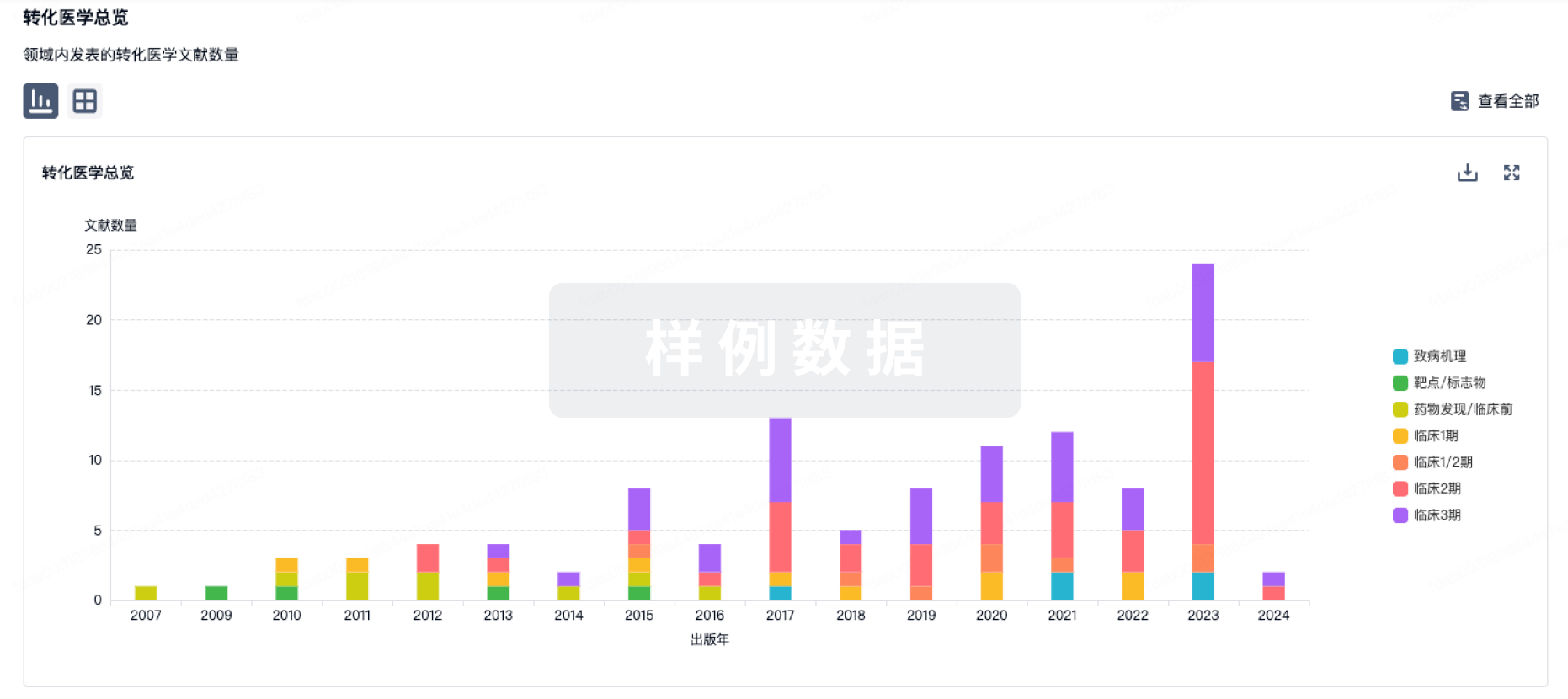
药物交易
使用我们的药物交易数据加速您的研究。
登录
或
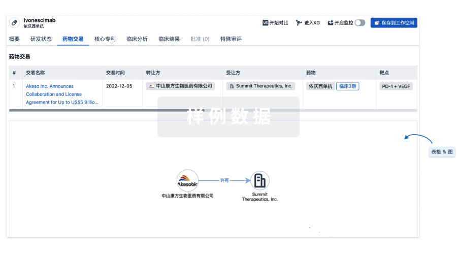
核心专利
使用我们的核心专利数据促进您的研究。
登录
或
临床分析
紧跟全球注册中心的最新临床试验。
登录
或
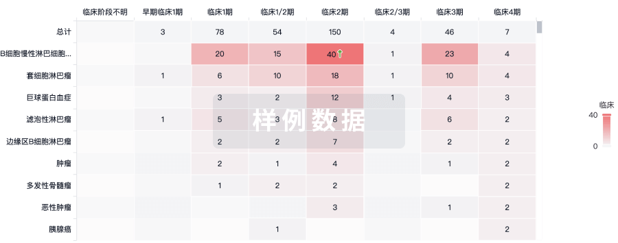
批准
利用最新的监管批准信息加速您的研究。
登录
或
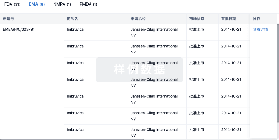
生物类似药
生物类似药在不同国家/地区的竞争态势。请注意临床1/2期并入临床2期,临床2/3期并入临床3期
登录
或
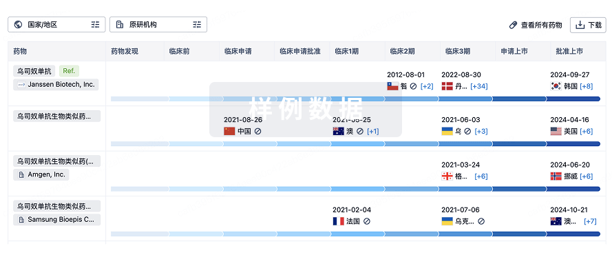
特殊审评
只需点击几下即可了解关键药物信息。
登录
或
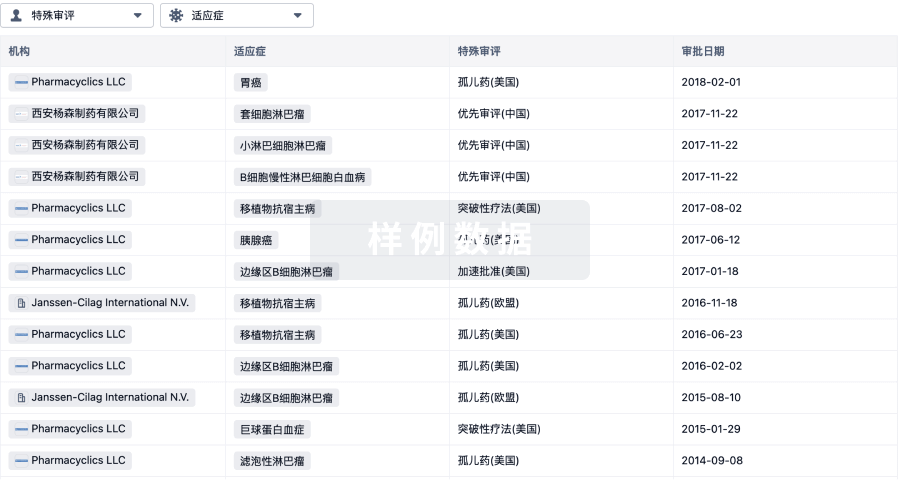
生物医药百科问答
全新生物医药AI Agent 覆盖科研全链路,让突破性发现快人一步
立即开始免费试用!
智慧芽新药情报库是智慧芽专为生命科学人士构建的基于AI的创新药情报平台,助您全方位提升您的研发与决策效率。
立即开始数据试用!
智慧芽新药库数据也通过智慧芽数据服务平台,以API或者数据包形式对外开放,助您更加充分利用智慧芽新药情报信息。
生物序列数据库
生物药研发创新
免费使用
化学结构数据库
小分子化药研发创新
免费使用