预约演示
更新于:2026-06-27
Xian-Janssen Pharmaceutical Ltd.
更新于:2026-06-27
概览
标签
肿瘤
免疫系统疾病
血液及淋巴系统疾病
小分子化药
单克隆抗体
酶
疾病领域得分
一眼洞穿机构专注的疾病领域

技术平台
公司药物应用最多的技术
靶点
公司最常开发的靶点
关联
靶点 |
作用机制 |
在研机构 |
原研机构 |
在研适应症 |
非在研适应症 |
最高研发阶段 |
首次获批国家/地区 |
首次获批日期 |


靶点 |
作用机制 |
在研机构 |
原研机构 |
在研适应症 |
非在研适应症 |
最高研发阶段 |
首次获批国家/地区 |
首次获批日期 |
靶点 |
作用机制 |
在研机构 |
原研机构 |
在研适应症 |
非在研适应症 |
最高研发阶段 |
首次获批国家/地区 |
首次获批日期 |


NCT07528040
An Observational, Multicenter Study to Evaluate the Effectiveness of Guselkumab for the Treatment of Participants With Crohn's Disease After Surgical Resection
NCT07310095
Efficacy of Guselkumab in Chinese Participants With Crohn's Disease Following Loss of Response to Ustekinumab
NCT07302360
Guselkumab Real-world Effectiveness Among Bio-NaÏve Patients With Moderate-to-severe Ulcerative Colitis in China: A Multicenter, Non-interventional, Prospective Study
100 项与 西安杨森制药有限公司 相关的临床结果
登录后查看更多信息
登录后查看更多信息
2025-08-01iScience
A randomized study of apalutamide in Chinese patients with non-metastatic castration-resistant prostate cancer
Article
作者: McCarthy, Sharon ; Liu, Yanmei ; Xu, Tao ; Xie, Liping ; Dong, Haiying ; Tian, Ye ; Li, Yanhui ; Guo, Jianming ; Luo, Hong ; Ye, Dingwei ; Li, Lei ; Liang, Hongchuan ; Lopez-Gitlitz, Angela ; Ma, Haocheng ; Chen, Na ; Jiang, Shusuan ; Guo, Hongqian ; Qiu, Mingxing ; Xue, Wei
This post-approval commitment study addressed the limited data on the safety and efficacy of apalutamide in Chinese patients with non-metastatic castration-resistant prostate cancer (NM-CRPC). Utilizing a double-blinded, placebo-controlled trial with pre-planned crossover design, 75 patients were randomized (2:1) to receive apalutamide 240 mg daily or placebo while continuing androgen deprivation therapy. Apalutamide significantly reduced the risk of prostate-specific antigen (PSA) progression by 76.7% compared with placebo (hazard ratio [HR] = 0.233, p = 0.0052), with confirmed PSA response rate of 92.0% versus 12.0%. The median metastasis-free survival with apalutamide was 36.8 months, while the median overall survival was not reached. Grade 3 and 4 treatment-emergent adverse events were reported in 43.1% and 4.2% of patients, respectively, with hypertension, pneumonia, and rash being the most frequently reported, and the safety profile was consistent with existing data on apalutamide. Overall, these findings indicate that apalutamide is both efficacious and safe for Chinese patients, providing a valuable treatment option for high-risk NM-CRPC.
2024-12-31ANNALS OF MEDICINE
Prognostic factors in Chinese patients with immunoglobulin light chain amyloidosis: a scoping review and meta-analysis
Article
作者: Wu, Yu ; Cai, Zhen ; Xu, Lingjie ; Gào, Xīn ; Wang, Bin ; Wang, Xiaohong
OBJECTIVE:
This scoping review and meta-analysis aimed to map the evidence regarding prognostic factors in Chinese patients with immunoglobulin light chain (AL) amyloidosis and to identify current research gaps.
METHODS:
We searched EMBASE, PubMed, and CNKI databases from their inception to 15 September 2021. All studies investigated the association between any prognostic factor and target outcomes, including overall survival (OS), progression-free survival (PFS), and end-stage renal disease (ESRD) in Chinese patients with AL amyloidosis.
RESULTS:
This scoping review included 52 studies, of which 44 with 6,432 patients contributed to the multivariate prognostic analysis. Multivariate analysis identified a total of 106 factors that correlated with OS, 16 factors with PFS, and 18 factors with ESRD. Five prognostic factors were significantly associated with PFS, and 11 prognostic factors were significantly associated with ESRD. Meta-analysis was only available for prognostic factors without heterogeneous cutoff values, for which hazard ratios (HRs) and their 95% confidence intervals (CIs) were reported. Meta-analysis showed that bone marrow plasma cells (BMCs) (HR: 1.96, 95% CI: 1.21-3.19, p < 0.05) and interventricular septal thickness (IVST) (HR: 1.23, 95% CI: 1.10-1.38, p < 0.05) were independently associated with OS.
CONCLUSION:
The significant prognostic factors associated with OS, PFS, and ESRD in Chinese patients with AL amyloidosis were related to plasma cell tumor load, biological characteristics, cardiac involvement, renal involvement, population characteristics, and treatment. Further studies should explore additional prognostic factors in patients with AL amyloidosis to develop prognostic models.
2024-11-01ANNALS OF HEMATOLOGY
Correction to: Daratumumab, bortezomib, melphalan, and prednisone versus bortezomib, melphalan, and prednisone alone in transplant-ineligible Asian patients with newly diagnosed multiple myeloma: final analysis of the phase 3 OCTANS study
作者: Huang, Honghui ; Li, Wei ; Wang, Yafei ; Carson, Robin ; Lee, Je-Jung ; Wang, Jianxiang ; An, Gang ; Zhao, Man ; Liu, Rui ; Kim, Kihyun ; Fu, Weijun ; Cai, Zhen ; Chim, Chor Sang ; Cui, Canchan ; Jin, Jie ; Bang, Soo-Mee ; Hou, Jian ; Chen, Xi
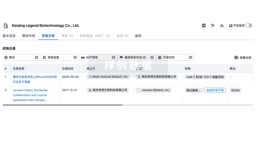
100 项与 西安杨森制药有限公司 相关的药物交易
登录后查看更多信息
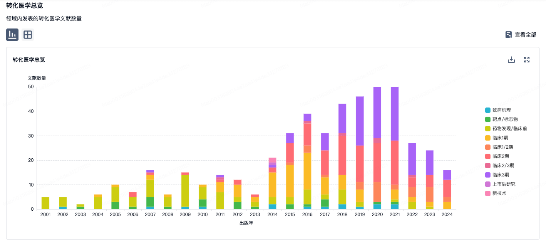
100 项与 西安杨森制药有限公司 相关的转化医学
登录后查看更多信息
组织架构
使用我们的机构树数据加速您的研究。
登录
或
管线布局
2026年07月21日管线快照
管线布局中药物为当前组织机构及其子机构作为药物机构进行统计,早期临床1期并入临床1期,临床1/2期并入临床2期,临床2/3期并入临床3期
临床2期
1
3
临床3期
申请上市
3
7
批准上市
其他
25
登录后查看更多信息
当前项目
登录后查看更多信息
药物交易
使用我们的药物交易数据加速您的研究。
登录
或
转化医学
使用我们的转化医学数据加速您的研究。
登录
或
营收
使用 Synapse 探索超过 36 万个组织的财务状况。
登录
或
科研基金(NIH)
访问超过 200 万项资助和基金信息,以提升您的研究之旅。
登录
或
投资
深入了解从初创企业到成熟企业的最新公司投资动态。
登录
或
融资
发掘融资趋势以验证和推进您的投资机会。
登录
或
芽仔
全新生物医药AI Agent 覆盖科研全链路,让突破性发现快人一步
立即开始免费试用!
智慧芽新药情报库是智慧芽专为生命科学人士构建的基于AI的创新药情报平台,助您全方位提升您的研发与决策效率。
立即开始数据试用!
智慧芽新药库数据也通过智慧芽数据服务平台,以API或者数据包形式对外开放,助您更加充分利用智慧芽新药情报信息。
生物序列数据库
生物药研发创新
免费使用
化学结构数据库
小分子化药研发创新
免费使用