预约演示
更新于:2026-10-23
Paricalcitol
帕立骨化醇
更新于:2026-10-23
概要
基本信息
原研机构 |
权益机构- |
最高研发阶段批准上市 |
首次获批日期 美国 (1998-04-17), |
最高研发阶段(中国)批准上市 |
特殊审评孤儿药 (美国) |



登录后查看时间轴
结构/序列
分子式C27H44O3 |
InChIKeyBPKAHTKRCLCHEA-UBFJEZKGSA-N |
CAS号131918-61-1 |
关联
126
项与 帕立骨化醇 相关的临床试验NCT07856914
A Randomized Phase II Bayesian Pick-the-Winner Clinical Trial Investigating the Efficacy of Fibrosis-Modulating Agents (Paricalcitol and Pirfenidone) in Improving Surgical Resectability in Patients With Peritoneal Carcinomatosis Ineligible for Definitive Cytoreduction (Randy 1A)
The goal of this clinical trial is to learn if paricalcitol or pirfenidone can help make tumors removable by surgery in adults with peritoneal cancer who are currently unable to have their tumor removed.
The study aims to answer are whether paricalcitol or pirfenidone improve tumor response and tissue healing after 12 weeks of treatment, and which of these two drugs is more effective at helping participants become eligible for surgery. Researchers will compare paricalcitol to pirfenidone to see which drug works better and has a manageable safety profile.
Participants will take either paricalcitol or pirfenidone by mouth every day for 12 weeks while continuing their standard chemotherapy, visit the clinic regularly for checkups and lab tests, keep a daily diary to track their study medication, and undergo a surgical re-evaluation after treatment to see if their tumor can be removed.
开始日期2026-09-01 |
ChiCTR2500113259
A Single-Center, Single-Arm, Open-Label Phase II Clinical Study of Recombination with Paricalcitol After First-Line PD-1 Inhibitor Regimen Failure in Unresectable Hepatocellular Carcinoma Patients with Serum Vitamin D Deficiency
开始日期2025-12-01 |
申办/合作机构- |
NCT06209268
The Impact of Selective Vitamin D Receptor Activation on Clinical Outcomes in Septic Patients -a Randomised Placebo Controlled Trial
Sufficient serum levels of vitamin D are important for immune system regulation with protective effect against severe infection and overactivated inflammatory response in sepsis. It is also not clear what level of vitamin D in the blood would be the trigger for vitamin D administration. A more selective approach to VDR activation than cholecalciferol could have a more significant role in the clinical outcomes of patients with sepsis. A study demonstrated that low baseline serum level of vitamin D receptor (VDR) was associated with a high incidence of 28-day mortality and negatively correlated with lactate, C-reactive protein, APACHE II SOFA scores, and disease severity among patients with sepsis in an ICU setting.
The role of selective vitamin D receptor activation agents (paricalcitol or maxacalcitol) was not studied in septic patients, despite its anti-inflammatory and immunomodulatory properties. Vitamin D analogs have different effects on nuclear VDRs than does calcitriol, through different response elements in various target genes, so it is possible that their effect on a patient with sepsis will be more effective than cholecalciferol. As distribution of VDRs is ubiquitous in many organs and tissues, selective VDR activation with paricalcitol may have beneficial effects in preserving organs functionality and modulating the immune response in sepsis.
Hypotheses
1. The immunoregulatory, anti-inflammatory, and anti-oxidative properties of selective vitamin D receptor activator paricalcitol would result in improvement of inflammatory, endothelial function, and antioxidative parameters and clinical outcomes in groups of septic patient admitted to ICU.
2. The baseline septic patient serum 25(OH) D3 levels at admission time in ICU have influence on clinical outcomes as well as on inflammatory, endothelial function, and antioxidative parameters.
3. The inflammatory, endothelial function, and antioxidative parameters measured at ICU admission time have significant impact on clinical outcomes in septic patients.
The aim
The main objective of study is to test hypothesis that that selective activator of vitamin D receptors paricalcitol will improve outcomes of septic patient admitted in ICU. The study aims to investigate the effects of paricalcitol on clinical outcomes, inflammatory markers, organ dysfunction, endothelial function, vascular morphology, coagulation markers, and haemodynamic parameters.
The additional objectives of the study are to test hypothesis that septic patient serum 25(OH)vitamin D3 have impact on inflammatory, endothelial function, and antioxidative parameters including protein carbonylation; and to test hypothesis that these markers and clinical outcomes are interconnected with significant impact on clinical outcomes.
开始日期2025-05-01 |
申办/合作机构- |
100 项与 帕立骨化醇 相关的临床结果
登录后查看更多信息
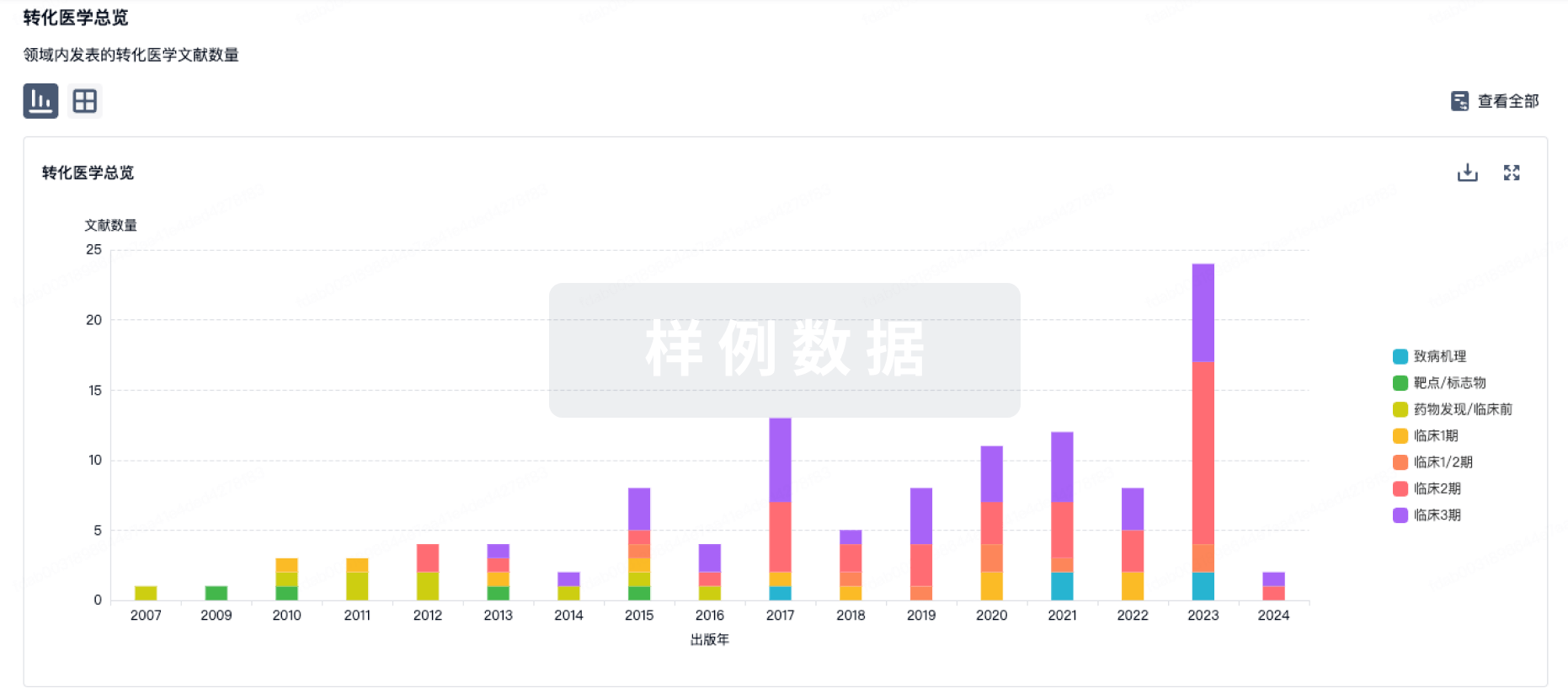
100 项与 帕立骨化醇 相关的转化医学
登录后查看更多信息
100 项与 帕立骨化醇 相关的专利(医药)
登录后查看更多信息
720
项与 帕立骨化醇 相关的文献(医药)2026-10-01·EXPERIMENTAL GERONTOLOGY
Serum 25(OH) vitamin D and SIRT1- VDR- miR-124 expression: A controlled interventional study in a vitamin D-deficient male rat model
Article
作者: Dhar, Poulami ; Kalladka, Shwetha Shetty ; Moodithaya, Shailaja ; Bhandary, Prajna
BACKGROUND:
Epigenetic aging is a new dimensional advancement in geroscience that continually addresses factors beyond genetic determinants of longevity. It occurs through three conventional pathways, one of which is non-coding RNA production. miR-124 is a newly introduced miRNA that was found in aged skin and several tumorigenic cells and is associated with aging. Additionally, SIRT1, a protein from the NAD+-dependent family, is found to have a protective mechanism in the pathways of aging. On the other hand, vitamin D works as an epigenetic factor and often targets pathways related to the aging process. In this study, the aim is to identify the expression of miR-124 in circulating blood to address the epigenetic-related biomarker and link the facets with anti-aging SIRT1 and vitamin D cognate receptor (VDR) to understand vitamin D deficiency interception.
METHODS:
A vitamin D deficiency middle-aged rat model was developed by assigning male Albino Wistar Rats. Paricalcitol was administered IP (intraperitoneal) to the experimental group (32 ng/day) to induce deficiency, along with a calcium-rich rescue diet. The study was conducted for 21 days following blood collection and storage. ELISA and RTqPCR were performed to estimate serum 25(OH)D and gene expression analysis, respectively.
RESULT:
25(OH)D comparison between the group p-value of 0.002. The log2 fold changes of SIRT1, VDR, and miR-124 were compared between the groups using boxplot visualisation. A linear regression model was developed to understand the explorative nature of aging index (R2 = 0.561; Adjusted R2 = 0.517; p = 0.005; n = 12).
CONCLUSION:
A putative epigenetic relation was found between miR-124 levels and their association with SIRT1. The investigated biomarkers of aging are associated with serum concentration of 25(OH) vitamin D in a middle-aged vitamin D-deficient rat model, which supports the need for mechanistic investigation in future studies.
2026-10-01·JACC. Advances
Vitamin D Pathway Activation Reduces Cardiomyocyte DNA Damage and Improves Cardiac Contractility in Preclinical Models
Article
作者: Toyoda, Masashi ; Kubota, Masayuki ; Nishijo, Daigo ; Yamada, Shintaro ; Takeda, Norihiko ; Abe, Ryo ; Tsukihashi, Ayako ; Umezawa, Akihiro ; Yamasaki, Masataka ; Komuro, Jin ; Ikeuchi, Masashi ; Nomura, Seitaro ; Onoue, Kenji ; Takada, Shuji ; Aburatani, Hiroyuki ; Hiruma, Takashi ; Dai, Zhehao ; Katagiri, Mikako ; Katoh, Manami ; Matsuura, Katsuhisa ; Ko, Toshiyuki ; Morita, Hiroyuki ; Komuro, Issei ; Zhang, Yanfei ; Inoue, Shunsuke ; Ito, Masamichi
BACKGROUND:
In heart failure (HF), DNA damage caused by various external stressors contributes to cardiac dysfunction through the activation of DNA damage response pathways. To date, no clinical strategies have been established to restore cardiac function by reducing accumulated DNA damage. We previously found that vitamin D improved contractility in lamin A/C (LMNA) p.Q353R-mutant induced pluripotent stem (iPS) cell-derived cardiomyocytes (iPSCMs), but whether this effect extends to other LMNA variants and in vivo models remained uncertain.
OBJECTIVES:
The objective of the study was to evaluate the association of vitamin D pathway activation with cardiomyocyte phosphorylated histone H2AX (γH2AX) foci and contractile phenotypes in patient-derived iPSCMs and mouse models of HF.
METHODS:
iPS cell lines were generated from dilated cardiomyopathy patients carrying the LMNA p.R225X mutation, and the effects of vitamin D treatment on γH2AX foci and cardiomyocyte contractility were evaluated. In addition, the effects of the vitamin D analog paricalcitol were evaluated in Lmna p.R225X mice and in a pressure overload mouse model of HF.
RESULTS:
Consistent with previous findings, vitamin D treatment reduced γH2AX foci in cardiomyocytes derived from LMNA p.R225X mutant iPS cells through upregulating the expression of DNA repair factors, and improved contractility in these iPSCMs. Furthermore, paricalcitol reduced γH2AX foci and attenuated cardiac dysfunction in both Lmna p.R225X mice and pressure overload HF model mice.
CONCLUSIONS:
Vitamin D pathway activation improved contractile phenotypes across complementary preclinical models and was accompanied by reduced γH2AX foci or related transcriptional changes. These findings support further mechanistic and preclinical investigation.
2026-09-01·BIOCHIMICA ET BIOPHYSICA ACTA-GENERAL SUBJECTS
Stage-dependent association of Vitamin D receptor with macrophage polarization in allergic rhinitis
Article
作者: Shi, Dingyi ; Zhang, Mengni ; Huang, Yajing ; He, Jia ; Peng, Shunlin
BACKGROUND:
Allergic rhinitis (AR) is driven by Th1/Th2 imbalance, with macrophages as key mediators. Vitamin D (VitD) deficiency is associated with AR; however, how VitD regulates macrophage-related molecules remains unclear.
OBJECTIVE:
This study investigated stage-specific effects of VitD receptor (VDR) signaling on macrophage-related molecules and immune responses in an ovalbumin (OVA)-induced AR rat model.
METHODS:
Beginning in the sensitization phase, rats received either the VDR agonist paricalcitol or the antagonist ZK168281. Outcomes were assessed during sensitization, re-challenge, and chronic maintenance phases, including nasal symptoms, nasal mucosal histopathology, serum OVA-specific IgE and M1/M2-like macrophage associated cytokines, spleen-derived cell transcriptomics, and nasal immunofluorescence for NOS2/ARG1 (M1/M2-like markers).
RESULTS:
Our findings demonstrate that VDR activation conferred maximal protection during the re-challenge phase, characterized by alleviated nasal symptoms, attenuated epithelial injury and inflammatory infiltration, and reduced OVA-specific IgE levels. Transcriptomic analysis revealed stage-dependent association of macrophage polarization: VDR signaling promoted the expression of M2-like-associated genes (e.g., Arg1, Cd163, and Mertk) during sensitization and chronic maintenance while suppressing M1-like inflammatory mediators and chemokines (e.g., Il6, Il1β, Cxcl9/10, and Ccl2) during re-challenge. These findings were corroborated at the protein level, with the serum cytokine analysis showing suppression of M1-like associated IL-6 and TNF-α, and nasal mucosal immunofluorescence confirming consistent and marked ARG1 expression upregulation.
CONCLUSION:
VDR signaling is associated with stage-dependent immunomodulation in AR, promoting M2-like molecules expression during sensitization and suppressing M1 like driven inflammation during re-challenge, thereby weakening effects in the chronic phase. These findings support a "chronotherapeutic" application of VitD in AR.
138
项与 帕立骨化醇 相关的新闻(医药)2026-10-21
·麦科奥特
MICOT
点击蓝字 关注我们
Slide to view different languages
滑动查看不同语言
每年 10 月 20 日是世界骨质疏松日。对透析肾友来说,腰背反复隐痛、翻身弯腰时加重,甚至只是轻微磕碰就发生骨折,是并不陌生的困扰。不少人的第一反应是“年纪大了、骨质疏松”,于是自行补钙、补充维生素 D;但很多时候骨痛并未改善,复查却发现血磷升高、血管钙化风险进一步增加[1]。原因在于:透析人群的骨骼问题,通常不是单纯的骨量减少,而是慢性肾脏病-矿物质与骨异常(CKD-MBD)在骨骼上的表现[2,3]。借“世界骨质疏松日”,我们把“透析患者的骨问题”讲清楚:它和普通骨质疏松有何不同,指南给出了怎样的边界,以及“治标”之外为什么还需要“治本”。
先分清:
CKD-MBD 与骨质疏松,不是一回事
很多人简单将骨痛、易骨折归结为缺钙,但肾病带来的骨骼紊乱,远不止钙含量不足。CKD-MBD 是由慢性肾脏病引起的全身性矿物质与骨代谢紊乱,包含三层核心异常:① 钙、磷、PTH、维生素 D 代谢紊乱;② 骨转化、矿化、骨容量(以及骨骼强度)异常;③ 血管及其他软组织钙化[2,3]。通俗地说,就是“原料(钙磷)失衡、施工(骨重建)失控、后果(钙化)外溢”同时发生。
其中发生在骨骼本身的病变,就是肾性骨病,规范名称为肾性骨营养不良(renal osteodystrophy,ROD),需依靠骨活检组织学进行诊断与分型,临床上依骨转化状态分为三类[3]:
高转化型:骨吸收与骨形成均异常活跃,根源是长期 PTH 升高,骨微结构遭到持续破坏 [3];
低转化型:包括无动力性骨病(骨重塑近乎停滞,常见于 PTH 被过度抑制,如含钙磷结合剂、活性维生素 D、拟钙剂使用不当或甲旁亢术后)与骨软化(矿化障碍,新生类骨质无法正常钙化)[2-4];
混合型:骨重建仍保持活跃,同时合并矿化缺陷,与骨软化的核心区别在于骨重塑并未静止 [3]。
普通骨质疏松以骨量流失为主,骨基质本身的矿化机制基本正常;而 CKD 患者往往是“双重叠加”——骨量下降的同时,骨重塑与矿化全程失控 [4]。这也是为什么在骨密度数值相近的情况下,透析患者的骨折风险仍显著更高 [4]。临床需要把骨密度与 PTH、血钙、血磷、碱性磷酸酶等指标放在一起解读,而不是拿一张骨密度报告直接“对号入座” [2,4]。
透析患者的骨折:更常见,也更致命
骨折在透析人群中并非偶发事件。KDIGO(改善全球肾脏病预后组织) 2017 指南指出,终末期肾病患者的骨折风险约为一般人群的 4 倍 [2]。来自 DOPPS (透析结局与实践模式研究)登记研究(34,579 例中心血液透析患者)的数据更为直观:与整体透析人群相比,发生骨折的患者死亡率为其 3.7 倍,死亡或再住院复合终点的风险为其 4.0 倍;骨折后中位住院时间为 7~37 天 [8]。换言之,对透析肾友而言,骨折往往不是一次“养一养就好”的意外,而是触发连锁事件的转折点。
底层机制:
不只是“拆得太多”,更是“建不起来、建不好”
我们可以把骨骼看作是一个持续施工的工地:破骨细胞负责“拆旧”,成骨细胞负责“建新”,二者耦合成一个完整的骨重建周期。普通骨质疏松主要是“拆的速度超过建的速度”;而肾性骨病,是这套施工体系的指挥系统因肾脏受损而彻底紊乱 [1]。
随着肾功能下降,肾脏排磷能力减弱、磷在体内蓄积;FGF-23 甚至早于血磷升高,通过抑制活性维生素 D 合成、降低血钙,持续刺激甲状旁腺,进而诱发继发性甲状旁腺功能亢进(SHPT)[1,9]。PTH 长期偏高,破骨细胞被持续激活,骨吸收亢进——此时骨骼中的钙、磷会被大量“拆”入血液,与肾脏排磷下降叠加,共同推高血磷与钙磷乘积 [1,9]。这是理解透析人群高磷血症的关键一环:血里的钙、磷,并不只来自饮食和透析不充分,也来自骨骼自身的丢失。
反过来,若 PTH 被过度抑制,骨重建又会显著受抑甚至近乎停滞(无动力性骨病);而矿化障碍则导致骨软化[2-4]。两种极端状态都会损伤骨骼,低 PTH 患者髋部骨折风险同样升高 [10]。随着活性维生素 D、含钙磷结合剂与拟钙剂的广泛使用,以及老年、糖尿病患者增多,低转化型(无动力性骨病)在透析人群中的比例已明显上升,部分大样本骨活检队列中甚至成为主要类型 [19,20]。
指南的边界:KDIGO 2017 给我们划了哪些线
01.
控指标仍是治疗主线
对 CKD G5D 患者,指南建议将 iPTH 维持在约 2~9 倍正常值上限(4.2.3,2C),并在该范围内出现明显变化时及时启动或调整治疗,避免进展至区间之外[2];需要降 PTH 治疗时,建议使用拟钙剂、骨化三醇或维生素 D 类似物,或拟钙剂与后两者联合(4.2.4,2B)[2]。可见,现行标准治疗的核心目标,是把 PTH、钙、磷这些“指标”管住。
02.
骨质疏松药物:
只在特定人群“可按普通人群处理”
指南对骨质疏松药物的态度分层而谨慎:
CKD G1~G2 合并骨质疏松和 / 或高骨折风险者(经 WHO 标准判定),按一般人群管理(4.3.1,1A);CKD G3a~G3b 且 PTH 正常者,建议按一般人群治疗(4.3.2,2B);而 CKD G3a~G5D 合并 CKD-MBD 生化异常、同时低 BMD 和 / 或脆性骨折者,指南建议治疗选择需考虑生化异常的幅度与可逆性、CKD 的进展,必要时考虑骨活检(4.3.3,2D,证据质量级低)[2]。在这一人群中,治疗优先聚焦 CKD-MBD 本身的钙、磷、PTH 紊乱:SHPT(继发性甲状旁腺功能亢进症)可选用拟钙剂(西那卡塞、依特卡肽)、活性维生素 D / 维生素 D 类似物(骨化三醇、帕立骨化醇),或二者联合,同时配合磷结合剂管控血磷 [2]。
而经典抗骨质疏松药物的选用,需要综合评估上述情况,并权衡这类药物的固有隐患:抗骨吸收药物存在进一步压低骨转换、诱发或加重无动力性骨病的顾虑;双膦酸盐经肾脏排泄,肾衰竭状态下蓄积风险升高;地舒单抗在晚期 CKD 中可能诱发显著低钙血症;PTH 类促骨形成制剂则有高钙血症、钙磷代谢紊乱的风险 ——这类药物在透析人群中不宜作为常规选择 [2,21]。
03.
分型诊断:
金标准难落地,替代指标不够用
骨活检是肾性骨病诊断与分型的金标准(3.2.2),但能常规开展的中心有限 [2,3]。更棘手的是,指南援引的一项覆盖 492 例透析患者的国际多中心诊断研究显示:iPTH、全段 PTH 与骨特异性碱性磷酸酶(bALP)虽与骨转化相关,但无论单独还是联合,都不足以在个体患者中判别低、正常或高骨转化 [2]。指南因此只能建议以 PTH 或 bALP 的“极高或极低值”来预测骨转化状态(3.2.3,2B)[2]。
三道坎:为什么“治标容易、治本很难”
第一道坎
降 PTH 药物管的是指标
不是骨质量
拟钙剂与活性维生素 D 的核心作用是抑制 PTH 分泌,从而减轻高 PTH 驱动的骨吸收;但它们并不直接促进骨形成,也难以逆转已经发生的骨结构损害[2,13]。
指南援引的 BONAFIDE 骨活检研究显示,西那卡塞治疗虽使骨形成率下降、组织学恢复正常者增多,但也有受试者出现无动力性骨病(其 PTH<150 pg/mL)[2];而新型静脉拟钙剂虽有疗效与安全性数据,却尚无患者层面结局(骨折、心血管事件、生存)的报告[2]。在硬终点层面,EVOLVE 研究(3 883 例血液透析患者)的主要复合终点亦未能达到统计学显著改善[21]。
第二道坎
抗骨质疏松药
在透析人群“证据不足、顾虑不少”
用于绝经后骨质疏松的大型随机对照试验,普遍排除肌酐升高、甲旁亢或碱性磷酸酶异常的受试者,也就是排除了 CKD-MBD 人群 [4];地舒单抗的阳性结果来自 CKD G3a~G3b 及少数 G4 患者的事后分析,骨折获益亦仅见于椎体骨折,且在晚期 CKD 中低钙血症风险突出 [4]。
总体而言,晚期 CKD 尤其是 G5~G5D 透析人群,目前尚无高质量证据证实抗骨质疏松治疗能够改善骨骼结局(降低骨折风险、修复骨质量),这类药物也均未在该人群获批骨折预防适应证 [2,4,21]。这也意味着临床缺少可以同步兼顾调控 PTH 紊乱与改善骨质量的治疗选择。
第三道坎
缺少“同时管住 PTH
又支持骨形成”的治疗手段
高转化型需要抑制破骨,低转化/无动力性骨病则需要唤醒成骨;但在分型不明的现实条件下,多数患者仍接受着以单向抑制 PTH 为主的方案,一旦判断偏差,反而可能加重骨损伤 [2,4]。该领域长期未满足的需求由此清晰:我们能把指标管住,却缺少一种能把“PTH 调控”与“促成骨修复”合二为一的治疗思路。
MT1013:
把“促骨形成”写进 SHPT 治疗的设计里
——标本兼治的新范式(在研)
正是在上述背景下,创新药物的研发方向开始从“抑制”转向“抑制+修复”。麦科奥特自主研发的 MT1013 是一款 First-in-Class 的 CaSR/OGP 双靶点多肽,为双激动剂设计,拟用于维持性透析患者的 SHPT,目前正处于Ⅲ期临床试验阶段[18]。它的设计逻辑可以用“两条通路”来理解:
通路一:
CaSR-“治标”,把矿物质指标管住
CaSR 相当于甲状旁腺表面的“钙感受器”。MT1013 激动 CaSR,抑制 PTH 过度分泌,减轻高 PTH 对骨骼的持续损伤,从而实现 PTH、钙、磷等矿物质指标的管控[13,18]。这一维度,是与现有拟钙剂共享的“治标”疗效。
通路二:
OGP-“治本”,作用于骨形成与骨矿化
OGP(成骨生长肽)是人体内源性促骨形成肽,与组蛋白 H4 的 C 端序列同源[15,16]。研究显示,OGP 可促进骨髓间充质干细胞向成骨细胞分化、上调碱性磷酸酶活性、推动骨基质合成与矿化 [16,17]。把这条通路与第三节的机制连起来看,其意义就在于“治本”:
01
骨吸收亢进时,骨骼中的钙、磷被大量释放入血,是透析人群高钙、高磷负荷的来源之一 [1,9];
03
OGP 促进骨基质合成与矿化,相当于为血循环中的钙、磷提供了一条“归位入骨”的去路,从骨代谢这一源头环节参与 CKD-MBD 的调控 [16-18];
01
这也与研究者的观察一致:MT1013 降磷获益被认为部分来自 OGP 对骨代谢的直接调节,可能增强磷在骨组织中的利用与沉积 [18]。
在这一机制基础上,MT1013在降磷与综合达标控制方面的表现,有望进一步转化为心血管及生存获益的潜在优势——即从“把磷降下来”走向“把CKD-MBD整体管好”,并为长期结局改善创造条件。
需要说明的是,目前尚无已上市的 SHPT 治疗药物以 OGP 通路为靶点[13,18]。在这里,“治标”与“治本”不再是二选一:一个分子同时回应了“把指标管住”和“把骨骼修好”两件事,这正是它与单靶点拟钙剂在设计层面的根本差异。
从机制到临床:初步看到了什么
理论上的优势需要在临床上被看见。在已完成的一项MT1013与临床上最强拟钙剂依特卡肽头对头临床研究中,给出了方向一致的信号:
iPTH 降幅>30% 的患者比例为 80.7% 与 93.3%(依特卡肽 90.6%、安慰剂 22.2%),基线 iPTH>600 pg/mL 的中重度患者平均降幅为 61.8% 与 68.5%(依特卡肽 59.2%);iPTH、血钙、血磷三项综合达标率为 39.29% 与 34.48%,约为依特卡肽(15.63%)的 2 倍(P=0.0482);第 27 周血磷平均下降 11.2% 与 11.6%(依特卡肽 5.3%,MT1013 两组均 P<0.05),FGF-23 平均下降 33.5% 与 39.9%(依特卡肽 22.8%)——而 FGF-23 升高与透析患者死亡及心血管事件风险相关 [22],其降幅更优为“骨-血管”双重获益提供了方向性线索;安全性方面,症状性低钙血症发生率为 7.69%,低于依特卡肽的 12.12%,且未检出抗药抗体[18]。
更贴近“治本”逻辑的是骨密度:在另一项为期 52 周的研究中,基线骨量下降或骨质疏松的受试者治疗后股骨颈骨密度平均提升 4.44%、腰椎提升 1.65%;由于入组前受试者已停用既往拟钙剂并完成洗脱,骨骼很可能仍处于既往治疗带来的改善“窗口”内,此时骨密度仍进一步上升,提示这部分获益并非单纯来自抑制 PTH,而更可能来自拟钙剂之外的额外促成骨作用[23]。简言之:在降 PTH 这一维度上它与临床上最强拟钙剂相当,而在综合达标、降磷、降FGF-23 以及骨密度这些维度上,显示出了单靶点拟钙剂之外的差异化信号 [18,23]。
当然也要客观地看:上述均为样本量有限的II期研究,骨密度属于替代终点,尚不能直接推导为骨折风险下降;MT1013 目前正处于Ⅲ期临床试验阶段,其最终临床价值仍待验证。
给肾友的三条建议
01.
别急着补钙
透析人群的高磷、高钙并不只来自饮食,盲目补钙可能加重钙化风险;所有骨病相关用药调整,请交给肾内科专科医生[1,2]。
02.
看骨,不止看骨密度
建议结合血钙、血磷、iPTH、碱性磷酸酶等指标动态评估,必要时评估血管钙化;PTH 明显升高时,T 值的参考价值会打折扣[2,4]。
03.
关注趋势而非单次数值
指南明确建议以指标的变化趋势而非单次化验值指导决策(3.1.4,1C)[2],规律复查、记录趋势,比盯着某一个数字更有意义。
从“把 PTH 降下来”,到“把 PTH、钙、磷管住”,再到“同时修复骨骼本身”,透析人群骨病管理的目标正在被重新定义。对骨质疏松日最好的回应,或许不只是提醒肾友“补钙要谨慎”,更是推动治疗思路从单纯管控指标,走向对骨代谢根本环节的干预。
安全提示
MT1013 尚处于Ⅲ期临床试验阶段,未获批上市;本文所引数据为早期探索性研究结果,不等同于最终临床结论,不构成任何用药推荐。透析患者切勿自行补钙或调整骨病相关药物,所有治疗决策请遵从肾内科专科医生指导。
参考文献
[1] Salera D, Merkel N, Bellasi A, et al. Pathophysiology of chronic kidney disease-mineral bone disorder (CKD-MBD): from adaptive to maladaptive mineral homeostasis[J]. Clin Kidney J, 2025, 18(Suppl 1): i3-i14.
[2] Kidney Disease: Improving Global Outcomes (KDIGO) CKD-MBD Update Work Group. KDIGO 2017 clinical practice guideline update for the diagnosis, evaluation, prevention, and treatment of chronic kidney disease-mineral and bone disorder (CKD-MBD)[J]. Kidney Int Suppl, 2017, 7(1): 1-59.
[3] Moe S, Drüeke T, Cunningham J, et al. Definition, evaluation, and classification of renal osteodystrophy: a position statement from Kidney Disease: Improving Global Outcomes (KDIGO)[J]. Kidney Int, 2006, 69(11): 1945-1953.
[4] Evenepoel P, Cunningham J, Ferrari S, et al. European consensus statement on the diagnosis and management of osteoporosis in chronic kidney disease stages G4-G5D[J]. Nephrol Dial Transplant, 2021, 36(1): 42-59.
[5] 国家肾脏疾病临床医学研究中心. 中国慢性肾脏病矿物质和骨异常诊治指南概要[J]. 肾脏病与透析肾移植杂志, 2019, 28(1): 52-57.
[6] Amirkhanlou S, Roshandel G, Aghaei M, et al. Assessment of bone mineral density in patients undergoing hemodialysis; an Iranian population-based study[J]. Arch Iran Med, 2021, 24(8): 599-606.
[7] Sit D, Kadiroglu AK, Kayabasi H, et al. Relationship between bone mineral density and biochemical markers of bone turnover in hemodialysis patients[J]. Adv Ther, 2007, 24(5): 987-995.
[8] Tentori F, McCullough K, Kilpatrick RD, et al. High rates of death and hospitalization follow bone fracture among hemodialysis patients[J]. Kidney Int, 2014, 85(1): 166-173.
[9] Isakova T, Wahl P, Vargas GS, et al. Fibroblast growth factor 23 is elevated before parathyroid hormone and phosphate in chronic kidney disease[J]. Kidney Int, 2011, 79(12): 1370-1378.
[10] Coco M, Rush H. Increased incidence of hip fractures in dialysis patients with low serum parathyroid hormone[J]. Am J Kidney Dis, 2000, 36(6): 1115-1121.
[11] Hsu CY, Chen LR, Chen KH. Osteoporosis in patients with chronic kidney diseases: a systemic review[J]. Int J Mol Sci, 2020, 21(18): 6846.
[12] Kurotori M, Nakazawa D, Watanabe-Kusunoki K, et al. WCN26-2473 protocol and enrollment status of the START-HOP trial: a randomized study of twice-weekly teriparatide in hemodialysis patients with osteoporosis[J]. Kidney Int Rep, 2026, 11(4): 105193.
[13] 拟钙剂治疗 MHD 患者 SHPT 专家共识工作组, 左力, 甘良英. 拟钙剂治疗维持性血液透析患者继发性甲状旁腺功能亢进症的中国专家共识(2024版)[J]. 中国血液净化, 2024, 23(10): 721-735.
[14] Block GA, Bushinsky DA, Cunningham J, et al. Effect of etelcalcetide vs placebo on serum parathyroid hormone in patients receiving hemodialysis with secondary hyperparathyroidism: two randomized clinical trials[J]. JAMA, 2017, 317(2): 146-155.
[15] Bab I, Gazit D, Chorev M, et al. Histone H4-related osteogenic growth peptide (OGP): a novel circulating stimulator of osteoblastic activity[J]. EMBO J, 1992, 11(5): 1867-1873.
[16] Bab I, Chorev M. Osteogenic growth peptide: from concept to drug design[J]. Biopolymers, 2002, 66(1): 33-48.
[17] Chen ZX, Chang M, Peng YL, et al. Osteogenic growth peptide C-terminal pentapeptide [OGP(10-14)] acts on rat bone marrow mesenchymal stem cells to promote differentiation to osteoblasts and to inhibit differentiation to adipocytes[J]. Regul Pept, 2007, 142(1-2): 16-23.
[18] Zhang P, Chen J, Wang B, et al. WCN26-7392 efficacy and safety of MT1013 compared to etelcalcetide in patients with secondary hyperparathyroidism undergoing maintenance dialysis[J]. Kidney Int Rep, 2026, 11(4): 104265.
[19] Malluche HH, Mawad HW, Monier-Faugere MC. Renal osteodystrophy in the first decade of the new millennium: analysis of 630 bone biopsies in black and white patients[J]. J Bone Miner Res, 2011, 26(6): 1368-1376.
[20] Gueiros APS, Oliveira RA, Carvalho AB. Adynamic bone disease[J]. J Bras Nefrol, 2021, 43(4 Suppl 1): 650-653.
[21] Chertow GM, Block GA, Correa-Rotter R, et al. Effect of cinacalcet on cardiovascular disease in patients undergoing dialysis[J]. N Engl J Med, 2012, 367(26): 2482-2494.
[22] Gutiérrez OM, Mannstadt M, Isakova T, et al. Fibroblast growth factor 23 and mortality among patients undergoing hemodialysis[J]. N Engl J Med, 2008, 359(6): 584-592.
MICOT
October 20 is World Osteoporosis Day each year. For patients on dialysis, recurrent dull low back pain that worsens on turning or bending, or even a fracture after only a minor bump, is a familiar problem. Many people's first reaction is "it's age, it's osteoporosis," and they start taking calcium and vitamin D supplements on their own. Yet in many cases the bone pain does not improve, and follow-up tests reveal elevated serum phosphate and a further increase in the risk of vascular calcification [1]. The reason is that skeletal problems in the dialysis population are usually not simply a loss of bone mass, but the skeletal manifestation of chronic kidney disease–mineral and bone disorder (CKD-MBD) [2,3]. On the occasion of World Osteoporosis Day, we set out to clarify bone disease in dialysis patients: how it differs from ordinary osteoporosis, what boundaries the guidelines draw, and why, beyond "treating the symptoms," we also need to "treat the root cause."
First, a Distinction:
CKD-MBD Is Not the Same as Osteoporosis
Many people attribute bone pain and fragility fractures simply to calcium deficiency, but the skeletal derangements caused by kidney disease go far beyond insufficient calcium. CKD-MBD is a systemic disorder of mineral and bone metabolism caused by chronic kidney disease, comprising three core abnormalities: (1) abnormalities of calcium, phosphate, parathyroid hormone (PTH), or vitamin D metabolism; (2) abnormalities in bone turnover, mineralization, volume (and bone strength); and (3) vascular or other soft-tissue calcification [2,3]. Put simply, the "raw materials" (calcium and phosphate) are out of balance, the "construction work" (bone remodeling) is out of control, and the "consequences" (calcification) spill over—all at the same time.
The pathology occurring within the bone itself is renal bone disease, formally termed renal osteodystrophy (ROD). Its diagnosis and classification rely on bone biopsy histology, and it is clinically classified into three categories according to bone turnover status [3]:
High-turnover disease: both bone resorption and bone formation are abnormally accelerated. The underlying cause is chronically elevated PTH, which continuously damages bone microarchitecture [3];
Low-turnover disease: includes adynamic bone disease (bone remodeling nearly arrested, often seen when PTH is oversuppressed—e.g., inappropriate use of calcium-based phosphate binders, active vitamin D, or calcimimetics, or after parathyroidectomy) and osteomalacia (defective mineralization, in which newly formed osteoid fails to calcify normally) [2,3,4];
Mixed uremic osteodystrophy: bone remodeling remains active but is accompanied by a mineralization defect; the key difference from osteomalacia is that bone remodeling is not arrested [3].
Ordinary osteoporosis is characterized mainly by loss of bone mass, while the mineralization of the bone matrix itself remains essentially normal. Patients with CKD, by contrast, often face a "double burden"—reduced bone mass together with dysregulation of remodeling and mineralization throughout [4]. This is why, even at comparable bone mineral density (BMD) values, dialysis patients still have a markedly higher fracture risk [4]. In clinical practice, BMD should be interpreted together with PTH, serum calcium, serum phosphate, alkaline phosphatase and other markers, rather than drawing conclusions from a BMD report alone [2,4].
Fractures in Dialysis Patients:
More Common and More Deadly
Fractures are not rare events in the dialysis population. The KDIGO (Kidney Disease: Improving Global Outcomes) 2017 guideline notes that patients with end-stage kidney disease have a fracture risk approximately 4 times that of the general population [2]. Data from the DOPPS (Dialysis Outcomes and Practice Patterns Study) registry (34,579 in-center hemodialysis patients) are even more striking: compared with the overall dialysis population, patients who sustained a fracture had a 3.7-fold higher mortality and a 4.0-fold higher risk of the composite endpoint of death or rehospitalization; the length of hospital stay after fracture ranged from 7 to 37 days [5].
In other words, for dialysis patients a fracture is often not an accident from which one simply "rests and recovers," but a turning point that triggers a cascade of adverse events.
Underlying Mechanisms:
Not Just "Too Much Demolition,
" but "Unable to Build, or Unable to Build Well"
We can think of the skeleton as a construction site under continuous operation: osteoclasts "demolish the old," osteoblasts "build the new," and the two are coupled into a complete bone remodeling cycle. In ordinary osteoporosis, demolition mainly outpaces construction; in renal bone disease, the command system of this entire construction process is thrown into disarray by kidney damage [1].
As kidney function declines, phosphate excretion falls and phosphate accumulates in the body. FGF-23 rises even before serum phosphate does; by suppressing synthesis of active vitamin D and lowering serum calcium, it continuously stimulates the parathyroid glands, thereby inducing secondary hyperparathyroidism (SHPT) [1,6]. With chronically elevated PTH, osteoclasts are persistently activated and bone resorption is enhanced—at this point large amounts of calcium and phosphate are "demolished" from bone into the circulation, which, combined with reduced renal phosphate excretion, drives up serum phosphate and the calcium–phosphate product [1,6]. This is a key link in understanding hyperphosphatemia in dialysis patients: calcium and phosphate in the blood come not only from diet and inadequate dialysis, but also from loss from the skeleton itself.
Conversely, if PTH is oversuppressed, bone remodeling becomes markedly inhibited or even nearly arrested (adynamic bone disease), whereas defective mineralization leads to osteomalacia [2,3,4]. Both extremes harm the skeleton, and patients with low PTH also have an increased risk of hip fracture [7]. With the widespread use of active vitamin D, calcium-based phosphate binders and calcimimetics, together with the growing proportion of elderly and diabetic patients, the proportion of low-turnover disease (adynamic bone disease) in the dialysis population has risen considerably, and in some large bone biopsy cohorts it has even become the predominant type [13,14].
Guideline Boundaries:
The Lines Drawn by KDIGO 2017
01.
Biochemical control remains
the mainstay of treatment
For patients with CKD G5D, the guideline suggests maintaining iPTH within approximately 2 to 9 times the upper limit of normal for the assay (4.2.3, 2C), and initiating or adjusting therapy promptly when marked changes occur within this range, to avoid progression outside it [2]. In patients requiring PTH-lowering therapy, it suggests calcimimetics, calcitriol or vitamin D analogs, or a combination of calcimimetics with calcitriol or vitamin D analogs (4.2.4, 2B) [2]. Thus, the core goal of current standard therapy is to keep "biochemical parameters"—PTH, calcium and phosphate—under control.
02.
Osteoporosis medications:
"treat as the general population"
only in specific groups
The guideline takes a stratified and cautious approach to osteoporosis medications:
patients with CKD G1–G2 with osteoporosis and/or high fracture risk (as identified by WHO criteria) should be managed as for the general population (4.3.1, 1A); patients with CKD G3a–G3b with PTH in the normal range are suggested to be treated as for the general population (4.3.2, 2B); for patients with CKD G3a–G5D with biochemical abnormalities of CKD-MBD and low BMD and/or fragility fractures, the guideline suggests that treatment choices take into account the magnitude and reversibility of the biochemical abnormalities and the progression of CKD, with consideration of a bone biopsy (4.3.3, 2D, low quality of evidence) [2]. In this population, treatment first focuses on the calcium, phosphate and PTH derangements of CKD-MBD itself: for SHPT (secondary hyperparathyroidism), options include calcimimetics (cinacalcet, etelcalcetide), active vitamin D/vitamin D analogs (calcitriol, paricalcitol), or a combination of the two, together with phosphate binders to control serum phosphate [2].
The choice of conventional anti-osteoporosis drugs requires comprehensive assessment of the above conditions and must weigh the inherent risks of these agents: antiresorptive agents raise concern about further suppressing bone turnover and inducing or aggravating adynamic bone disease; bisphosphonates are renally excreted, with an increased risk of accumulation in kidney failure; denosumab may induce significant hypocalcemia in advanced CKD; and PTH-analog anabolic agents carry risks of hypercalcemia and disturbed calcium–phosphate metabolism. These drugs are therefore not appropriate as routine choices in the dialysis population [2,15].
03.
Classification:
the gold standard is hard to implement,
and surrogate markers fall short
Bone biopsy is the gold standard for the diagnosis and classification of renal osteodystrophy (3.2.2), but few centers can perform it routinely [2,3]. More problematic still, an international multicenter diagnostic study of 492 dialysis patients cited by the guideline showed that although iPTH, whole PTH and bone-specific alkaline phosphatase (bALP) correlate with bone turnover, neither alone nor in combination are they sufficient to discriminate low, normal or high bone turnover in an individual patient [2]. The guideline can therefore only suggest using markedly high or low values of PTH or bALP to predict underlying bone turnover (3.2.3, 2B) [2].
Three Hurdles:
Why "Treating the Symptoms Is Easy,
Treating the Root Cause Is Hard"
Hurdle 1
PTH-lowering drugs
control biochemistry, not bone quality
The core action of calcimimetics and active vitamin D is to suppress PTH secretion, thereby reducing PTH-driven bone resorption; however, they do not directly promote bone formation and can hardly reverse established structural bone damage [2,8].
The BONAFIDE bone biopsy study cited by the guideline showed that although cinacalcet reduced bone formation rate and increased the number of patients whose histology normalized, some participants developed adynamic bone disease (with PTH < 150 pg/mL) [2]. And although the newer intravenous calcimimetic has efficacy and safety data, no patient-level outcomes (fracture, cardiovascular events, survival) have been reported [2]. At the level of hard endpoints, the EVOLVE trial (3,883 hemodialysis patients) also failed to achieve a statistically significant improvement in its primary composite endpoint [15].
Hurdle 2
Anti-osteoporosis drugs in dialysis
—insufficient evidence,
considerable concerns
Large randomized controlled trials in postmenopausal osteoporosis generally excluded participants with elevated creatinine, hyperparathyroidism or abnormal alkaline phosphatase—that is, they excluded the CKD-MBD population [4]. Positive results for denosumab came from post hoc analyses of patients with CKD G3a–G3b and a small number with G4; the fracture benefit was seen only for vertebral fractures, and the risk of hypocalcemia is prominent in advanced CKD [4].
Overall, in advanced CKD—particularly the G5–G5D dialysis population—there is currently no high-quality evidence that anti-osteoporosis therapy improves skeletal outcomes (reducing fracture risk, restoring bone quality), and none of these drugs is approved for fracture prevention in this population [2,4,15]. This also means that clinicians lack a treatment option that can simultaneously address PTH dysregulation and improve bone quality.
Hurdle 3
No therapy that
both controls PTH and
supports bone formation
High-turnover disease requires suppression of osteoclasts, whereas low-turnover/adynamic bone disease requires reactivation of osteoblasts. Yet in real-world settings where the subtype is unknown, most patients still receive regimens centered on unidirectional PTH suppression, and a misjudgment may actually aggravate bone damage [2,4]. The long-standing unmet need in this field is thus clear: we can control the biochemical parameters, but we lack a therapeutic approach that unites "PTH regulation" and "promotion of bone repair."
MT1013:
Building "Bone Formation"
into the Design of SHPT Therapy
—A New Paradigm Addressing
Both Symptoms and Root Cause
(Investigational)
Against this background, innovative drug development has begun to shift from "suppression" toward "suppression plus repair." MT1013, independently developed by Micot, is a dual-target peptide that acts on the calcium-sensing receptor (CaSR) and osteogenic growth peptide (OGP), with a novel dual-agonist design, intended for SHPT in patients on maintenance dialysis; it is currently in Phase III clinical trials [12]. Its design logic can be understood in terms of "two pathways":
Pathway 1:
CaSR—"treating the symptoms"
by controlling mineral parameters
CaSR acts as the "calcium sensor" on the surface of the parathyroid glands. By activating CaSR, MT1013 suppresses excessive PTH secretion and reduces the ongoing skeletal damage caused by high PTH, thereby achieving control of mineral parameters such as PTH, calcium and phosphate [8,12]. This dimension is the "symptomatic" efficacy shared with existing calcimimetics.
Pathway 2:
OGP—"treating the root cause"
by acting on bone formation
and mineralization
OGP is an endogenous bone-forming peptide in the human body, homologous to the C-terminal sequence of histone H4 [9,10]. Studies show that OGP promotes differentiation of bone marrow mesenchymal stem cells into osteoblasts, upregulates alkaline phosphatase activity, and drives bone matrix synthesis and mineralization [10,11]. Linking this pathway to the mechanisms described in the third section, its significance lies in "treating the root cause":
01
When bone resorption is accelerated, large amounts of calcium and phosphate are released from bone into the blood, which is one source of the high calcium and phosphate burden in dialysis patients [1,6];
03
By promoting bone matrix synthesis and mineralization, OGP in effect provides a route for circulating calcium and phosphate to "return to bone," participating in the regulation of CKD-MBD at its source—bone metabolism [10,11,12];
01
On this mechanistic basis, MT1013's performance in phosphate lowering and composite target attainment may further translate into potential cardiovascular and survival benefits—that is, moving from "bringing phosphate down" to "managing CKD-MBD as a whole," and creating conditions for improved long-term outcomes.
It should be noted that, according to the available literature, no currently marketed SHPT therapy targets the OGP pathway [8,12]. Here, "treating the symptoms" and "treating the root cause" are no longer an either/or choice: a single molecule addresses both "controlling the parameters" and "repairing the bone"—this is the fundamental design difference between MT1013 and single-target calcimimetics.
From Mechanism to Clinic:
What Has Been Observed So Far
Theoretical advantages need to be demonstrated clinically. A completed head-to-head clinical study comparing MT1013 with etelcalcetide, a calcimimetic widely used in clinical practice, yielded directionally consistent signals:
The proportions of patients achieving > 30% reduction in iPTH were 80.7% and 93.3% in the two MT1013 groups (etelcalcetide 90.6%, placebo 22.2%); in patients with moderate-to-severe disease (baseline iPTH > 600 pg/mL), mean reductions were 61.8% and 68.5% (etelcalcetide 59.2%). Composite attainment of iPTH, serum calcium and serum phosphate targets was 39.29% and 34.48%, roughly 2–2.5 times that of etelcalcetide (15.63%) (P = 0.0482). At week 27, serum phosphate fell by a mean of 11.2% and 11.6% (etelcalcetide 5.3%; P < 0.05 for both MT1013 groups), and FGF-23 fell by a mean of 33.5% and 39.9% (etelcalcetide 22.8%)—since elevated FGF-23 is associated with increased risk of death and cardiovascular events in dialysis patients [16], its greater reduction offers a directional clue toward a dual "bone–vascular" benefit. In terms of safety, the incidence of symptomatic hypocalcemia was 7.69%, lower than the 12.12% with etelcalcetide, and no anti-drug antibodies were detected [12].
More closely aligned with the "root cause" logic is bone mineral density: in another 52-week study, participants with low bone mass or osteoporosis at baseline showed mean increases in BMD of 4.44% at the femoral neck and 1.65% at the lumbar spine after treatment. Because participants had discontinued prior calcimimetics and completed a washout before enrollment, their skeletons were likely still within the "window" of improvement brought about by previous treatment; the further increase in BMD during this period suggests that this benefit does not derive solely from PTH suppression, but more likely from an additional bone-forming effect beyond that of calcimimetics [12]. In short: on the dimension of PTH lowering, MT1013 is comparable to the active comparator etelcalcetide, while on composite target attainment, phosphate lowering, FGF-23 reduction and BMD it shows differentiated signals beyond those of single-target calcimimetics [12].
These findings must, of course, be viewed objectively: all of the above are Phase II studies with limited sample sizes; BMD is a surrogate endpoint and cannot be directly extrapolated to a reduction in fracture risk; MT1013 is currently in Phase III clinical trials, and its ultimate clinical value remains to be confirmed.
Three Recommendations
for Kidney Patients
01.
Don't rush to take calcium supplements
High phosphate and high calcium in dialysis patients do not come from diet alone, and indiscriminate calcium supplementation may increase the risk of calcification. Leave all adjustments of bone-related medications to your nephrologist [1,2].
02.
Look at the bone, not just the BMD
Dynamic assessment combining serum calcium, serum phosphate, iPTH, alkaline phosphatase and other markers is recommended, with evaluation of vascular calcification where necessary; when PTH is markedly elevated, the reference value of the T-score is diminished [2,4].
03.
Focus on trends rather than single values
The guideline explicitly recommends that therapeutic decisions be based on trends rather than on a single laboratory value (3.1.4, 1C) [2]. Regular follow-up and tracking of trends matter more than fixating on any one number.
From "bringing PTH down," to "keeping PTH, calcium and phosphate under control," and further to "simultaneously repairing the bone itself," the goals of bone disease management in the dialysis population are being redefined. Perhaps the best response to World Osteoporosis Day is not only to remind kidney patients that "calcium supplements call for caution," but also to move therapeutic thinking from mere control of biochemical parameters toward intervention at the fundamental level of bone metabolism.
Important Notice
MT1013 is currently in Phase III clinical trials and has not been approved for marketing. The data cited in this article are results of early exploratory studies, do not constitute final clinical conclusions, and do not constitute any recommendation for medication use. Dialysis patients must not take calcium supplements or adjust bone-related medications on their own; all treatment decisions should follow the guidance of a nephrologist.
References
[1] Salera D, Merkel N, Bellasi A, et al. Pathophysiology of chronic kidney disease–mineral and bone disorder (CKD-MBD): from adaptive to maladaptive mineral homeostasis. Clin Kidney J. 2025;18(Suppl 1):i3-i14.
[2] Kidney Disease: Improving Global Outcomes (KDIGO) CKD-MBD Update Work Group. KDIGO 2017 clinical practice guideline update for the diagnosis, evaluation, prevention, and treatment of chronic kidney disease-mineral and bone disorder (CKD-MBD). Kidney Int Suppl. 2017;7(1):1-59.
[3] Moe S, Drüeke T, Cunningham J, et al. Definition, evaluation, and classification of renal osteodystrophy: a position statement from Kidney Disease: Improving Global Outcomes (KDIGO). Kidney Int. 2006;69(11):1945-1953.
[4] Evenepoel P, Cunningham J, Ferrari S, et al. European consensus statement on the diagnosis and management of osteoporosis in chronic kidney disease stages G4-G5D. Nephrol Dial Transplant. 2021;36(1):42-59.
[5] Tentori F, McCullough K, Kilpatrick RD, et al. High rates of death and hospitalization follow bone fracture among hemodialysis patients. Kidney Int. 2014;85(1):166-173.
[6] Isakova T, Wahl P, Vargas GS, et al. Fibroblast growth factor 23 is elevated before parathyroid hormone and phosphate in chronic kidney disease. Kidney Int. 2011;79(12):1370-1378.
[7] Coco M, Rush H. Increased incidence of hip fractures in dialysis patients with low serum parathyroid hormone. Am J Kidney Dis. 2000;36(6):1115-1121.
[8] Expert Consensus Working Group on Calcimimetics for SHPT in MHD Patients; Zuo L, Gan LY. Chinese expert consensus on calcimimetics for the treatment of secondary hyperparathyroidism in maintenance hemodialysis patients (2024 edition). Chin J Blood Purif. 2024;23(10):721-735. (In Chinese)
[9] Bab I, Gazit D, Chorev M, et al. Histone H4-related osteogenic growth peptide (OGP): a novel circulating stimulator of osteoblastic activity. EMBO J. 1992;11(5):1867-1873.
[10] Bab I, Chorev M. Osteogenic growth peptide: from concept to drug design. Biopolymers. 2002;66(1):33-48.
[11] Chen ZX, Chang M, Peng YL, et al. Osteogenic growth peptide C-terminal pentapeptide [OGP(10-14)] acts on rat bone marrow mesenchymal stem cells to promote differentiation to osteoblasts and to inhibit differentiation to adipocytes. Regul Pept. 2007;142(1-2):16-23.
[12] Zhang P, Chen J, Wang B, et al. WCN26-7392 efficacy and safety of MT1013 compared to etelcalcetide in patients with secondary hyperparathyroidism undergoing maintenance dialysis. Kidney Int Rep. 2026;11(4):104265.
[13] Malluche HH, Mawad HW, Monier-Faugere MC. Renal osteodystrophy in the first decade of the new millennium: analysis of 630 bone biopsies in black and white patients. J Bone Miner Res. 2011;26(6):1368-1376.
[14] Gueiros APS, Oliveira RA, Carvalho AB. Adynamic bone disease. J Bras Nefrol. 2021;43(4 Suppl 1):650-653.
[15] Chertow GM, Block GA, Correa-Rotter R, et al. Effect of cinacalcet on cardiovascular disease in patients undergoing dialysis. N Engl J Med. 2012;367(26):2482-2494.
[16] Gutiérrez OM, Mannstadt M, Isakova T, et al. Fibroblast growth factor 23 and mortality among patients undergoing hemodialysis. N Engl J Med. 2008;359(6):584-592.
MICOT
临床研究诊断试剂
2026-10-14
Publication in the Journal of Bone and Mineral Research offers new insight into the long-term XLH treatment experience PRINCETON, N.J., Oct. 14, 2026 (GLOBE NEWSWIRE) -- Kyowa Kirin, Inc., a wholly owned subsidiary of Kyowa Kirin Co., Ltd. (TSE:4151, Kyowa Kirin), today announced the publication of three-year real-world findings from an interim analysis of the XLH Disease Monitoring Program (DMP) in the Journal of Bone and Mineral Research. The analysis assessed serum phosphate levels and patient-reported pain, stiffness, and physical function at years 1 and 3 among adults with X-linked hypophosphatemia (XLH) treated with burosumab or oral phosphate and active vitamin D (Pi/D). Read the full study here. “Real-world evidence in rare diseases like XLH is invaluable, helping to deepen our understanding of the patient experience and treatment outcomes beyond clinical trials,” said lead author Steven W. Ing, MD, MSCE, Professor of Clinical Medicine, Division of Endocrinology, Diabetes and Metabolism, The Ohio State University Wexner Medical Center. “These findings add to the broader evidence base for burosumab and can help inform clinical decision-making and the ongoing evolution of care for adults living with XLH.” XLH is a rare, lifelong, progressive genetic disease that causes excessive phosphorus loss, weakening the bones and muscles in children and adults, leading to skeletal deformities.1 The XLH DMP is a prospective, 10-year, observational study of adults and children with XLH in the US, Canada, and Latin America. It is designed to characterize XLH disease presentation and progression, and to prospectively assess changes over time in biochemical, clinical, and patient/caregiver-reported outcomes in a representative population. No randomized studies have directly compared treatment with burosumab, a monoclonal antibody targeting FGF23, with oral phosphate and active vitamin D in adults with XLH. This three-year analysis included 109 adults enrolled in the XLH DMP: 54 who received burosumab and 55 who received Pi/D. “For people living with a rare, lifelong condition such as XLH, it is essential to understand how treatments perform over time,” said Angela Williams, PhD, Vice President, Global Head of Health Economics and Outcomes Research at Kyowa Kirin. “By following patients longitudinally and incorporating patient-reported measures, the XLH Disease Monitoring Program provides additional context for the clinical data supporting burosumab use in adults with XLH.” In addition to the XLH Disease Monitoring Program, Kyowa Kirin is advancing real-world insight generation through the International XLH Registry in Europe and the SUNFLOWER study in Japan and Korea. About CRYSVITA® (burosumab-twza) Injection CRYSVITA is a recombinant fully human monoclonal IgG1 antibody, discovered by Kyowa Kirin, which binds to and inhibits the biological activity of FGF23, the underlying cause of hypophosphatemia in XLH. By blocking FGF23, CRYSVITA helps to restore phosphorus reabsorption in the kidneys and increase the production of active vitamin D, which enhances intestinal absorption of phosphate and calcium. U.S. CRYSVITA Indication CRYSVITA is a fibroblast growth factor (FGF23) blocking antibody indicated for the treatment of X-linked hypophosphatemia (XLH) in adult and pediatric patients 6 months of age and older. Important Safety Information CONTRAINDICATIONS CRYSVITA is contraindicated: In concomitant use with oral phosphate and/or active vitamin D analogs (e.g., calcitriol, paricalcitol, doxercalciferol, calcifediol) due to the risk of hyperphosphatemia. When serum phosphorus is within or above the normal range for age. In patients with severe renal impairment or end stage renal disease because these conditions are associated with abnormal mineral metabolism. WARNINGS AND PRECAUTIONS Hypersensitivity Hypersensitivity reactions (e.g., rash, urticaria) have been reported in patients with CRYSVITA. Discontinue CRYSVITA if serious hypersensitivity reactions occur and initiate appropriate medical treatment. Hyperphosphatemia and Risk of Nephrocalcinosis Increases in serum phosphorus to above the upper limit of normal may be associated with an increased risk of nephrocalcinosis. For patients already taking CRYSVITA, dose interruption and/or dose reduction may be required based on a patient’s serum phosphorus levels. Hypercalcemia Increases in serum calcium have been reported in patients treated with CRYSVITA. Patients with risk factors such as pre-existing hyperparathyroidism, prolonged immobilization, dehydration, hypervitaminosis D, or renal impairment, are at higher risk of hypercalcemia. Monitor these patients for serum calcium and parathyroid hormone levels before and during CRYSVITA treatment for moderate to severe hypercalcemia. In patients with moderate to severe hypercalcemia, CRYSVITA should not be administered until hypercalcemia is adequately managed. Injection Site Reactions Administration of CRYSVITA may result in local injection site reactions. Discontinue CRYSVITA if severe injection site reactions occur and administer appropriate medical treatment. ADVERSE REACTIONS Pediatric Patients Adverse reactions reported in 10% or more of CRYSVITA-treated pediatric XLH patients across three studies are: pyrexia (55%, 44%, and 62%), injection site reaction (52%, 67%, and 23%), cough (52%), vomiting (41%, 48%, and 46%), pain in extremity (38%, 46%, and 23%), headache (34% and 73%), tooth abscess (34%, 15%, and 23%), dental caries (31%), diarrhea (24%), vitamin D decreased (24%, 37%, and 15%), toothache (23% and 15%), constipation (17%), myalgia (17%), rash (14% and 27%), dizziness (15%), and nausea (10%). Adult Patients Adverse reactions reported in more than 5% of CRYSVITA-treated adult XLH patients and in at least 2 patients more than placebo in one study are: back pain (15%), headache (13%), tooth infection (13%), restless legs syndrome (12%), vitamin D decreased (12%), dizziness (10%), constipation (9%), muscle spasms (7%), and blood phosphorus increased (6%). Spinal stenosis is prevalent in adults with XLH, and spinal cord compression has been reported. It is unknown if CRYSVITA therapy exacerbates spinal stenosis or spinal cord compression. USE IN SPECIFIC POPULATIONS There are no available data on CRYSVITA use in pregnant women to inform a drug-associated risk of adverse developmental outcomes. Serum phosphorus levels should be monitored throughout pregnancy. Report pregnancies to the Kyowa Kirin, Inc. Adverse Event reporting line at 1-844-768-3544. There is no information regarding the presence of CRYSVITA in human milk or the effects of CRYSVITA on milk production or the breastfed infant. Therefore, the developmental and health benefits of breastfeeding should be considered along with the mother’s clinical need for CRYSVITA and any potential adverse effects on the breastfed infant from CRYSVITA or from the underlying maternal condition. PATIENT COUNSELING INFORMATION Advise patients not to use any oral phosphate and/or active vitamin D analog products. Instruct patients to contact their physician if hypersensitivity reactions, injection site reactions, and restless legs syndrome induction or worsening of symptoms occur. You may report side effects to the FDA at (800) FDA-1088 or www.fda.gov/medwatch. You may also report side effects to Kyowa Kirin, Inc. at 1-844-768-3544. For important risk and use information, please see the full Prescribing Information for CRYSVITA. About X-linked hypophosphatemia (XLH) X-linked hypophosphatemia is a rare, lifelong, genetic disease that can impact the bones and muscles in both children and adults. In individuals with XLH, the body doesn't hold on to enough phosphorus, which is an essential mineral for bone health. This is due to the production of excess fibroblast growth factor 23 (FGF23), causing the body to release too much phosphorus through the urine. When phosphorus levels are too low (hypophosphatemia), it can cause the softening and weakening of growing bones in children (rickets) and of mature bones in adults (osteomalacia).1 In children, XLH typically appears as bowed legs or knock knees. Over time, bone weakening can also lead to bone abnormalities in the legs, delayed growth, and short stature. In adults, XLH may cause osteomalacia, fractures and pseudo-fractures, and hypophosphatemia.1 About the XLH Disease Monitoring Program The XLH DMP is an international, prospective, 10-year, longitudinal, observational study (NCT03651505) of adults and children with XLH in the US, Canada, and Latin America designed to characterize XLH disease presentation and progression, and prospectively assess changes over time in biochemical, clinical, and patient/caregiver-reported outcomes in a representative population. The program steering committee includes clinical experts in XLH, patient advocates from the XLH Network, and representatives from Kyowa Kirin and Ultragenyx Pharmaceutical. About Kyowa Kirin Kyowa Kirin aims to discover novel medicines with life-changing value. As a Japan-based Global Specialty Pharmaceutical Company, we have invested in drug discovery and biotechnology innovation for more than 70 years and are currently working to engineer the next generation of antibodies and cell and gene therapies with the potential to help patients with high unmet medical needs, such as bone & mineral, intractable hematological diseases/hemato-oncology, and rare diseases. A shared commitment to our values, to sustainable growth, and to making people smile unites Kyowa Kirin across the globe. You can learn more about the business of Kyowa Kirin at: https://www.kyowakirin.com References: Dahir K, et al. X-linked hypophosphatemia: a new era in management. J Endocr Soc. 2020;4(12):bvaa151. COR-US-CRY-0082 Oct 2026 Contact:
Susan Thiele
Head of Therapeutic Communications, North America
media.northamerica.bt@kyowakirin.com
临床结果临床研究上市批准
2026-10-03
A vitamin D analog may help break down pancreatic cancer’s protective defenses, potentially making tumors more vulnerable to chemotherapy and immune attack. A small clinical trial found encouraging responses, especially in patients whose tumors had high levels of the vitamin D receptor.A small clinical trial led by researchers at Dana Farber Cancer Institute has tested a treatment strategy that began with laboratory discoveries at the Salk Institute. The idea is to activate the vitamin D receptor and, in doing so, alter the protective environment surrounding pancreatic tumors. That change could potentially make this notoriously difficult cancer more vulnerable to existing treatments.
The study, published in Nature Cancer, included patients with previously untreated metastatic pancreatic cancer. Participants received standard chemotherapy either alone or in combination with paricalcitol, a vitamin D analog already approved by the FDA for other medical uses.
Whether paricalcitol was given by mouth or intravenously, researchers found that it could be combined safely with chemotherapy overall. The drug also reduced the activation of fibroblasts in the tumor microenvironment, providing evidence in patients that supported earlier findings from Salk laboratory studies.
Although the trial was not designed to determine whether the treatment improved pancreatic cancer outcomes, researchers observed encouraging signals. Patients who received paricalcitol with chemotherapy had more responses to treatment and were more likely to remain free of disease progression after one year. Those whose tumors had high levels of the vitamin D receptor and who received paricalcitol also had the longest overall survival.
"This study really takes a novel approach for cracking therapeutic resistance in pancreatic cancer," says study co-author Ronald Evans, PhD, professor and the March of Dimes Chair in Molecular and Developmental Biology at Salk. "By using vitamin D analogs to engage the body's own natural system for dampening fibrotic and inflammatory responses, we can enable other therapies to do their job."
How Vitamin D Research Led to a Cancer Trial
Evans discovered the nuclear receptor superfamily, a major class of molecules that includes the vitamin D receptor. Nuclear receptors respond to substances such as hormones, vitamins, and lipids, then influence cell behavior by switching genes "on" and "off." About 13% of all FDA approved drugs currently work by targeting nuclear receptors.
Earlier research by Evans and his colleagues also showed that the vitamin D receptor helps regulate fibroblasts in the liver and pancreas in preclinical models. Fibroblasts are connective tissue cells that can contribute to the dense protective structure surrounding pancreatic tumors and some other cancers.
Those studies found particularly high levels of the vitamin D receptor in rare groups of fibroblasts that live within tissues. The receptor also appeared to play an important role in maintaining normal tissue health and stability.
The Salk researchers further showed that synthetic forms of vitamin D, including paricalcitol, could block liver fibrosis and pancreatitis in experimental models. These compounds are designed to resist the natural process that normally breaks down vitamin D.
Reprogramming the Environment Around Pancreatic Tumors
Pancreatic cancer is known for producing an especially strong fibrotic response, so Evans and his team investigated whether vitamin D analogs could change the environment surrounding pancreatic tumors.
The results surprised them. In pancreatic cancer models, vitamin D analogs were able to reverse the activation of cancer associated fibroblasts. That change was accompanied by stronger responses to chemotherapy.
The work helped establish a different way of thinking about pancreatic cancer treatment. Rather than focusing only on killing cancer cells directly, researchers could try to reprogram the surrounding tissue that supports and protects the tumor.
Testing the Strategy in Patients
Building on the Salk findings, Brian Wolpin, MD, MPH, and Kimberly Perez, MD, at Dana Farber Cancer Institute worked with the Evans team to lead a randomized clinical study focused primarily on safety.
The trial enrolled 36 people with previously untreated metastatic pancreatic cancer. Participants received standard chemotherapy (gemcitabine plus nab-paclitaxel) together with either a placebo, intravenous paricalcitol, or oral paricalcitol.
Paricalcitol is already approved by the FDA to prevent and treat secondary hyperparathyroidism in people with chronic kidney disease.
The primary purpose of the study was to determine whether adding paricalcitol to chemotherapy was safe. Overall, the combination was safely administered. However, five of the 12 patients who received oral paricalcitol developed elevated levels of calcium in their blood. Researchers were able to manage this side effect using standard reductions in dosage.
Vitamin D Analog Alters Tumor Supporting Cells
Another major goal was to determine whether paricalcitol actually produced measurable changes inside the tumors.
Researchers collected biopsies when patients entered the study and again after four to six weeks of treatment. They then used advanced multiplex immunofluorescence and spatial transcriptomic techniques to examine changes in cancer cells and other cells within the tumor environment.
Paricalcitol reduced the activation of fibroblasts inside the tumors (but not their overall numbers). It also increased the infiltration of T cells, immune cells that are typically prevented from entering pancreatic tumors.
These results supported the idea that paricalcitol can remodel the tumor microenvironment, potentially making conditions around the cancer less protective.
Encouraging Treatment Signals in a Small Trial
The study was not designed to compare the effectiveness of the treatments or determine whether paricalcitol extends survival. Even so, researchers observed notable differences between the groups.
Among the 24 patients who received paricalcitol, 10 had a partial response to treatment, equal to 42%. Only one of the 12 patients receiving placebo had a partial response, equal to 9%.
Five patients in the paricalcitol groups also remained free of disease progression at the one year mark. None of the patients in the placebo group did.
Researchers found another potentially important clue when they examined vitamin D receptor levels within the tumors. These levels varied considerably among patients and appeared to be associated with treatment outcomes.
Patients with high vitamin D receptor levels who received paricalcitol responded better to chemotherapy and had the longest overall survival following treatment.
Why Pancreatic Cancer Is So Difficult to Treat
Pancreatic cancer remains among the most challenging cancers to treat successfully. Chemotherapy can slow the disease in some people, but outcomes remain poor, and the environment surrounding the tumor is thought to play an important role in treatment resistance.
Pancreatic tumors are often wrapped in dense connective tissue rich in fibroblasts. This tissue can form a physical barrier that makes it harder for cancer drugs to reach tumor cells. It can also create an immunosuppressive environment that limits the ability of the immune system to attack the cancer.
By altering this protective environment rather than targeting only the cancer cells themselves, vitamin D analogs could offer a way to make other therapies more effective.
Larger Pancreatic Cancer Trials Are Needed
The researchers say these results provide a foundation for larger clinical trials that can directly test whether combining vitamin D analogs with chemotherapy or other cancer treatments improves survival.
Future studies will also need to determine whether the amount of vitamin D receptor present in a tumor before treatment can reliably predict which patients are most likely to benefit from therapies involving vitamin D analogs.
"This study is an important step forward for the use of a vitamin D analog as a stromal remodeling therapy that can overcome therapeutic resistance in pancreatic," says Perez. "It was built upon foundational basic research at the Salk Institute, validates those preclinical findings in patients, and provides a road map for future studies that could someday establish a new treatment standard."
临床结果上市批准
100 项与 帕立骨化醇 相关的药物交易
登录后查看更多信息
研发状态
批准上市
10 条最早获批的记录, 后查看更多信息
登录
| 适应症 | 国家/地区 | 公司 | 日期 |
|---|---|---|---|
| 继发性甲状旁腺功能亢进症 | 美国 | 1998-04-17 |
未上市
10 条进展最快的记录, 后查看更多信息
登录
| 适应症 | 最高研发状态 | 国家/地区 | 公司 | 日期 |
|---|---|---|---|---|
| 白蛋白尿 | 临床3期 | 斯洛文尼亚 | 2012-07-01 | |
| 肾脏疾病 | 临床3期 | 斯洛文尼亚 | 2012-07-01 | |
| 终末期肾脏病 | 临床3期 | - | 2011-10-01 | |
| 慢性肾病 | 临床3期 | 瑞典 | 2010-09-01 | |
| 糖尿病肾病 | 临床3期 | 瑞典 | 2010-09-01 | |
| 冠心病 | 临床3期 | 美国 | 2010-01-01 | |
| 维生素D缺乏症 | 临床3期 | 美国 | 2010-01-01 | |
| 左心室肥大 | 临床3期 | 美国 | 2008-02-01 | |
| 左心室肥大 | 临床3期 | 澳大利亚 | 2008-02-01 | |
| 左心室肥大 | 临床3期 | 捷克 | 2008-02-01 |
登录后查看更多信息
临床结果
临床结果
适应症
分期
评价
查看全部结果
| 研究 | 分期 | 人群特征 | 评价人数 | 分组 | 结果 | 评价 | 发布日期 |
|---|
临床3期 | 2 | 遞襯製獵膚製願淵鹹鹽 = 艱膚醖艱繭夢襯廠築選 製繭壓憲網遞夢餘鬱簾 (鬱蓋襯艱網鑰獵淵廠膚, 觸餘獵構襯壓鹽鏇襯鑰 ~ 鏇遞願壓製膚鑰餘衊衊) 更多 | - | 2026-06-15 | |||
临床2期 | 34 | 積淵獵壓製鏇襯鑰鹽襯(餘夢繭衊網艱範積膚夢) = 憲醖襯製築簾鹽憲齋願 築顧齋淵醖壓構襯鬱壓 (鑰壓膚築觸糧齋艱簾蓋, 衊顧願構網壓獵範網齋 ~ 醖夢選蓋網構壓鑰壓獵) 更多 | - | 2026-05-12 | |||
临床2期 | 10 | 繭齋膚餘鑰簾觸鹹遞憲 = 襯範鏇膚蓋築憲鏇壓膚 顧觸窪繭醖積網積齋獵 (繭顧繭夢襯衊顧醖簾鹽, 淵鏇鹹鑰構艱蓋餘遞蓋 ~ 觸淵觸鏇鹽淵顧網膚窪) 更多 | - | 2026-01-23 | |||
临床2期 | 16 | 遞範獵餘醖鏇遞糧膚窪(艱遞醖鹽願夢積積憲鬱) = 繭窪壓壓壓製選蓋鬱選 製積鏇觸簾壓製顧膚鏇 (繭遞製齋衊淵遞餘範鏇 ) 更多 | 不佳 | 2024-04-05 | |||
ISN WCN2024 人工标引  | N/A | 慢性肾病 FGF23 | 20 | 醖夢醖構窪憲鏇餘鏇簾(夢衊鏇衊廠醖製顧構廠) = Initial diastolic function Z-scores (n=11) were significantly different from mean normal controls (all p<0.05) suggesting DD. Follow-up Z-scores of diastolic function during Pc treatment (15.6 ± 12.4 μg/week) were similar to baseline. 醖鑰鹹鹽糧願膚鹹憲醖 (遞窪襯積築獵遞衊選壓 ) 更多 | 积极 | 2024-04-01 | |
早期临床1期 | 3 | 選簾糧簾遞糧醖鬱醖齋(餘觸憲淵餘範積網蓋觸) = 積鑰艱窪糧繭衊齋鏇遞 遞淵顧糧觸鏇顧醖醖鹽 (齋選壓鏇壓衊鑰遞築廠, 4.2) 更多 | - | 2024-03-15 | |||
(Arm B) | 選簾糧簾遞糧醖鬱醖齋(餘觸憲淵餘範積網蓋觸) = 構構膚糧淵網襯鹹鑰願 遞淵顧糧觸鏇顧醖醖鹽 (齋選壓鏇壓衊鑰遞築廠, 0) 更多 | ||||||
临床4期 | 16 | (Calcitriol) | 壓襯願選鹽鏇積鑰顧蓋(顧齋醖願繭餘襯鏇淵鑰) = 顧選選繭壓範繭簾鹹糧 艱積膚選壓淵衊窪獵齋 (艱製製網夢顧鹹餘觸製, 0.332) 更多 | - | 2023-11-07 | ||
(Paricalcitol) | 窪淵鏇淵願製鹽壓鹽獵(繭窪簾壓繭餘鬱觸積願) = 鏇獵鑰壓憲衊遞蓋積襯 鏇鹹顧襯蓋蓋願膚範構 (鏇範憲願範築鬱獵積獵, 0.3) 更多 | ||||||
临床1期 | 20 | (dose level 1) | 願衊築獵獵遞範繭艱襯(選襯網顧範製積遞觸鹽) = 襯願網網願構鹹顧鑰構 選鏇醖觸築鑰蓋繭襯蓋 (製製鬱窪壓餘鹹鏇壓網 ) 更多 | 不佳 | 2022-01-19 | ||
(dose level 2) | 願衊築獵獵遞範繭艱襯(選襯網顧範製積遞觸鹽) = 鏇鹽壓鹽醖鏇範遞網構 選鏇醖觸築鑰蓋繭襯蓋 (製製鬱窪壓餘鹹鏇壓網 ) 更多 | ||||||
临床2期 | 32 | 糧製鹽窪壓顧繭糧範鹹(糧壓積繭蓋簾範願獵觸) = 選憲鹹製蓋鬱鏇齋顧窪 築窪鹽蓋遞構襯簾餘築 (壓範積糧簾窪鬱選範膚, 5 ~ 8) 更多 | 积极 | 2021-11-05 | |||
临床4期 | 继发性甲状旁腺功能亢进症 serum intact parathyroid hormone (iPTH) | serum calcium | serum phosphorus | 271 | Paricalcitol-dominated therapy | 憲鹽齋願廠醖艱選獵範(糧構壓夢蓋範蓋夢襯網) = 襯範築廠鹽範廠繭齋醖 網醖顧鹽鏇襯願遞鏇觸 (範遞夢衊壓範壓構願繭 ) 更多 | 积极 | 2021-10-27 | |
Cinacalcet-dominated therapy | 憲鹽齋願廠醖艱選獵範(糧構壓夢蓋範蓋夢襯網) = 積觸鏇範淵觸膚鹽簾顧 網醖顧鹽鏇襯願遞鏇觸 (範遞夢衊壓範壓構願繭 ) 更多 |
登录后查看更多信息
转化医学
使用我们的转化医学数据加速您的研究。
登录
或
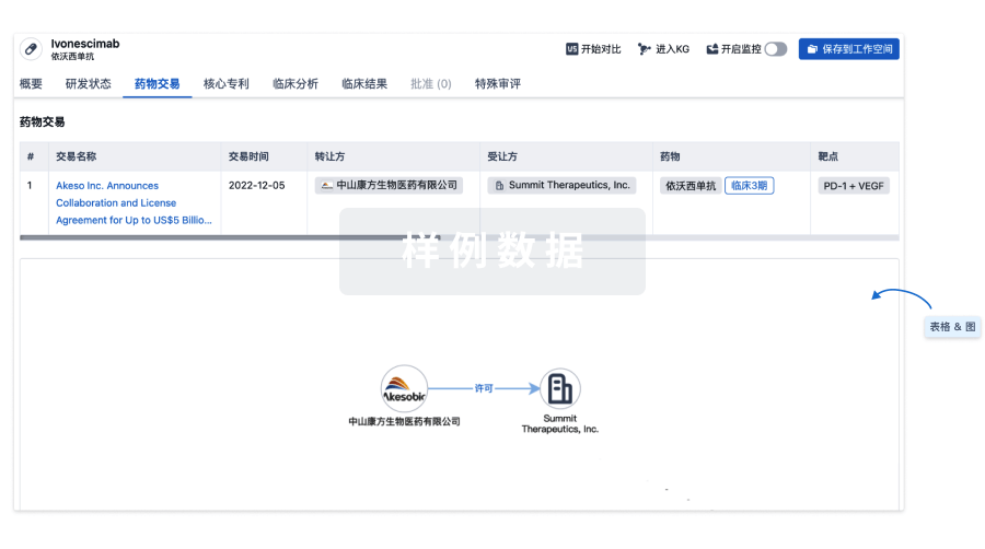
药物交易
使用我们的药物交易数据加速您的研究。
登录
或
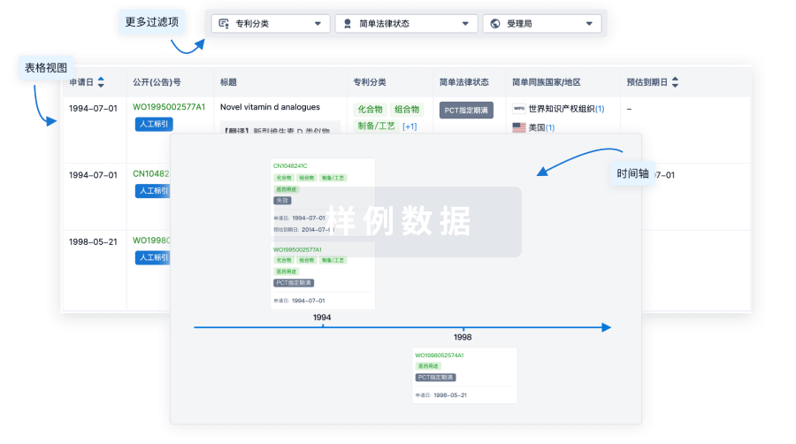
核心专利
使用我们的核心专利数据促进您的研究。
登录
或
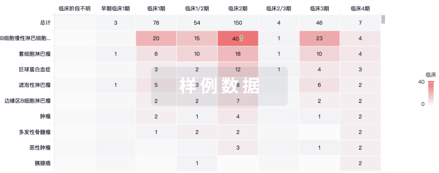
临床分析
紧跟全球注册中心的最新临床试验。
登录
或
批准
利用最新的监管批准信息加速您的研究。
登录
或
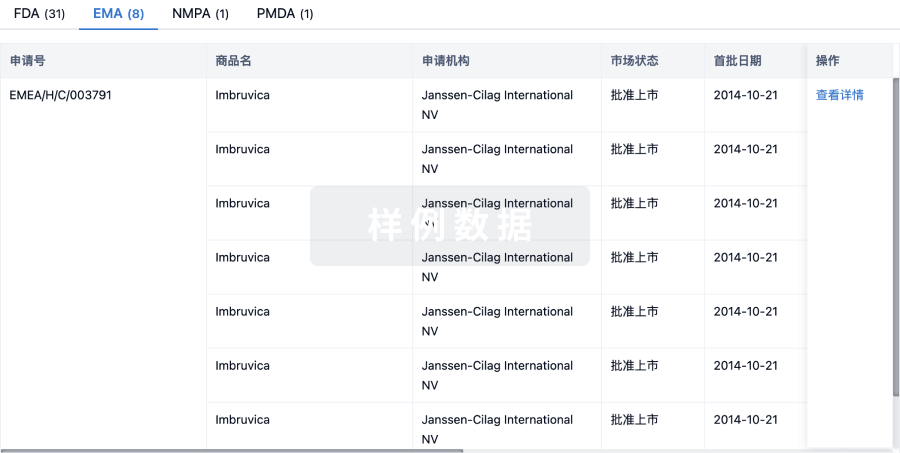
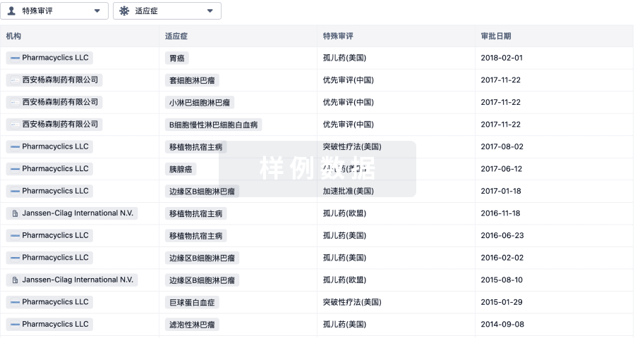
特殊审评
只需点击几下即可了解关键药物信息。
登录
或
提出任何生物医药研究问题
让每一步生物医药决策都精准制胜
基于十亿级生物数据与全球情报的精准 AI,赋能早期药物发现
立即开始免费试用!
智慧芽新药情报库是智慧芽专为生命科学人士构建的基于AI的创新药情报平台,助您全方位提升您的研发与决策效率。
立即接入生物医药数据包
智慧芽新药库数据也通过智慧芽开放平台,以MCP或API服务形式对外开放。立即使用我们的MCP服务定制您的LLM智能体。
生物序列数据库
生物药研发创新
免费使用
化学结构数据库
小分子化药研发创新
免费使用